Presentation1.pptx, radiological imaging of anteversion angle.
•Download as PPTX, PDF•
21 likes•4,156 views


Recommended
More Related Content
What's hot
What's hot (20)
Viewers also liked
Viewers also liked (20)
Similar to Presentation1.pptx, radiological imaging of anteversion angle.
Similar to Presentation1.pptx, radiological imaging of anteversion angle. (20)
More from Abdellah Nazeer
More from Abdellah Nazeer (20)
Presentation1.pptx, radiological imaging of anteversion angle.
- 1. Dr/ ABD ALLAH NAZEER. MD. Radiological Imaging of Anteversion angle.
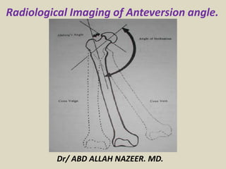
- 2. Femoral neck anteversion is defined as the angle between an imaginary transverse line that runs medially to laterally through the knee joint and an imaginary transverse line passing through the center of the femoral head and neck . In adults without pathology, the femur is twisted so the head and neck of the femur are angled forward between 15 and 20 degrees from the frontal plane of the body. In some instances, the FNA angle is directed forward or backward well beyond this angle. Some researchers suggest that FNA angles outside this 15- to 20-degree average are a contributing factor in many different orthopedic problems in the lower extremity that are commonly seen by physical therapists. The purpose of this Update is to describe how FNA is related to hip rotation, how hip rotation range of motion can be used to predict abnormal FNA, and how asymmetries in hip rotation may be used to identify patients who may be at risk for developing various orthopedic problems in the hip and lower extremity.
- 4. What causes femoral anteversion? Femoral anteversion can be the result of stiff hip muscles due to the position of the baby in the uterus. It also has a tendency to run in families. Typically, a child's walking style looks like that of his or her parents. When the child is first learning how to walk, femoral anteversion can create an intoeing appearance. As the knees and feet turn in, the legs look like they are bowed. The bowed leg stance actually helps the child achieve greater balance as they stand. Balance is not as steady when they try to stand and walk with their feet close together or with their feet turned out. This may cause them to trip and fall. How is femoral anteversion diagnosed? The diagnosis of femoral anteversion is made by a history and physical examination by your child's doctor. During the examination, the doctor obtains a complete prenatal and birth history of the child and asks if other family members are known to have femoral anteversion. Generally, no X-rays are necessary.
- 5. CT protocols: Femoral anteversion: This limited study allow the radiologist to measure the angle of rotation of femoral neck relative to the femoral condyles bilaterally. A secondary measurement is femoral lengths, made by calculating the difference in table position at the ends of the bones. Radiographic Scanogram: This is a radiographic study in which coned-down images of both hips, knees &ankles are shot on a single conventional-sized film(or CR plate) with a radiopaque ruler in place. The sole purpose of this study is to measure leg- length. This is a radiographic study in which both leg are images in their entities from hip to ankle on a single long film using a scoliosis cassette. Typically, these are used by orthopedic surgeons for planning purposes.
- 8. CT protocol for femoral anteversion measurement. Measure right and left side individually, Find the slice that reveals the alignment of the femoral neck, Measure the neck-Horizontal angle, Find the slice that best reveals the alignment of the femoral condyles, measure the condyle- Horizontal angle and calculate the angle of the neck relative to the condyles.
- 10. Thank You.