Design of artificial respiratory model
•Download as PPTX, PDF•
0 likes•92 views

Design of Artificial Respiratory Model.. Know about the respiratory system. The respiratory system consists of the upper respiratory tract (nasal passages), the airway conduction system (larynx, trachea, bronchi, bronchioles and terminal bronchioles), and the lower respiratory tract (alveolar ducts and alveoli). Not all segments of the respiratory system mature at the same pace. The olfactory epithelium matures earliest by PND 7. The lung, however, is not considered mature until PND 21, when alveolarization and microvascular maturation are complete. This chapter will discuss the embryological development (briefly), adult histomorphology, and postnatal histologic development of each major component of the respiratory system.

Recommended
Recommended
More Related Content
What's hot
What's hot (20)
Similar to Design of artificial respiratory model
Similar to Design of artificial respiratory model (20)
Recently uploaded
Recently uploaded (20)
Design of artificial respiratory model
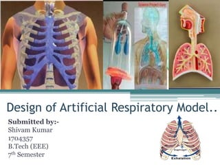
- 1. Design of Artificial Respiratory Model.. Submitted by:- Shivam Kumar 1704357 B.Tech (EEE) 7th Semester
- 2. Content. . • Introduction of Respiratory System • Artificial Respiratory System • Working of Respiratory System • Parts of the respiratory system • What does the respiratory system do? • Comparison between Spontaneous Respiration and Artificial Ventilation • Conclusion • References
- 3. Introduction of Respiratory System The respiratory system consists of the upper respiratory tract (nasal passages), the airway conduction system (larynx, trachea, bronchi, bronchioles and terminal bronchioles), and the lower respiratory tract (alveolar ducts and alveoli). Not all segments of the respiratory system mature at the same pace. The olfactory epithelium matures earliest by PND 7. The lung, however, is not considered mature until PND 21, when alveolarization and microvascular maturation are complete. This chapter will discuss the embryological development (briefly), adult histomorphology, and postnatal histologic development of each major component of the respiratory system.
- 4. Artificial Respiratory System Artificial respiration, breathing induced by some manipulative technique when natural respiration has ceased or is faltering. Such techniques, if applied quickly and properly, can prevent some deaths from drowning, choking, strangulation, suffocation, carbon monoxide poisoning, and electric shock. Resuscitation by inducing artificial respiration consists chiefly of two actions: • Establishing and maintaining an open air passage from the upper respiratory tract (mouth, throat, and pharynx) to the lungs • Exchanging air and carbon dioxide in the terminal air sacs of the lungs while the heart is still functioning.
- 5. Working of Respiratory System • The primary organs of the respiratory system are the lungs, which function to take in oxygen and expel carbon dioxide as we breathe. • The gas exchange process is performed by the lungs and respiratory system. Air, a mix of oxygen and other gases, is inhaled. • In the throat, the trachea, or windpipe, filters the air. The trachea branches into two bronchi, tubes that lead to the lungs. • Once in the lungs, oxygen is moved into the bloodstream. Blood carries the oxygen through the body to where it is needed. • Red blood cells collect carbon dioxide from the body’s cells and transports it back to the lungs. • An exchange of oxygen and carbon dioxide takes place in the alveoli, small structures within the lungs. The carbon dioxide, a waste gas, is exhaled and the cycle begins again with the next breath.
- 6. parts of the respiratory system The respiratory system has many different parts that work together to help you breathe. Each group of parts has many separate components. Your airways deliver air to your lungs. Your airways are a complicated system that includes your: • Mouth and nose: Openings that pull air from outside your body into your respiratory system. • Sinuses: Hollow areas between the bones in your head that help regulate the temperature and humidity of the air you inhale. • Pharynx (throat): Tube that delivers air from your mouth and nose to the trachea (windpipe). • Trachea: Passage connecting your throat and lungs. • Bronchial tubes: Tubes at the bottom of your windpipe that connect into each lung. • Lungs: Two organs that remove oxygen from the air and pass it into your blood.
- 7. From your lungs, your bloodstream delivers oxygen to all your organs and other tissues. Muscles and bones help move the air you inhale into and out of your lungs. Some of the bones and muscles in the respiratory system include your: • Diaphragm: Muscle that helps your lungs pull in air and push it out • Ribs: Bones that surround and protect your lungs and heart When you breathe out, your blood carries carbon dioxide and other waste out of the body. Other components that work with the lungs and blood vessels include: • Alveoli: Tiny air sacs in the lungs where the exchange of oxygen and carbon dioxide takes place. • Bronchioles: Small branches of the bronchial tubes that lead to the alveoli. • Capillaries: Blood vessels in the alveoli walls that move oxygen and carbon dioxide.
- 8. • Lung lobes: Sections of the lungs – three lobes in the right lung and two in the left lung. • Pleura: Thin sacs that surround each lung lobe and separate your lungs from the chest wall. Some of the other components of your respiratory system include: • Cilia: Tiny hairs that move in a wave-like motion to filter dust and other irritants out of your airways. • Epiglottis: Tissue flap at the entrance to the trachea that closes when you swallow to keep food and liquids out of your airway. • Larynx (voice box): Hollow organ that allows you to talk and make sounds when air moves in and out.
- 9. What does the respiratory systemdo? The respiratory system has many functions. Besides helping you inhale (breathe in) and exhale (breathe out), it: • Allows you to talk and to smell. • Brings air to body temperature and moisturizes it to the humidity level your body needs. • Delivers oxygen to the cells in your body. • Removes waste gases, including carbon dioxide, from the body when you exhale. • Protects your airways from harmful substances and irritants.
- 10. Comparison between Spontaneous Respiration and Artificial Ventilation Spontaneous respiration and artificial ventilation have been compared in five supine healthy subjects. • Ventilation volumes, gaseous exchange volumes and arterial blood gas tensions were first measured with the subject breathing spontaneously. The measurements were then repeated with the subject anaesthetized, paralyzed and ventilated artificially via an endotracheal tube at a rate and depth determined by the findings during spontaneous breathing. • Ventilation-blood flow relationships were assessed by measuring the dead space and the alveolar-arterial (A-a) O3 tension gradient. • When ventilated artificially the group as a whole showed a highly significant increase in dead space but little increase in A-a PO, gradient, implying over ventilation of parts of the lungs which have a small pulmonary blood flow. There were, however, considerable individual differences suggesting that, even in normal supine subjects with intact thoracic and abdominal walls, various abnormal patterns of ventilation- blood flow distribution may occur during artificial ventilation. • Measurements of compliance and metabolic rate were in agreement with those previously reported. • The clinical implications of the findings are discussed, and the simple mathematical relationships between ventilation, dead space and arterial Pco2 are emphasized.
- 11. Conclusion. . There is increasing demand for technologies that can assist an injured or recently transplanted lung or completely replace the native lung. Such a device should allow for ambulation in chronic or acute respiratory failure, either as a bridge to lung transplantation or as a true destination therapy. Extracorporeal lung support devices have undergone significant advancement in the last several years. These changes have led to more miniaturized circuits that are increasingly efficient at gas exchange while decreasing the complication rates. Hollow fiber membrane gas exchange enables longer term wearable systems to be used in ambulatory and awake patients. Biomimetic devices based on micro channel technologies may even enable intracorporeal implantation. The continuous progress in the development of bioartificial lungs includes advances in both tissue engineering and cell‐based technologies. The future trials may start testing transplantation of one single bioengineered lung lobe using it as a transient therapy. The final goal is to achieve a truly implantable artificial lung, it might be also a regenerated lung, or both, that is applicable for destination therapy.
- 12. References. . Web Articles/Journals:- • A comparison of artificial ventilation and spontaneous respiration with particular reference to ventilation blood flow relationship by E. M. J. Campbell – 1958 • Artificial lungs––Where are we going with the lung replacement therapy by Norihisa Shigemur- 23 oct 220 Web-sites:- • https://my.clevelandclinic.org/health/articles/21205-respiratory- system Links:- ▫ https://www.google.com/search?q=respiratory%20system%20model&tbm=isch&rlz= 1C1CHBD_enIN917IN917&hl=en- GB&sa=X&ved=0CCMQtI8BKAFqFwoTCKD2pJauxu0CFQAAAAAdAAAAABAP&biw =1349&bih=695#imgrc=APesI_XMhdzdxM ▫ https://my.clevelandclinic.org/health/articles/21205-respiratory- system#:~:text=The%20respiratory%20system%20is%20the,waste%20gases%20like %20carbon%20dioxide.