SlideShare a Scribd company logo
1 of 18
Sri Siddhartha Medical
College Agalkote,
Tumakuru.
Department of Orthopaedics.
SUBJECT SEMINAR ON: Flexors &
extensor tendon injuries of hand.
Chairperson& Moderator: Prof, & HOD: Dr Kiran kalaiah
Presented by: Dr Yashavardhan T.M
On: 30/03/2017
Introduction.
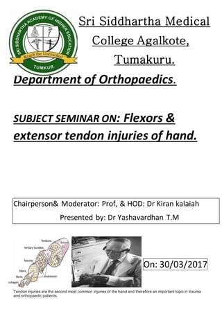
Tendon injuries are the second most common injuries of the hand and therefore an important topic in trauma
and orthopaedic patients.
“A glistening structure between muscle & bone which transmit force from muscle to the bone’”
Paratenon: Loose areolar tissue encasing tendon in low mechanical stress area
Tendon sheath: a dense fibrous tissue tunnel enclosing tendon in high mechanical stress area
70% collagen (Type I)
Extracellular components
 Elastin
 Mucopolysaccharides (enhance water-binding capability)
 Endotenon – around collagen bundles
 Epitenon – covers surface of tendon
 Paratenon – visceral/parietal adventitia surrounding tendons in hand.
Most injuries are open injuries to the flexor or extensor tendons, but less frequent injuries, e.g., damage to the
functional system tendon sheath and pulley or dull avulsions, also need to be considered.
After clinical examination, ultrasound and magnetic resonance imaging have proved to be important diagnostic
tools. Tendon injuries mostly require surgical repair, dull avulsions of the distal phalanges extensor tendon can
receive conservative therapy.
Injuries of the flexor tendon sheath or single pulley injuries are treated conservatively and multiple pulley injuries
receive surgical repair. In the postoperative course of flexor tendon injuries, the principle of early passive
movement is important to trigger an "intrinsic" tendon healing to guarantee a good outcome.
Many substances were evaluated to see if they improved tendon healing; however, little evidence was found.
Nevertheless, hyaluronic acid may improve intrinsic tendon healing.
Anatomical position
Flexors muscles: FDP and FDS tendons have fibrous sheaths on the palmar aspect of the digits
Fibrous arches and cruciate (cross-shaped) ligaments, which are attached posteriorly to the margins of the
phalanges and to the palmar ligaments hold the tendons to the bony plane and prevent the tendons from bowing
when the digits are flexed. The tendons are surrounded by a synovial sheath.
Flexor tendon system consists of intrinsic and extrinsic components
Extrinsics:
FDP: flexing the DIP joint
FDS: Flexing the PIP Joint
FPL: Flexing the IP joint of the thumb
Intrinsics:
Lumbricals: Flex the MCP joints and Extend the IP joints
FDP inserts on base of distal phalanx
FDS inserts on sides of middle phalanx
FPL inserts on proximal portion of the distal phalanx
INTEROSSEIUS
BLOOD SUPPLY
Pulleysof flexiours
Synovial sheathisreinforcedbyasystemof fibrouspulleys
5 annular pulleys(A) and
3 Cruciformpulleys (C)
A1: 8-10 mm overMCPJ
A2: 18-20mm overproximal phalanx
A3: 2-4 mm overPIPJ
A4: 10-12mm overmiddle phalanx
A5: 2-4 mm overDIPJ
C1, C2, C3 proximal toA3, A4, A5
Allowshorteningof the pulleysysteminflexion
A2 and A4 are consideredmostimportant.Theirdisruption
leadstobowstringing,reducedmechanical efficiencyanddecreasedflexion.
Function:increase the mechanical efficiencybypreventingbowstringing
PULLEY BIOMECHANICS
ZONES OF FLEXOR TENDON INJURY
Zone I: Betweeninsertionof FDPandFDS
Zone II: Frominsertionof FDSto A1 Pulley
Zone III:BetweenA1pulleyanddistal limitof carpal tunnel
Zone IV:Withinthe carpal tunnel
Zone V: Betweenthe entrance of Carpal tunnel andmusculo-tendinousjunction.
FDS decussation at A1 pulley FDS slips rotate 180° around FDP
Slips rejoin at PIP – Camper’s Chiasma Insert on P2
Tendon nutrition
Parietal paratenon
Passive nutrition by diffusion
Vincula and bony attachments
Direct nutrition
Segmental nutrition
Vincula may prevent retraction
Vascularity dominance is deep surface of tendon
Consider with suture placement
Biomechanically superior to place suture deep
TENDON HEALING
Tendonsare capable of activelyparticipatinginthe repairprocessthroughIntrinsicHealing
Tendonhealingoccursinthree phases:
1. Inflammation 2. Active repair 3. Remodelling
 Early tendon motion has significant role in modifying the repair response
 Mobilized tendons showed progressively greater ultimate load compared with immobilized tendons
 Studies confirm “Wolff’s law” which states that the strength of a healing tendon is proportional to
the controlled stress applied to it
ETIOLOGY
 sharp object direct laceration (broken glass, kitchen knives or table saws)
 crush injury
 avulsions
 burns
 animal or human bites
 suicide attempts
 motor vehicle accidents
ZONE 1: ZONE OF FDP AVULSIONINJURIES
 Region b/w middle aspects of middle phalanx to finger tips
 Contains only one tendon-fdp
 Tendon laceration occurs close to its insertion
 Tendon to bone repair is required than tendon repair
Leddy classificationofzoneI flexortendon injuries
Type I: tendonretractedintopalm(fullnessinpalm)
Type II: tendontrappedinthe sheathat PIP(unable toflex PIP)
Type III:tendontrappedinA4 pully
ZONE II-NOMANS LAND
 From metacarpal head to middle phalanx
 Called so because initial attempts for tendon repair here produced poor results
 FDS and FDP within one sheath
 Adhesion formation risk is amplified at campers chiasma
ZONE III-DISTAL PALMAR CREASE
 B/w transverse carpal ligament and proximal margin of tendon sheath formation
 Lumbricals origin here prevents profundus tendons from over acting
 Delayedtendon repairs are succesfull evenafter several weeks of injury
ZONE IV-TRANSVERSE CARPAL LIGAMENT
Liesdeepto deeptransverse ligament. Tendoninjuriesare rare.
ZONE V LIES PROXIMAL TO TRANSVERSE CARPAL LIGAMENT
SIGNS& SYMPTOMS
 Unable to bend one or more finger joints
 Pain when bending finger/s
 Open injury to hand (e.g., cut on palm side of hand, particularly in area where skin folds as fingers
bend)
 Mild swelling over joint closest to fingertip
 Tenderness along effected finger/s on palm side of hand
 Lies deep to deep transverse ligament Tendon injuries are rare
EXAMINATION:
 INSPECTION
There isa normal arcade to hand withindex fingershowingleastandlittle fingershowingmax flexion If
affectedfingershowsmore extensionthanotherdigits,chance of tendoninjuriesare high
 Goals of reconstruction:
 Coaptation of tendons
 anatomical repair
 multiple strand repair to permit active range of motion rehabilitation
 Pully reconstruction to minimize bow-stringing
 atraumatic surgical technique to minimize adhesions
 strict adherence to rehabilitation protocol.
Timingof flexortendon repair: Primary:repairwithin24 hours(contraindicatedincase of highgrade contamination
i.e.humanbites,infection)
 Delayed Primary: 1-10 days when the wound can be still pulled open without incision
 Early Secondary: 2-4 weeks.
 Late Secondary : after 4 weeks
 No repair if less than <25% laceration,
 only epitenon repair in 25-50% lacerations,
 core suture plus epitenon repair when >50% laceration
 Dorsal blocking splint for 6-8 weeks as conservative measure
Tendon sheathrepair:
Advantages:
 barrier to the formation of extrinsic adhesions
 quicker return of synovial nutrition
 better tendon-sheath biomechanics
Disadvantages:
 technically difficult.
 may narrow and restrict tendon gliding.
ZONE 1 REPAIR
 WOUND EXTENDED PROXIMALLY AND DISTALLY
 PROXIMAL TENDON RETRIEVED, CORE SUTURES ARE PLACED
 KEITH NEEDLES USED TO PASS THE SUTURES AROUND THE DISTAL PHALANX
EXITING THROUGH NAIL PLATE DISTALLY
 REMAINING DISTAL END OF TENDON SUTURED TO THE RE-ATTACHED PROXIMAL
PORTION.
Directrepair: if lacerationismore than1 cm fromFDP insertion
Tendonadvancement: if the lacerationislessthen1cm frominsertion
ZONE II REPAIRS
 REPAIR BOTH TENDON LACERATIONS
 TENDON SHEATH MAY BE OPENED FOR EXPOSURE
BUT A2 AND A4 ARE PRESERVED AS MUCH AS POSSIBLE
 FDS IS REPAIRED FIRST FOLLOWED BY FDP
 Try to milk the tendon with the wrist flexed
 Morris and Martin. single skin hook is carefully inserted into sheath, then the hook is then turned
toward the tendons and when it is secured to the tendon, withdrawal of the hook should retrieve both
tendons
 Sourmelis and McGrouther. a small catheter is passed into the sheath and is delivered proximally
into a small wound in the palm, just proximal to the A1 pulley, the catheter is sutured to both tendons
2 cm proximal to A1 pulley, which is then pulled distally to deliver the tendons into the synovial
window
ZONE III REPAIRS
 If both tendons are lacerated, both are repaired, end to end with
circumferential re-enforcing sutures
 May affect lumbricals in addition to flexor tendons
 Damaged lumbrical is either repaired or excised depending on severity of injury and the location of
the laceration
ZONE IV REPAIR
 Lacerations of flexor tendons within the carpal canal are typically associated with partial or complete
laceration of median nerve
 Here median nerves should be repaired first and the tendons last.
ZONE V REPAIR
 In this area there may be concomitant ulnar nerve & artery damage as well as radial artery & median
nerve damage.
 Primary repair of the arteries is usually indicated
 If wound is contaminated, arteries are repaired and delayed repair of tendons and nerves is planned
Zone 3 injuries
Lumbrical muscle belliesusuallyare notsuturedbecause thiscanincrease the tension
of these musclesandresultina“lumbrical plus”finger(paradoxical proximal
interphalangeal extensiononattemptedactive fingerflexion).
Quadriga effect
Tendonadvancementshortensthe FDP& completesthe gripbefore the normal fingers,if the tensionontendon
graft issettoo high,and limittheirflexionandthusweekgrip
Tendon Repair:
Silfverskiöld
Fish-MouthEnd-to-End Suture (Pulvertaft)
End-to-Side method
Most of them involve active motion exercises. Then the suture strength has to increase
 Active Extention-Rubber Band Flexion Method: e.g. Kleinert , and Brooke-Army
 Immobilization
 Controlled Passive Motion Methods: e.g. Duran’s protocol
 Strickland: Early active ROM
Kleinert Protocol
 Combines dorsal extension block with rubber-band traction proximal to wrist
 Originally, included a nylon loop placed through the nail, and around the nail is placed a rubber band
 This passively flexes fingers, & the patient actively extends within the limits of the splint
Duran protocol
At surgery, a dorsal extension-block splint is applied with the wrist at 20-30° of flexion, the MCP joints at
50-60° of flexion, and the IP joints straight
Active range of motion rehabilitation
Kleinert !!
Complications
 Joint contracture
 Adhesions
 Rupture
 Bowstringing
 Infection
Adhesions&stiffnessrequirestenolysisin18-25% cases
Tenolysisis indicatedafter3monthsif no improvementisnoted For1-2 months extensivephysiotherapy
DONOR TENDONS FOR GRAFTING
Palmaris Longus: Tendon of choice (fulfils requirement of length, diam & availability)
Plantaris Tendon: Equally satisfactory & advantageof being almost twice as long, but is not
accessible. Others: FDS, EDC
Extensor Tendons
1. Extrinsic System radial N innervated
2. Intrinsic System ulnar and median N innervated
Extrinsic Extensors
 Wrist Extensors: ECRL, ECRB, ECU
 Finger Extensors: EDC, EIP, EDQM
 Thumb Extensors: APL, EPL, EPB
EDC has a common muscle bellywithmultipletendons
EIP & EDM lie onthe ulnarside of the respective EDCtendon
Thumb Extensors
APL inserts on the metacarpal and radially abducts it
EPB inserts on proximal phalanx and extends MCP Joint
EPL inserts on distal phalanx and extends IP Joint
Testing the Extrinsics:
APL:Palpate with thumb abduction
EPB:MP extension with IP flexion, palpate tendon
EPL:Palpate tendon with retropulsed thumb
EDC:Test with wrist in neutral-extension
Testing the Extrinsics
Vascularity & Innervation
 Volar and dorsal metacarpalvessels
 Median nerve supplies radial one or 2 lumbricals
 Ulnar nerve supplies ulnar 2 lumbricals
Extensor Apparatus
EDC tendon trifurcates into central slip & 2 lateral slips
Intrinsic extensor tendons join the lateral slips to form the lateral bands
Winslow’sRhombus
The central slip inserting on the baseof the middle-phalanx
and two lateral slips inserting to the distal-phalanx.
Lateral band:
Juncturae Tendinium
Functional roles:
spacing of ED tendons force redistribution coordinate extension MP stabilization
Ring finger has least independent extension due to the orientation of the juncturae
The most common patterns single extensor indicis proprius inserting to the ulnar side of the index extensor
digitorum communis a single extensor digitorum communis to the index finger a single extensor digitorum
communis to the long finger, a double extensor digitorum communis to the ring finger, an absent extensor
digitorum communis to the small finger, and a double extensor digiti quinti with double insertions.
SAGITTAL BANDS
Stabilize the common extensor during digital flexion over MCPJ
Limit the excursion of the common extensor tendon during digital extension
EDC allows extension of MP joint via insertion onto the sagittal bands
There is usually no tendinous insertion of EDC to the dorsal base of the proximal phalanx
No MP joint hyperextension: EDC extends MP, PIP, and DIP joints even in the absence of intrinsic muscle
function.
INTRINSIC PARALYSIS: “slack” develops in EDC systemdistal to the sagittal bands all producing a flexion
posture at PIP and DIP joints, the “claw” finger.
Transverse &oblique fibres of Interosseous Hood
1) EDC Tendon
2) Central Slip
3) Lateral Slip
4) Intertendinous Connection
5) Volar Interosseous Muscle
6) Lumbrical Muscle
Triangular Ligament
Connectsbothlateral bandsoverthe middle phalanx.
 Limits the volar and lateral shifting of the
lateral conjoined extensor tendon during
digital flexion
 In boutonniere deformity; elongated
 In fixed swan neck deformity; retracted
Retinacular Ligament
Lateral continuation of the triangular ligament extending from the lateral margin of the lateral conjoined
extensor tendon to PIPJ articular volar plat
Extensor Tendon Injury.
Extensor apparatusExtrinsic muscles (ED, EI, EDM)
Intrinsic Muscles ( Lumbricals and Interossei)
Fixed fibrous structures.
Zone 1
 Mallet finger – persistent flexon of distal phalanx
 Closed: splinting 6-8 weeks
 Open: suture repair, Soft tissue reconstruction
Zone II injury- Middle Phalanx Level:
 Repair by interrupted suture.
 Immobilization for 5-6 weeks
 DIP joint in extension
 PIP joint left free
Zone III injury- PIP joint level
Most complex anatomically and physiologically
Causes two deformities
Boutonniere disruption of central tendon
Closed: splinting MCP and PIP in hyperextension for 6 weeks
Open: suture repair (figure of 8 suture)
Swan Neck excessive traction of central tendon
Closed: splinting DIP & Open: suture repair
Zone IV injury- shaft of proximal phalanx level
Repair relatively easy Adhesion is the problem.
Zone V injury – MP joint level.
Closed: splinting, 45 extension at wrist and 20
flexion at MCP & Open: suture repair by 5.0 prolene
Zone VI injury- Metacarpal level
Better prognosis than in fingers
All structures, even inter-tendinous band should be repaired.
Core type suture possible. Delayed suture is possible.
Zone VII- wrist level
Extensor tendons are under dorsal retinaculum. Retinaculum should be repaired
or partially preserved. Adhesion is the problem Grasping core suture should be
used. Immobilization for 5-6 weeks.
IMMOBILIZATION
INJURIES IN ZONES PROXIMAL TO MCPs
 May be immobilized for 3 weeks.
 Afterwards, finger may be placed in removable volar splint between exercise periods for 2 weeks
 Progressive ROM after 3 weeks
 If full flexion is not regained rapidly, dynamic flexion may be started after 6 weeks
INJURIES IN ZONES DISTAL TO MCPs
 Require a longer period of immobilization (usually 6 weeks)
 A progressive exercise program is initiated
 Dynamic splinting during day and static splinting at night to maintain extension
Injury to Thumb Extensor
Zone I and II
Mallet injuries are rare
Operative treatment is a good option
Zone V – VII
MCP area is designated zone V
Extensor lag usually minimal
Proximal to zone V, EPL retracts far
Repair >1mo requires rerouting EPL from Listers tubercle.
REFERENCES.
1. Campbel's 0perative 0rthopaedics 13th.
2. GREENS HAND TEXTBOOK.
3. AO-ASIF MANUAL OF ORTHOPAEDICS.
4. GRAYS HUMAN ANOTOMY TEXTBOOK.
THANK YOU!!!

More Related Content

What's hot

Hand rehabilitation after flexor tendon repair
Hand rehabilitation after flexor tendon repairHand rehabilitation after flexor tendon repair
Hand rehabilitation after flexor tendon repairMohammed Aljodah
 
RADIAL NERVE PALSY AND TENDON TRANSFER
RADIAL NERVE PALSY AND TENDON TRANSFERRADIAL NERVE PALSY AND TENDON TRANSFER
RADIAL NERVE PALSY AND TENDON TRANSFERBenthungo Tungoe
 
ULNAR NERVE PALSY AND TENDON TRANSFERS
ULNAR NERVE PALSY AND TENDON TRANSFERSULNAR NERVE PALSY AND TENDON TRANSFERS
ULNAR NERVE PALSY AND TENDON TRANSFERSBenthungo Tungoe
 
Extensor tendon injuries
Extensor tendon injuriesExtensor tendon injuries
Extensor tendon injuriesmhmad farooq
 
Flexor tendon injuries
Flexor tendon injuriesFlexor tendon injuries
Flexor tendon injuriesMohamed Eldeeb
 
Hand rehabilitation following flexor tendon injuries
Hand rehabilitation following flexor tendon injuriesHand rehabilitation following flexor tendon injuries
Hand rehabilitation following flexor tendon injuriesAbey P Rajan
 
Extensor tendons injury repair and rehabilitation
 Extensor tendons injury repair and rehabilitation Extensor tendons injury repair and rehabilitation
Extensor tendons injury repair and rehabilitationMohammed Aljodah
 
Flexor tendon injuries.m
Flexor tendon injuries.mFlexor tendon injuries.m
Flexor tendon injuries.mMansoor Khan
 
Flexor tendon injuries
Flexor tendon injuriesFlexor tendon injuries
Flexor tendon injuriesDeepak Kumar
 
Phalangeal fractures of hand
Phalangeal fractures of handPhalangeal fractures of hand
Phalangeal fractures of handDarshan K Swamy
 
Extensor tendon injuries hand
Extensor tendon injuries handExtensor tendon injuries hand
Extensor tendon injuries handNousfierNuchu
 
Flexor tendon injuries(1)
Flexor tendon injuries(1)Flexor tendon injuries(1)
Flexor tendon injuries(1)orthoprince
 
Tendon transfer for radial nerve palsy
Tendon transfer for radial nerve palsyTendon transfer for radial nerve palsy
Tendon transfer for radial nerve palsyMohammed Aljodah
 

What's hot (20)

Hand rehabilitation after flexor tendon repair
Hand rehabilitation after flexor tendon repairHand rehabilitation after flexor tendon repair
Hand rehabilitation after flexor tendon repair
 
Flexor tendon repair
Flexor tendon repairFlexor tendon repair
Flexor tendon repair
 
05 hand tendon
05 hand tendon05 hand tendon
05 hand tendon
 
RADIAL NERVE PALSY AND TENDON TRANSFER
RADIAL NERVE PALSY AND TENDON TRANSFERRADIAL NERVE PALSY AND TENDON TRANSFER
RADIAL NERVE PALSY AND TENDON TRANSFER
 
ULNAR NERVE PALSY AND TENDON TRANSFERS
ULNAR NERVE PALSY AND TENDON TRANSFERSULNAR NERVE PALSY AND TENDON TRANSFERS
ULNAR NERVE PALSY AND TENDON TRANSFERS
 
Extensor tendon injuries
Extensor tendon injuriesExtensor tendon injuries
Extensor tendon injuries
 
Flexor tendon injuries
Flexor tendon injuriesFlexor tendon injuries
Flexor tendon injuries
 
Hand rehabilitation following flexor tendon injuries
Hand rehabilitation following flexor tendon injuriesHand rehabilitation following flexor tendon injuries
Hand rehabilitation following flexor tendon injuries
 
Extensor tendons injury repair and rehabilitation
 Extensor tendons injury repair and rehabilitation Extensor tendons injury repair and rehabilitation
Extensor tendons injury repair and rehabilitation
 
management of claw hand
management of claw handmanagement of claw hand
management of claw hand
 
Pollicization
PollicizationPollicization
Pollicization
 
Swan neck-deformity
Swan neck-deformitySwan neck-deformity
Swan neck-deformity
 
Flexor tendon injuries.m
Flexor tendon injuries.mFlexor tendon injuries.m
Flexor tendon injuries.m
 
Flexor tendon injuries
Flexor tendon injuriesFlexor tendon injuries
Flexor tendon injuries
 
Phalangeal fractures of hand
Phalangeal fractures of handPhalangeal fractures of hand
Phalangeal fractures of hand
 
Extensor tendon injuries hand
Extensor tendon injuries handExtensor tendon injuries hand
Extensor tendon injuries hand
 
Tendon transfer- principles and techniques
Tendon transfer- principles and techniquesTendon transfer- principles and techniques
Tendon transfer- principles and techniques
 
Flexor tendon injuries(1)
Flexor tendon injuries(1)Flexor tendon injuries(1)
Flexor tendon injuries(1)
 
Tendon repair
Tendon repairTendon repair
Tendon repair
 
Tendon transfer for radial nerve palsy
Tendon transfer for radial nerve palsyTendon transfer for radial nerve palsy
Tendon transfer for radial nerve palsy
 

Similar to Tendon injury by dr yash

Flexor tendon injury final edit with pictures
Flexor tendon injury final edit with picturesFlexor tendon injury final edit with pictures
Flexor tendon injury final edit with picturesGautam Kalra
 
Tendon injuries of hand
Tendon injuries of handTendon injuries of hand
Tendon injuries of handrohit raj
 
Jc flexor tendon injury, repair &amp; rehabilitaion
Jc flexor tendon injury, repair &amp; rehabilitaionJc flexor tendon injury, repair &amp; rehabilitaion
Jc flexor tendon injury, repair &amp; rehabilitaionLove2jaipal
 
Hand Flexor tendon injury.pdf
Hand Flexor tendon injury.pdfHand Flexor tendon injury.pdf
Hand Flexor tendon injury.pdfmhmad farooq
 
Extensorapparatusofhandinjuries
Extensorapparatusofhandinjuries Extensorapparatusofhandinjuries
Extensorapparatusofhandinjuries Dr. Mohit Sharma
 
Acute flexor tendon injuries z 1
Acute flexor tendon injuries z 1Acute flexor tendon injuries z 1
Acute flexor tendon injuries z 1Ashraf Abdelaziz
 
Principle of Fracture Management
Principle of Fracture ManagementPrinciple of Fracture Management
Principle of Fracture ManagementQubezo
 
CRUSH INJURIES OF THE HAND.ppt
CRUSH INJURIES OF THE HAND.pptCRUSH INJURIES OF THE HAND.ppt
CRUSH INJURIES OF THE HAND.pptAmina Rajah
 
CRUSH INJURIES OF THE HAND.ppt
CRUSH INJURIES OF THE HAND.pptCRUSH INJURIES OF THE HAND.ppt
CRUSH INJURIES OF THE HAND.pptAminaRajah1
 
Lis Franc Injury
Lis Franc InjuryLis Franc Injury
Lis Franc Injuryjfreshour
 
Fracture of shaft and distal part of Femoral bone by Dr. Ammar Alsabae
Fracture of shaft and distal part  of Femoral bone by Dr. Ammar AlsabaeFracture of shaft and distal part  of Femoral bone by Dr. Ammar Alsabae
Fracture of shaft and distal part of Femoral bone by Dr. Ammar AlsabaeAmmar Alsbae
 
Extensor apparatus of hand injuries
Extensor apparatus of hand injuriesExtensor apparatus of hand injuries
Extensor apparatus of hand injuriesShamendra Sahu
 
Thumb reconstruction by conventional technique.
Thumb reconstruction by conventional technique.Thumb reconstruction by conventional technique.
Thumb reconstruction by conventional technique.Dr. Suiyibangbe
 
Hand Injuries & Their Managements
Hand Injuries & Their ManagementsHand Injuries & Their Managements
Hand Injuries & Their ManagementsRobert G. Mulbah
 
The effect of intact fibula on functional outcome of reamed intramedullary in...
The effect of intact fibula on functional outcome of reamed intramedullary in...The effect of intact fibula on functional outcome of reamed intramedullary in...
The effect of intact fibula on functional outcome of reamed intramedullary in...Love2jaipal
 
Sports related injuries to hand sagar
Sports related injuries to hand   sagarSports related injuries to hand   sagar
Sports related injuries to hand sagarSagar Savsani
 

Similar to Tendon injury by dr yash (20)

Flexor tendon injury final edit with pictures
Flexor tendon injury final edit with picturesFlexor tendon injury final edit with pictures
Flexor tendon injury final edit with pictures
 
Tendon reconstruction
Tendon reconstructionTendon reconstruction
Tendon reconstruction
 
Tendon injuries of hand
Tendon injuries of handTendon injuries of hand
Tendon injuries of hand
 
Jc flexor tendon injury, repair &amp; rehabilitaion
Jc flexor tendon injury, repair &amp; rehabilitaionJc flexor tendon injury, repair &amp; rehabilitaion
Jc flexor tendon injury, repair &amp; rehabilitaion
 
Extensor 1
Extensor 1Extensor 1
Extensor 1
 
Hand Flexor tendon injury.pdf
Hand Flexor tendon injury.pdfHand Flexor tendon injury.pdf
Hand Flexor tendon injury.pdf
 
Extensorapparatusofhandinjuries
Extensorapparatusofhandinjuries Extensorapparatusofhandinjuries
Extensorapparatusofhandinjuries
 
Acute flexor tendon injuries z 1
Acute flexor tendon injuries z 1Acute flexor tendon injuries z 1
Acute flexor tendon injuries z 1
 
Principle of Fracture Management
Principle of Fracture ManagementPrinciple of Fracture Management
Principle of Fracture Management
 
CRUSH INJURIES OF THE HAND.ppt
CRUSH INJURIES OF THE HAND.pptCRUSH INJURIES OF THE HAND.ppt
CRUSH INJURIES OF THE HAND.ppt
 
CRUSH INJURIES OF THE HAND.ppt
CRUSH INJURIES OF THE HAND.pptCRUSH INJURIES OF THE HAND.ppt
CRUSH INJURIES OF THE HAND.ppt
 
Lis Franc Injury
Lis Franc InjuryLis Franc Injury
Lis Franc Injury
 
Fracture of shaft and distal part of Femoral bone by Dr. Ammar Alsabae
Fracture of shaft and distal part  of Femoral bone by Dr. Ammar AlsabaeFracture of shaft and distal part  of Femoral bone by Dr. Ammar Alsabae
Fracture of shaft and distal part of Femoral bone by Dr. Ammar Alsabae
 
Flexor tendon injury
Flexor tendon injuryFlexor tendon injury
Flexor tendon injury
 
Extensor apparatus of hand injuries
Extensor apparatus of hand injuriesExtensor apparatus of hand injuries
Extensor apparatus of hand injuries
 
Thumb reconstruction by conventional technique.
Thumb reconstruction by conventional technique.Thumb reconstruction by conventional technique.
Thumb reconstruction by conventional technique.
 
Hand Injuries & Their Managements
Hand Injuries & Their ManagementsHand Injuries & Their Managements
Hand Injuries & Their Managements
 
The effect of intact fibula on functional outcome of reamed intramedullary in...
The effect of intact fibula on functional outcome of reamed intramedullary in...The effect of intact fibula on functional outcome of reamed intramedullary in...
The effect of intact fibula on functional outcome of reamed intramedullary in...
 
Fracture proximal humerus
Fracture proximal humerusFracture proximal humerus
Fracture proximal humerus
 
Sports related injuries to hand sagar
Sports related injuries to hand   sagarSports related injuries to hand   sagar
Sports related injuries to hand sagar
 

More from yashavardhan yashu

antibiotic coated nails in orthopedic, antibiotic nail
antibiotic coated nails in orthopedic, antibiotic nail antibiotic coated nails in orthopedic, antibiotic nail
antibiotic coated nails in orthopedic, antibiotic nail yashavardhan yashu
 
CRPS (Complex regional pain syndrome)
CRPS (Complex regional pain syndrome) CRPS (Complex regional pain syndrome)
CRPS (Complex regional pain syndrome) yashavardhan yashu
 
10 seminar arthroscopy of knee and meniscectomy
10 seminar arthroscopy of knee and meniscectomy10 seminar arthroscopy of knee and meniscectomy
10 seminar arthroscopy of knee and meniscectomyyashavardhan yashu
 
paediatric injuries around the elbow.
paediatric injuries around the elbow. paediatric injuries around the elbow.
paediatric injuries around the elbow. yashavardhan yashu
 

More from yashavardhan yashu (8)

antibiotic coated nails in orthopedic, antibiotic nail
antibiotic coated nails in orthopedic, antibiotic nail antibiotic coated nails in orthopedic, antibiotic nail
antibiotic coated nails in orthopedic, antibiotic nail
 
CRPS (Complex regional pain syndrome)
CRPS (Complex regional pain syndrome) CRPS (Complex regional pain syndrome)
CRPS (Complex regional pain syndrome)
 
10 seminar arthroscopy of knee and meniscectomy
10 seminar arthroscopy of knee and meniscectomy10 seminar arthroscopy of knee and meniscectomy
10 seminar arthroscopy of knee and meniscectomy
 
08 seminar by yash on thr
08 seminar by yash on thr08 seminar by yash on thr
08 seminar by yash on thr
 
paediatric injuries around the elbow.
paediatric injuries around the elbow. paediatric injuries around the elbow.
paediatric injuries around the elbow.
 
06 ppt gait seminar
06 ppt gait seminar06 ppt gait seminar
06 ppt gait seminar
 
04 ankle fractures ppt
04 ankle fractures ppt04 ankle fractures ppt
04 ankle fractures ppt
 
03 traction ppt
03 traction ppt03 traction ppt
03 traction ppt
 

Recently uploaded

Basics of Anatomy- Language of Anatomy.pptx
Basics of Anatomy- Language of Anatomy.pptxBasics of Anatomy- Language of Anatomy.pptx
Basics of Anatomy- Language of Anatomy.pptxAyush Gupta
 
VIP Call Girl Sector 32 Noida Just Book Me 9711199171
VIP Call Girl Sector 32 Noida Just Book Me 9711199171VIP Call Girl Sector 32 Noida Just Book Me 9711199171
VIP Call Girl Sector 32 Noida Just Book Me 9711199171Call Girls Service Gurgaon
 
Call Girls Service Chandigarh Gori WhatsApp ❤9115573837 VIP Call Girls Chandi...
Call Girls Service Chandigarh Gori WhatsApp ❤9115573837 VIP Call Girls Chandi...Call Girls Service Chandigarh Gori WhatsApp ❤9115573837 VIP Call Girls Chandi...
Call Girls Service Chandigarh Gori WhatsApp ❤9115573837 VIP Call Girls Chandi...Niamh verma
 
Call Girl Raipur 9873940964 Book Hot And Sexy Girls
Call Girl Raipur 9873940964 Book Hot And Sexy GirlsCall Girl Raipur 9873940964 Book Hot And Sexy Girls
Call Girl Raipur 9873940964 Book Hot And Sexy Girlsddev2574
 
Vip sexy Call Girls Service In Sector 137,9999965857 Young Female Escorts Ser...
Vip sexy Call Girls Service In Sector 137,9999965857 Young Female Escorts Ser...Vip sexy Call Girls Service In Sector 137,9999965857 Young Female Escorts Ser...
Vip sexy Call Girls Service In Sector 137,9999965857 Young Female Escorts Ser...Call Girls Noida
 
VIP Call Girl Sector 88 Gurgaon Delhi Just Call Me 9899900591
VIP Call Girl Sector 88 Gurgaon Delhi Just Call Me 9899900591VIP Call Girl Sector 88 Gurgaon Delhi Just Call Me 9899900591
VIP Call Girl Sector 88 Gurgaon Delhi Just Call Me 9899900591adityaroy0215
 
Call Girls Secunderabad 7001305949 all area service COD available Any Time
Call Girls Secunderabad 7001305949 all area service COD available Any TimeCall Girls Secunderabad 7001305949 all area service COD available Any Time
Call Girls Secunderabad 7001305949 all area service COD available Any Timedelhimodelshub1
 
Vip Kolkata Call Girls Cossipore 👉 8250192130 ❣️💯 Available With Room 24×7
Vip Kolkata Call Girls Cossipore 👉 8250192130 ❣️💯 Available With Room 24×7Vip Kolkata Call Girls Cossipore 👉 8250192130 ❣️💯 Available With Room 24×7
Vip Kolkata Call Girls Cossipore 👉 8250192130 ❣️💯 Available With Room 24×7Miss joya
 
Call Girls Hyderabad Kirti 9907093804 Independent Escort Service Hyderabad
Call Girls Hyderabad Kirti 9907093804 Independent Escort Service HyderabadCall Girls Hyderabad Kirti 9907093804 Independent Escort Service Hyderabad
Call Girls Hyderabad Kirti 9907093804 Independent Escort Service Hyderabaddelhimodelshub1
 
Russian Escorts Aishbagh Road * 9548273370 Naughty Call Girls Service in Lucknow
Russian Escorts Aishbagh Road * 9548273370 Naughty Call Girls Service in LucknowRussian Escorts Aishbagh Road * 9548273370 Naughty Call Girls Service in Lucknow
Russian Escorts Aishbagh Road * 9548273370 Naughty Call Girls Service in Lucknowgragteena
 
hyderabad call girl.pdfRussian Call Girls in Hyderabad Amrita 9907093804 Inde...
hyderabad call girl.pdfRussian Call Girls in Hyderabad Amrita 9907093804 Inde...hyderabad call girl.pdfRussian Call Girls in Hyderabad Amrita 9907093804 Inde...
hyderabad call girl.pdfRussian Call Girls in Hyderabad Amrita 9907093804 Inde...delhimodelshub1
 
Dehradun Call Girls Service 7017441440 Real Russian Girls Looking Models
Dehradun Call Girls Service 7017441440 Real Russian Girls Looking ModelsDehradun Call Girls Service 7017441440 Real Russian Girls Looking Models
Dehradun Call Girls Service 7017441440 Real Russian Girls Looking Modelsindiancallgirl4rent
 
Russian Call Girls Hyderabad Saloni 9907093804 Independent Escort Service Hyd...
Russian Call Girls Hyderabad Saloni 9907093804 Independent Escort Service Hyd...Russian Call Girls Hyderabad Saloni 9907093804 Independent Escort Service Hyd...
Russian Call Girls Hyderabad Saloni 9907093804 Independent Escort Service Hyd...delhimodelshub1
 
Hot Call Girl In Chandigarh 👅🥵 9053'900678 Call Girls Service In Chandigarh
Hot  Call Girl In Chandigarh 👅🥵 9053'900678 Call Girls Service In ChandigarhHot  Call Girl In Chandigarh 👅🥵 9053'900678 Call Girls Service In Chandigarh
Hot Call Girl In Chandigarh 👅🥵 9053'900678 Call Girls Service In ChandigarhVip call girls In Chandigarh
 
Leading transformational change: inner and outer skills
Leading transformational change: inner and outer skillsLeading transformational change: inner and outer skills
Leading transformational change: inner and outer skillsHelenBevan4
 
Call Girls Service Chandigarh Grishma ❤️🍑 9907093804 👄🫦 Independent Escort Se...
Call Girls Service Chandigarh Grishma ❤️🍑 9907093804 👄🫦 Independent Escort Se...Call Girls Service Chandigarh Grishma ❤️🍑 9907093804 👄🫦 Independent Escort Se...
Call Girls Service Chandigarh Grishma ❤️🍑 9907093804 👄🫦 Independent Escort Se...High Profile Call Girls Chandigarh Aarushi
 

Recently uploaded (20)

Basics of Anatomy- Language of Anatomy.pptx
Basics of Anatomy- Language of Anatomy.pptxBasics of Anatomy- Language of Anatomy.pptx
Basics of Anatomy- Language of Anatomy.pptx
 
VIP Call Girl Sector 32 Noida Just Book Me 9711199171
VIP Call Girl Sector 32 Noida Just Book Me 9711199171VIP Call Girl Sector 32 Noida Just Book Me 9711199171
VIP Call Girl Sector 32 Noida Just Book Me 9711199171
 
Call Girls Service Chandigarh Gori WhatsApp ❤9115573837 VIP Call Girls Chandi...
Call Girls Service Chandigarh Gori WhatsApp ❤9115573837 VIP Call Girls Chandi...Call Girls Service Chandigarh Gori WhatsApp ❤9115573837 VIP Call Girls Chandi...
Call Girls Service Chandigarh Gori WhatsApp ❤9115573837 VIP Call Girls Chandi...
 
Call Girl Raipur 9873940964 Book Hot And Sexy Girls
Call Girl Raipur 9873940964 Book Hot And Sexy GirlsCall Girl Raipur 9873940964 Book Hot And Sexy Girls
Call Girl Raipur 9873940964 Book Hot And Sexy Girls
 
Call Girl Guwahati Aashi 👉 7001305949 👈 🔝 Independent Escort Service Guwahati
Call Girl Guwahati Aashi 👉 7001305949 👈 🔝 Independent Escort Service GuwahatiCall Girl Guwahati Aashi 👉 7001305949 👈 🔝 Independent Escort Service Guwahati
Call Girl Guwahati Aashi 👉 7001305949 👈 🔝 Independent Escort Service Guwahati
 
Vip sexy Call Girls Service In Sector 137,9999965857 Young Female Escorts Ser...
Vip sexy Call Girls Service In Sector 137,9999965857 Young Female Escorts Ser...Vip sexy Call Girls Service In Sector 137,9999965857 Young Female Escorts Ser...
Vip sexy Call Girls Service In Sector 137,9999965857 Young Female Escorts Ser...
 
VIP Call Girl Sector 88 Gurgaon Delhi Just Call Me 9899900591
VIP Call Girl Sector 88 Gurgaon Delhi Just Call Me 9899900591VIP Call Girl Sector 88 Gurgaon Delhi Just Call Me 9899900591
VIP Call Girl Sector 88 Gurgaon Delhi Just Call Me 9899900591
 
Call Girls Secunderabad 7001305949 all area service COD available Any Time
Call Girls Secunderabad 7001305949 all area service COD available Any TimeCall Girls Secunderabad 7001305949 all area service COD available Any Time
Call Girls Secunderabad 7001305949 all area service COD available Any Time
 
Vip Kolkata Call Girls Cossipore 👉 8250192130 ❣️💯 Available With Room 24×7
Vip Kolkata Call Girls Cossipore 👉 8250192130 ❣️💯 Available With Room 24×7Vip Kolkata Call Girls Cossipore 👉 8250192130 ❣️💯 Available With Room 24×7
Vip Kolkata Call Girls Cossipore 👉 8250192130 ❣️💯 Available With Room 24×7
 
Call Girls Hyderabad Kirti 9907093804 Independent Escort Service Hyderabad
Call Girls Hyderabad Kirti 9907093804 Independent Escort Service HyderabadCall Girls Hyderabad Kirti 9907093804 Independent Escort Service Hyderabad
Call Girls Hyderabad Kirti 9907093804 Independent Escort Service Hyderabad
 
Russian Escorts Aishbagh Road * 9548273370 Naughty Call Girls Service in Lucknow
Russian Escorts Aishbagh Road * 9548273370 Naughty Call Girls Service in LucknowRussian Escorts Aishbagh Road * 9548273370 Naughty Call Girls Service in Lucknow
Russian Escorts Aishbagh Road * 9548273370 Naughty Call Girls Service in Lucknow
 
Model Call Girl in Subhash Nagar Delhi reach out to us at 🔝9953056974🔝
Model Call Girl in Subhash Nagar Delhi reach out to us at 🔝9953056974🔝Model Call Girl in Subhash Nagar Delhi reach out to us at 🔝9953056974🔝
Model Call Girl in Subhash Nagar Delhi reach out to us at 🔝9953056974🔝
 
hyderabad call girl.pdfRussian Call Girls in Hyderabad Amrita 9907093804 Inde...
hyderabad call girl.pdfRussian Call Girls in Hyderabad Amrita 9907093804 Inde...hyderabad call girl.pdfRussian Call Girls in Hyderabad Amrita 9907093804 Inde...
hyderabad call girl.pdfRussian Call Girls in Hyderabad Amrita 9907093804 Inde...
 
Dehradun Call Girls Service 7017441440 Real Russian Girls Looking Models
Dehradun Call Girls Service 7017441440 Real Russian Girls Looking ModelsDehradun Call Girls Service 7017441440 Real Russian Girls Looking Models
Dehradun Call Girls Service 7017441440 Real Russian Girls Looking Models
 
Russian Call Girls Hyderabad Saloni 9907093804 Independent Escort Service Hyd...
Russian Call Girls Hyderabad Saloni 9907093804 Independent Escort Service Hyd...Russian Call Girls Hyderabad Saloni 9907093804 Independent Escort Service Hyd...
Russian Call Girls Hyderabad Saloni 9907093804 Independent Escort Service Hyd...
 
Call Girl Lucknow Gauri 🔝 8923113531 🔝 🎶 Independent Escort Service Lucknow
Call Girl Lucknow Gauri 🔝 8923113531  🔝 🎶 Independent Escort Service LucknowCall Girl Lucknow Gauri 🔝 8923113531  🔝 🎶 Independent Escort Service Lucknow
Call Girl Lucknow Gauri 🔝 8923113531 🔝 🎶 Independent Escort Service Lucknow
 
Call Girl Dehradun Aashi 🔝 7001305949 🔝 💃 Independent Escort Service Dehradun
Call Girl Dehradun Aashi 🔝 7001305949 🔝 💃 Independent Escort Service DehradunCall Girl Dehradun Aashi 🔝 7001305949 🔝 💃 Independent Escort Service Dehradun
Call Girl Dehradun Aashi 🔝 7001305949 🔝 💃 Independent Escort Service Dehradun
 
Hot Call Girl In Chandigarh 👅🥵 9053'900678 Call Girls Service In Chandigarh
Hot  Call Girl In Chandigarh 👅🥵 9053'900678 Call Girls Service In ChandigarhHot  Call Girl In Chandigarh 👅🥵 9053'900678 Call Girls Service In Chandigarh
Hot Call Girl In Chandigarh 👅🥵 9053'900678 Call Girls Service In Chandigarh
 
Leading transformational change: inner and outer skills
Leading transformational change: inner and outer skillsLeading transformational change: inner and outer skills
Leading transformational change: inner and outer skills
 
Call Girls Service Chandigarh Grishma ❤️🍑 9907093804 👄🫦 Independent Escort Se...
Call Girls Service Chandigarh Grishma ❤️🍑 9907093804 👄🫦 Independent Escort Se...Call Girls Service Chandigarh Grishma ❤️🍑 9907093804 👄🫦 Independent Escort Se...
Call Girls Service Chandigarh Grishma ❤️🍑 9907093804 👄🫦 Independent Escort Se...
 

Tendon injury by dr yash

  • 1. Sri Siddhartha Medical College Agalkote, Tumakuru. Department of Orthopaedics. SUBJECT SEMINAR ON: Flexors & extensor tendon injuries of hand. Chairperson& Moderator: Prof, & HOD: Dr Kiran kalaiah Presented by: Dr Yashavardhan T.M On: 30/03/2017 Introduction. Tendon injuries are the second most common injuries of the hand and therefore an important topic in trauma and orthopaedic patients.
  • 2. “A glistening structure between muscle & bone which transmit force from muscle to the bone’” Paratenon: Loose areolar tissue encasing tendon in low mechanical stress area Tendon sheath: a dense fibrous tissue tunnel enclosing tendon in high mechanical stress area 70% collagen (Type I) Extracellular components  Elastin  Mucopolysaccharides (enhance water-binding capability)  Endotenon – around collagen bundles  Epitenon – covers surface of tendon  Paratenon – visceral/parietal adventitia surrounding tendons in hand. Most injuries are open injuries to the flexor or extensor tendons, but less frequent injuries, e.g., damage to the functional system tendon sheath and pulley or dull avulsions, also need to be considered. After clinical examination, ultrasound and magnetic resonance imaging have proved to be important diagnostic tools. Tendon injuries mostly require surgical repair, dull avulsions of the distal phalanges extensor tendon can receive conservative therapy. Injuries of the flexor tendon sheath or single pulley injuries are treated conservatively and multiple pulley injuries receive surgical repair. In the postoperative course of flexor tendon injuries, the principle of early passive movement is important to trigger an "intrinsic" tendon healing to guarantee a good outcome. Many substances were evaluated to see if they improved tendon healing; however, little evidence was found. Nevertheless, hyaluronic acid may improve intrinsic tendon healing. Anatomical position Flexors muscles: FDP and FDS tendons have fibrous sheaths on the palmar aspect of the digits Fibrous arches and cruciate (cross-shaped) ligaments, which are attached posteriorly to the margins of the phalanges and to the palmar ligaments hold the tendons to the bony plane and prevent the tendons from bowing when the digits are flexed. The tendons are surrounded by a synovial sheath. Flexor tendon system consists of intrinsic and extrinsic components Extrinsics: FDP: flexing the DIP joint FDS: Flexing the PIP Joint FPL: Flexing the IP joint of the thumb Intrinsics: Lumbricals: Flex the MCP joints and Extend the IP joints FDP inserts on base of distal phalanx FDS inserts on sides of middle phalanx FPL inserts on proximal portion of the distal phalanx INTEROSSEIUS
  • 3. BLOOD SUPPLY Pulleysof flexiours Synovial sheathisreinforcedbyasystemof fibrouspulleys
  • 4. 5 annular pulleys(A) and 3 Cruciformpulleys (C) A1: 8-10 mm overMCPJ A2: 18-20mm overproximal phalanx A3: 2-4 mm overPIPJ A4: 10-12mm overmiddle phalanx A5: 2-4 mm overDIPJ C1, C2, C3 proximal toA3, A4, A5 Allowshorteningof the pulleysysteminflexion A2 and A4 are consideredmostimportant.Theirdisruption leadstobowstringing,reducedmechanical efficiencyanddecreasedflexion. Function:increase the mechanical efficiencybypreventingbowstringing PULLEY BIOMECHANICS ZONES OF FLEXOR TENDON INJURY Zone I: Betweeninsertionof FDPandFDS Zone II: Frominsertionof FDSto A1 Pulley Zone III:BetweenA1pulleyanddistal limitof carpal tunnel Zone IV:Withinthe carpal tunnel Zone V: Betweenthe entrance of Carpal tunnel andmusculo-tendinousjunction. FDS decussation at A1 pulley FDS slips rotate 180° around FDP Slips rejoin at PIP – Camper’s Chiasma Insert on P2 Tendon nutrition Parietal paratenon Passive nutrition by diffusion Vincula and bony attachments Direct nutrition Segmental nutrition Vincula may prevent retraction Vascularity dominance is deep surface of tendon Consider with suture placement Biomechanically superior to place suture deep TENDON HEALING Tendonsare capable of activelyparticipatinginthe repairprocessthroughIntrinsicHealing Tendonhealingoccursinthree phases: 1. Inflammation 2. Active repair 3. Remodelling
  • 5.  Early tendon motion has significant role in modifying the repair response  Mobilized tendons showed progressively greater ultimate load compared with immobilized tendons  Studies confirm “Wolff’s law” which states that the strength of a healing tendon is proportional to the controlled stress applied to it ETIOLOGY  sharp object direct laceration (broken glass, kitchen knives or table saws)  crush injury  avulsions  burns  animal or human bites  suicide attempts  motor vehicle accidents
  • 6.
  • 7. ZONE 1: ZONE OF FDP AVULSIONINJURIES  Region b/w middle aspects of middle phalanx to finger tips  Contains only one tendon-fdp  Tendon laceration occurs close to its insertion  Tendon to bone repair is required than tendon repair Leddy classificationofzoneI flexortendon injuries Type I: tendonretractedintopalm(fullnessinpalm) Type II: tendontrappedinthe sheathat PIP(unable toflex PIP) Type III:tendontrappedinA4 pully ZONE II-NOMANS LAND  From metacarpal head to middle phalanx  Called so because initial attempts for tendon repair here produced poor results  FDS and FDP within one sheath  Adhesion formation risk is amplified at campers chiasma ZONE III-DISTAL PALMAR CREASE  B/w transverse carpal ligament and proximal margin of tendon sheath formation  Lumbricals origin here prevents profundus tendons from over acting  Delayedtendon repairs are succesfull evenafter several weeks of injury ZONE IV-TRANSVERSE CARPAL LIGAMENT Liesdeepto deeptransverse ligament. Tendoninjuriesare rare. ZONE V LIES PROXIMAL TO TRANSVERSE CARPAL LIGAMENT SIGNS& SYMPTOMS  Unable to bend one or more finger joints  Pain when bending finger/s  Open injury to hand (e.g., cut on palm side of hand, particularly in area where skin folds as fingers bend)  Mild swelling over joint closest to fingertip  Tenderness along effected finger/s on palm side of hand  Lies deep to deep transverse ligament Tendon injuries are rare EXAMINATION:  INSPECTION There isa normal arcade to hand withindex fingershowingleastandlittle fingershowingmax flexion If affectedfingershowsmore extensionthanotherdigits,chance of tendoninjuriesare high
  • 8.  Goals of reconstruction:  Coaptation of tendons  anatomical repair  multiple strand repair to permit active range of motion rehabilitation  Pully reconstruction to minimize bow-stringing  atraumatic surgical technique to minimize adhesions  strict adherence to rehabilitation protocol. Timingof flexortendon repair: Primary:repairwithin24 hours(contraindicatedincase of highgrade contamination i.e.humanbites,infection)  Delayed Primary: 1-10 days when the wound can be still pulled open without incision  Early Secondary: 2-4 weeks.  Late Secondary : after 4 weeks  No repair if less than <25% laceration,  only epitenon repair in 25-50% lacerations,  core suture plus epitenon repair when >50% laceration  Dorsal blocking splint for 6-8 weeks as conservative measure Tendon sheathrepair: Advantages:  barrier to the formation of extrinsic adhesions  quicker return of synovial nutrition  better tendon-sheath biomechanics Disadvantages:  technically difficult.  may narrow and restrict tendon gliding.
  • 9. ZONE 1 REPAIR  WOUND EXTENDED PROXIMALLY AND DISTALLY  PROXIMAL TENDON RETRIEVED, CORE SUTURES ARE PLACED  KEITH NEEDLES USED TO PASS THE SUTURES AROUND THE DISTAL PHALANX EXITING THROUGH NAIL PLATE DISTALLY  REMAINING DISTAL END OF TENDON SUTURED TO THE RE-ATTACHED PROXIMAL PORTION. Directrepair: if lacerationismore than1 cm fromFDP insertion Tendonadvancement: if the lacerationislessthen1cm frominsertion ZONE II REPAIRS  REPAIR BOTH TENDON LACERATIONS  TENDON SHEATH MAY BE OPENED FOR EXPOSURE BUT A2 AND A4 ARE PRESERVED AS MUCH AS POSSIBLE  FDS IS REPAIRED FIRST FOLLOWED BY FDP  Try to milk the tendon with the wrist flexed  Morris and Martin. single skin hook is carefully inserted into sheath, then the hook is then turned toward the tendons and when it is secured to the tendon, withdrawal of the hook should retrieve both tendons  Sourmelis and McGrouther. a small catheter is passed into the sheath and is delivered proximally into a small wound in the palm, just proximal to the A1 pulley, the catheter is sutured to both tendons 2 cm proximal to A1 pulley, which is then pulled distally to deliver the tendons into the synovial window ZONE III REPAIRS  If both tendons are lacerated, both are repaired, end to end with circumferential re-enforcing sutures  May affect lumbricals in addition to flexor tendons  Damaged lumbrical is either repaired or excised depending on severity of injury and the location of the laceration ZONE IV REPAIR  Lacerations of flexor tendons within the carpal canal are typically associated with partial or complete laceration of median nerve  Here median nerves should be repaired first and the tendons last. ZONE V REPAIR  In this area there may be concomitant ulnar nerve & artery damage as well as radial artery & median nerve damage.  Primary repair of the arteries is usually indicated  If wound is contaminated, arteries are repaired and delayed repair of tendons and nerves is planned
  • 10. Zone 3 injuries Lumbrical muscle belliesusuallyare notsuturedbecause thiscanincrease the tension of these musclesandresultina“lumbrical plus”finger(paradoxical proximal interphalangeal extensiononattemptedactive fingerflexion). Quadriga effect Tendonadvancementshortensthe FDP& completesthe gripbefore the normal fingers,if the tensionontendon graft issettoo high,and limittheirflexionandthusweekgrip Tendon Repair: Silfverskiöld Fish-MouthEnd-to-End Suture (Pulvertaft) End-to-Side method
  • 11. Most of them involve active motion exercises. Then the suture strength has to increase  Active Extention-Rubber Band Flexion Method: e.g. Kleinert , and Brooke-Army  Immobilization  Controlled Passive Motion Methods: e.g. Duran’s protocol  Strickland: Early active ROM Kleinert Protocol  Combines dorsal extension block with rubber-band traction proximal to wrist  Originally, included a nylon loop placed through the nail, and around the nail is placed a rubber band  This passively flexes fingers, & the patient actively extends within the limits of the splint Duran protocol At surgery, a dorsal extension-block splint is applied with the wrist at 20-30° of flexion, the MCP joints at 50-60° of flexion, and the IP joints straight Active range of motion rehabilitation Kleinert !!
  • 12. Complications  Joint contracture  Adhesions  Rupture  Bowstringing  Infection Adhesions&stiffnessrequirestenolysisin18-25% cases Tenolysisis indicatedafter3monthsif no improvementisnoted For1-2 months extensivephysiotherapy DONOR TENDONS FOR GRAFTING Palmaris Longus: Tendon of choice (fulfils requirement of length, diam & availability) Plantaris Tendon: Equally satisfactory & advantageof being almost twice as long, but is not accessible. Others: FDS, EDC Extensor Tendons 1. Extrinsic System radial N innervated 2. Intrinsic System ulnar and median N innervated Extrinsic Extensors  Wrist Extensors: ECRL, ECRB, ECU  Finger Extensors: EDC, EIP, EDQM  Thumb Extensors: APL, EPL, EPB EDC has a common muscle bellywithmultipletendons EIP & EDM lie onthe ulnarside of the respective EDCtendon Thumb Extensors APL inserts on the metacarpal and radially abducts it EPB inserts on proximal phalanx and extends MCP Joint EPL inserts on distal phalanx and extends IP Joint Testing the Extrinsics: APL:Palpate with thumb abduction EPB:MP extension with IP flexion, palpate tendon EPL:Palpate tendon with retropulsed thumb EDC:Test with wrist in neutral-extension Testing the Extrinsics
  • 13. Vascularity & Innervation  Volar and dorsal metacarpalvessels  Median nerve supplies radial one or 2 lumbricals  Ulnar nerve supplies ulnar 2 lumbricals Extensor Apparatus EDC tendon trifurcates into central slip & 2 lateral slips Intrinsic extensor tendons join the lateral slips to form the lateral bands Winslow’sRhombus The central slip inserting on the baseof the middle-phalanx and two lateral slips inserting to the distal-phalanx.
  • 15. Juncturae Tendinium Functional roles: spacing of ED tendons force redistribution coordinate extension MP stabilization Ring finger has least independent extension due to the orientation of the juncturae The most common patterns single extensor indicis proprius inserting to the ulnar side of the index extensor digitorum communis a single extensor digitorum communis to the index finger a single extensor digitorum communis to the long finger, a double extensor digitorum communis to the ring finger, an absent extensor digitorum communis to the small finger, and a double extensor digiti quinti with double insertions. SAGITTAL BANDS Stabilize the common extensor during digital flexion over MCPJ Limit the excursion of the common extensor tendon during digital extension EDC allows extension of MP joint via insertion onto the sagittal bands There is usually no tendinous insertion of EDC to the dorsal base of the proximal phalanx No MP joint hyperextension: EDC extends MP, PIP, and DIP joints even in the absence of intrinsic muscle function. INTRINSIC PARALYSIS: “slack” develops in EDC systemdistal to the sagittal bands all producing a flexion posture at PIP and DIP joints, the “claw” finger. Transverse &oblique fibres of Interosseous Hood 1) EDC Tendon 2) Central Slip 3) Lateral Slip 4) Intertendinous Connection 5) Volar Interosseous Muscle 6) Lumbrical Muscle
  • 16. Triangular Ligament Connectsbothlateral bandsoverthe middle phalanx.  Limits the volar and lateral shifting of the lateral conjoined extensor tendon during digital flexion  In boutonniere deformity; elongated  In fixed swan neck deformity; retracted Retinacular Ligament Lateral continuation of the triangular ligament extending from the lateral margin of the lateral conjoined extensor tendon to PIPJ articular volar plat
  • 17. Extensor Tendon Injury. Extensor apparatusExtrinsic muscles (ED, EI, EDM) Intrinsic Muscles ( Lumbricals and Interossei) Fixed fibrous structures. Zone 1  Mallet finger – persistent flexon of distal phalanx  Closed: splinting 6-8 weeks  Open: suture repair, Soft tissue reconstruction Zone II injury- Middle Phalanx Level:  Repair by interrupted suture.  Immobilization for 5-6 weeks  DIP joint in extension  PIP joint left free Zone III injury- PIP joint level Most complex anatomically and physiologically Causes two deformities Boutonniere disruption of central tendon Closed: splinting MCP and PIP in hyperextension for 6 weeks Open: suture repair (figure of 8 suture) Swan Neck excessive traction of central tendon Closed: splinting DIP & Open: suture repair Zone IV injury- shaft of proximal phalanx level Repair relatively easy Adhesion is the problem. Zone V injury – MP joint level. Closed: splinting, 45 extension at wrist and 20 flexion at MCP & Open: suture repair by 5.0 prolene Zone VI injury- Metacarpal level Better prognosis than in fingers All structures, even inter-tendinous band should be repaired. Core type suture possible. Delayed suture is possible. Zone VII- wrist level Extensor tendons are under dorsal retinaculum. Retinaculum should be repaired or partially preserved. Adhesion is the problem Grasping core suture should be used. Immobilization for 5-6 weeks.
  • 18. IMMOBILIZATION INJURIES IN ZONES PROXIMAL TO MCPs  May be immobilized for 3 weeks.  Afterwards, finger may be placed in removable volar splint between exercise periods for 2 weeks  Progressive ROM after 3 weeks  If full flexion is not regained rapidly, dynamic flexion may be started after 6 weeks INJURIES IN ZONES DISTAL TO MCPs  Require a longer period of immobilization (usually 6 weeks)  A progressive exercise program is initiated  Dynamic splinting during day and static splinting at night to maintain extension Injury to Thumb Extensor Zone I and II Mallet injuries are rare Operative treatment is a good option Zone V – VII MCP area is designated zone V Extensor lag usually minimal Proximal to zone V, EPL retracts far Repair >1mo requires rerouting EPL from Listers tubercle. REFERENCES. 1. Campbel's 0perative 0rthopaedics 13th. 2. GREENS HAND TEXTBOOK. 3. AO-ASIF MANUAL OF ORTHOPAEDICS. 4. GRAYS HUMAN ANOTOMY TEXTBOOK. THANK YOU!!!