Hypothyroidism: from a surgical perspecctive
•Download as PPTX, PDF•
0 likes•53 views

Presentation intended for Undergraduate Seminar. Department of General Surgery, Viswabharathi Medical College, Kurnool.

Recommended
More Related Content
What's hot
What's hot (20)
Similar to Hypothyroidism: from a surgical perspecctive
Similar to Hypothyroidism: from a surgical perspecctive (20)
More from Vighnesh D
More from Vighnesh D (10)
Recently uploaded
Recently uploaded (20)
Hypothyroidism: from a surgical perspecctive
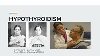
- 1. HYPOTHYROIDISM - D. VIGHNESH, 4th Year MBBS, Dept. of General Surgery, VMCKNL
- 2. INTRODUCTION Hypothyroidism results from low levels of thyroid hormone with varied etiology and manifestations. Untreated hypothyroidism increases morbidity and mortality. The patient presentation can vary from asymptomatic disease to myxedema coma. Today, the diagnosis of hypothyroidism is easily made with simple blood tests and can be treated with exogenous thyroid hormone.
- 5. CRETINISM Cretinism is defined as thyroid hormone deficiency present at fetal / neonatal development It must be diagnosed promptly because delay in treatment can lead to irreversible neurological deficits. Before the newborn screening program, it was one of the most common preventable causes of intellectual disability. Newborn screening (NBS) programs have led to earlier diagnosis and treatment, resulting in improved neurodevelopmental outcomes.
- 7. MANAGEMENT Treatment with levothyroxine (L-T4) must be started immediately after the diagnosis of congenital hypothyroidism (CH). NBS programs and early L-T4 treatment initiation (prior to 2 weeks of life) can prevent intellectual deficits and optimize neurodevelopmental outcomes. L-T4 alone is the treatment of choice. The initial dose depends upon the severity of CH. A higher initial L-T4 dose of 10 to 15 micrograms (ug)/kg/day (50 ug/day for full-term infants with severe CH) is recommended, especially for neonates with a very low pretreatment T4 level. A high initial L-T4 dose can normalize serum T4 in 3 days and TSH by two weeks of therapy.
- 10. TREATMENT
- 11. MYXOEDEMA Myxedema is a serious disorder with high mortality. Most patients die from GI bleeding, sepsis, or respiratory failure despite optimal treatment. Poor prognostic factors include advanced age, persistent hypothermia, and altered mental status. Treatment: Bolus of 0,5 mcg T4 or 10 mcg T3, preferably IV over every 4 to 6 hours. Additionally, IV Broad spectrum Antibiotics and Hydrocortisone are recommended DYSHORMONOGENESIS Thyroid dyshormonogenesis continues to be a significant cause of congenital hypothyroidism because of genetic deficiencies in enzymes overlooking hormone synthesis. Common family history. Most commonly affects Thyroid Peroxidase (TPO) and Thyroglobulin activity. Pendred syndrome is a classical example of the condition.
- 12. BENIGN TUMORS OF THE THYROID
- 14. FOLLICULAR ADENOMA OF THYROID
- 15. INTRODUCTION Follicular adenomas are one subset of benign neoplasms that can occur in the thyroid gland or ectopic thyroid tissue. They typically present as a solitary thyroid nodule or in association with nodular hyperplasia or thyroiditis. Thyroid nodules are palpable in 4 to 7 % of individuals, but the prevalence of nodules detected incidentally by ultrasound shows a higher prevalence of 19 to 67 percent. The majority of thyroid nodules are asymptomatic.. Most of these are benign, although 5% exhibit malignant features. Although the distinguishing line between the adenoma itself and its malignant counterpart is tricky, this is for all practical purposes a benign neoplasm.
- 17. ETIOLOGY Although follicular adenomas are mostly sporadic, multiple other etiological factors have been identified Other causes implicated in the development of a follicular adenoma include: ● Iodine deficiency: This is a known risk factor seen in individuals consuming non-iodized sea salt regularly. ● Genetic alterations: PTEN hamartoma tumor syndrome (PHTS) includes several syndromes like Cowden syndrome and BRRS. Multiple follicular adenomas may occur along with other clinical manifestations. PTHS is due to mutations causing inactivation of the PTEN tumor suppressor gene. Other genetic mutations in BRAF, NRAS, RET, and KRAS can also cause unexplained follicular adenomas. Follicular adenomas are also part of various syndromes like Familial adenomatous polyposis(FAP), Carney Complex syndrome. Genetic rearrangement of the PAX8-PPAR gene causes loss of follicular growth inhibition, thus facilitating the development of follicular neoplasms. ● Prior I-131 radiation exposure also increases the risk of malignant lesions of the thyroid.
- 19. HISTOPATHOLOGY Follicular adenoma is grossly described as a solitary, encapsulated nodule; the size can be extremely variable, ranging from a few millimeters to 10-15 cm. The color vary from tan to light brown with solid and fleshy appearance. It can resemble multinodular goiter due to secondary changes in hemorrhage and cystic degeneration The two key characteristics that make the follicular lesion malignant is the evidence of capsular invasion and angioinvasion. Histological examination of the nodule reveals a follicular architecture present in the entire or nearly entire lesion. The nodule can be described as either microfollicular or macrofollicular growth pattern, and the thyrocytes have a normal cytological appearance.
- 21. CLINICAL PRESENTATION ● Most patients with a follicular adenoma present with solitary thyroid nodule in an otherwise normal thyroid gland, however, it may occur in association with thyroiditis or nodular hyperplasia. while taking history and performing a physical of a patient with neck swelling, it is imperative to keep in mind the features of hyperthyroidism or hypothyroidism that could accompany the presentation. ● Family history of Hashimoto disease, Grave disease thyroid carcinoma or familial syndromes like Gardners all are valid points to consider. Commonly patients present with and visually describe a slowly growing mass in the neck, pressure sensation over the neck.
- 22. CLINICAL PRESENTATION ● Pain seldom accompanies a thyroid nodule unless spontaneous hemorrhage or cystic degeneration has occurred within the nodule. Pressure symptoms could cause dyspnea due to tracheal compression, increased coughing, voice hoarseness, and choking spells due to recurrent laryngeal nerve irritation and dysphagia secondary to esophagus compression ● Patients with a follicular adenoma present with a thyroid nodule that's palpable on examination or identified on an imaging study. Nodules less than 1 cm are usually challenging to palpate unless located anteriorly on the gland. There may be more than one nodule that is palpated. However, even after neck palpation, nearly half of these nodules go undetected and can only be picked up on ultrasonography
- 23. INVESTIGATIONS ● The four main parameters to assess in a thyroid nodule evaluation are detailed patient history, physical examination, baseline serum TSH assay followed by an ultrasound. ● 5% of microfollicular adenomas, when subjected to histopathological examination, are reported as follicular cancers ● CT scan and MRI have a limited role in the initial evaluation f solitary thyroid nodule. Indications for these imaging techniques include suspected tracheal involvement, either by invasion or compression, extension into the mediastinum, or recurrent disease. ● FNA is performed to provide a cytological examination of the nodule, and it remains the mainstay for assessing these lesions, although it may not provide a diagnosis in all cases. The success rate of FNA is improved when the procedure is done under ultrasound guidance.
- 24. MANAGEMENT Medical Management ● If the patient is not clinically euthyroid, medical therapy done to achieve a clinically euthyroid state. ● In such lesions, additional, free T3, T4 assay must be done. As mentioned above, an iodine-123 thyroid scan may help determine the functionality of the nodule. These patients must be appropriately treated with medication. ● The patient may undergo observation or levothyroxine suppression therapy as an initial treatment modality. Levothyroxine is administered for six months to determine if the nodule decreases in size.
- 25. MANAGEMENT Surgical Management ● In the case of follicular neoplasms determined by FNA, the risk of malignancy is less than 1% in a hyperfunctioning nodule, with a higher 20% risk if the nodule is hypo functioning. ● If the FNA result shows a follicular neoplasm, surgical management in the form of a thyroid lobectomy with isthmusectomy is the norm. If the ultrasound shows suspicious features, the surgeon should keep in mind the likelihood of malignancy. ● After due consideration of additional risk factors like family history, the presence of other comorbidities, previous history of neck/head radiation, a decision of performing a total thyroidectomy may be taken. ● Patients having a solitary toxic nodule, after determination of the functionality of the same, could undergo therapeutic iodine-131 therapy. Surgically, a unilateral thyroid lobectomy is adequate. ● If the histopathological examination (HPE) examination confirms the Follciular neoplasm as an adenoma, no further intervention is required.
- 26. Main Advantages of Surgical Therapy ● Relief from pressure-compressive symptoms like dyspnea, dysphagia, hoarseness, etc ● Removal of the lesion helps alleviate the patient's anxiety ● Resolves the issues of thyrotoxicosis in a toxic follicular adenoma ● Avoids unnecessary radiation exposure to the healthy part of the thyroid
- 27. PROGNOSIS ● Follicular adenomas are benign neoplasms. They are slow-growing and can progress to a size that may cause compressive symptoms. The patient should ideally be reassured that these do not signify a malignant process but rather that the mass is causing compression on another structure ● 20% of nonfunctioning follicular adenomas have oncogene mutations that may progress to develop into a follicular carcinoma. ● Thyroxine supplementation is not recommended to suppress the gland unless hypothyroidism occurs after gland lobectomy.
- 28. THANK YOU