SlideShare a Scribd company logo
1 of 73
PHACO & BEHCET’S DISEASE
ESLAM MOTAWEA
INTRODUCTION
First described in 1937 by Hulusi Behcet,
• As classic trisymptom complex of hypopyon, iritis, and
orogenital aphthosis.
• Turkey has the highest prevalence: 80 to 370 cases per
100,000 population.
• Chronic, relapsing-remitting, occlusive vasculitis
affecting multiple organ systems
• Characterized by oral and genital ulcerations (sores),
uveitis, skin rashes, arthritis, thrombophlebitis, colitis,
and neurologic symptoms.
• Histologically, there is a combination of a perivascular
neutrophilic or lymphocytic infiltration, endothelial
cell damage or swelling, fibrinoid necrosis coupled
with a pro-thrombotic tendency
BEHCET’S DISEASE
• Predominant age: 3rd to 4th decades
• Predominant sex: M > F and with more severe
disease in some Mediterranean areas.
• In Iran, M:F ratio was 24:1 among 1712 patients.
• In Turkey, ratio was 16:1 among 427 patients.
Mortality/Morbidity
• Chronic morbidity is typical; leading cause is
ophthalmic involvement, which can result in
blindness.
• Effects of disease may be cumulative, especially with
neurologic, vascular, and ocular involvement.
• Mortality rate is low, but death can occur from
neurologic involvement, vascular disease, bowel
perforation, cardiopulmonary disease, or as a
complication of immunosuppressive therapy.
PATHOPHYSIOLOGY
• Cause is not known
• Immunogenetics, immune regulation, vascular
abnormalities, or bacterial and viral infection may
have a role
History
• Signs and symptoms may be recurrent, may precede onset of mucosal
membrane ulcerations by 6 months to 5 years.
• Prior to onset of disease, patients may experience a variety of
symptoms.
• Malaise
• Anorexia
• Weight loss
• Generalized weakness
• Headache
• Perspiration
• Decreased or elevated temperature
• Lymphadenopathy
• Pain of the substernal and temporal regions
• A history of repeated sore throats, tonsillitis, myalgias, and migratory
erythralgias without overt arthritis is common.
spectrum of organ involvement in behçet’s disease.
Diagnostic criteria from the Behçet syndrome research
committee of Japan (1987 revision)
Diagnosis
a) Complete –
Four major features
b) Incomplete –
(1) 3 major features,
(2) 2 major and 2 minor
features,
(3) Typical ocular symptom
and 1 major or 2 minor
features
c) Possible –
(1) 2 major features
(2) 1 major and 2 minor
features
• Major features
• Recurrent aphthous ulceration of oral mucous
membrane
• Skin lesions -Erythema nodosum – like lesions,
subcutaneous thrombophlebitis, folliculitis
(acnelike lesions), cutaneous hypersensitivity
• Eye lesions - Iridocyclitis, chorioretinitis,
retinouveitis, definite history of chorioretinitis
or retinouveitis
• Genital ulcers
• Minor features
• Arthritis without deformity and ankylosis
• Gastrointestinal lesions characterized by
ileocecal ulcers
• Epididymitis
• Vascular lesions
• Central nervous system symptoms
• Recurrent oral ulceration - Minor aphthous or major aphthous or
herpetiform ulceration recurs at least 3 times in one 12-month period
plus 2 of the following:
• Recurrent genital ulceration –
Recurrent genital aphthous ulceration or scarring, especially males, observed
by a physician or reliably reported by a patient
• Eye lesions –
(1) Anterior uveitis, posterior uveitis, and cells in vitreous upon slit-lamp
examination or (2) retinal vasculitis observed by physician (ophthalmologist)
• Skin lesions –
(1) Erythema nodosum–like lesions observed by physician or reliably reported
by a patient, pseudofolliculitis, and papulopustular lesions or (2) acneiform
nodules consistent with Behçet disease, observed by a physician, and in
postadolescent patients not receiving corticosteroids
• Positive pathergy test –
An erythematous papule larger than 2 mm at prick site 48 hours after
application of a 20- to 22-gauge sterile needle, which obliquely penetrated
avascular skin to a depth of 5 mm as read by a physician at 48 hours
Findings are applicable if no other clinical explanation is present.
International criteria for the classification of Behçet disease (1990)
Physical
• Oral ulcers (100%)
• Indistinguishable from common aphthae (canker sores).
• Aphthae may be more extensive, more painful, more frequent,
and evolve quickly from a pinpoint flat ulcer to a large sore.
• Lesions can be shallow or deep (2-30 mm in diameter) and
usually have a central, yellowish, necrotic base and a punched-
out, clean margin.
• Appear singly or in crops, located anywhere in the oral cavity,
persist for 1-2 weeks, and subside without leaving scars.
• M/c sites - tongue, lips, buccal mucosa, and gingiva;
• Tonsils, palate, and pharynx are less common sites.
• Interval b/w recurrences ranges from weeks to months.
• 3 types:
Minor ulcer
Consists of 1-5 small, moderately
painful ulcers persisting for 4- 14
days
Major ulcer
1-10 very painful ulcers,
measuring 10-30 mm, persisting
up to 6 weeks, and possibly
leaving a scar upon healing
Herpetiform ulcer
Recurrent crop of as many as
1000 small and painful ulcers
Genital manifestations
• Genital ulcers (90%, M>F)
resemble their oral
counterparts but cause greater
scarring.
• In males, usually occur on
scrotum, penis, and groin.
• In females, occur on vulva,
vagina, groin, and cervix
• Ulcers may found in urethral
orifice and perianal area.
• Epididymitis may arise in men
• Orchiepididymitis,10.8% of
men.
Cutaneous manifestations(58.6-97%)
 A variety of skin lesions may appear,
 Eg.-
Sweet syndrome–like
lesion.
Papulopustular
eruptions (55-83%, M>F)
Erythema nodosum–like
lesions (44-62%, F>M)
Cutaneous manifestations
• Erythema multiforme–like lesions
• Thrombophlebitis
• Ulcers
• Bullous necrotizing vasculitis
• Pyoderma gangrenosum
• Pathergy reaction
• Erythematous papule larger than 2
mm develops at prick site after 48
hours
• Higher positivity (84-98%) is found
in Mediterranean areas and Middle
East than in Far East (40-70%),
• Western countries having
significantly lower positivity
Typical positive
pathergy reaction at
injection site
Ocular manifestations(70/90%)
The frequency of ocular involvement in patients with BD is approximately 70%-90%
The characteristic ocular involvement is a relapsing remitting panuveitis
The ocular disease manifests approximately in 2-3 years after the initial symptoms noted
Morever, eye involvement may be the first presenting
manifestation of BD in approximately 10% to 20 % of the patients.
The mean age at onset of uveitis is around 30 years.
Males are more frequently involved, had an earlier disease onset, and had a more severe
disease.
The frequency of bilateral involvement ranges between 78% and 95% in in most of the
series
Bilateral but asymmetric ocular inflammation is a characteristic feature
Behçet panuveitis is a medical emergency,
which must be treated immediately. Ocular involvement in patients with BD, especially
posterior uveitis presence, is the primary indication of immunosuppressive
treatment.
Decreased visual acuity is a result of secondary glaucoma, cataracts, or vitreous hemorrhage.
Blindness has been reported to occur within 4-5 years from onset of ocular symptoms.
Posterior segment involvement is a poor prognostic sign of ocular BD and is seen in up to
93% of patients with ocular disease
The most common and universal posterior segment finding are vitritis and retinal
perivasculitis involving both the arteries (periarteritis) and veins (periphlebitis).
The most common complication of ocular BD is cystoid macular edema (CME), which
observed in approximately in half of the patients with uveitis.
It may resolve with appropriate treatment or if untreated progress to persistent chronic
macular damage and
sequel of CME, with structural changes after recurrences resulting in permanent visual loss.
In some cases, it may lead to form a partial or full-thickness macular hole formation
.
Vascular involvement
• Occurs in 30-50%, mostly men.
• H/P- media thickening, elastic fiber splitting, and
perivascular round cell infiltration.
• 4 types of vascular lesions –
• Aterial and venous occlusions, aneurysms, and varices.
• Venous inv. M/C occlusion, rarely varices.
• M/C sites - SVC,IVC, deep femoral vein, and
subclavian vein.
• Gastrointestinal involvement (>10%)
• Clinical spectrum is enormously varied
• Anorexia, vomiting, dyspepsia, diarrhea, abdominal
distention, and abdominal pain
• Cardiac manifestations (5-17%)
• Coronary vasculitis and thrombosis, pericarditis,
myocarditis, endocarditis with granulomatous changes
or fibrosis, regurgitation, and diastolic dysfunction
• Lung involvement (up to 18%)
• Pulmonary vasculitis, hypertension, pleural effusions
and Aneurysms
Joint manifestations (30 - 40%)
• More than half patients develop signs or symptoms of
synovitis, arthritis, and/or arthralgia during course of
disease.
• Most frequent minor feature in childhood-onset BD is
reported to be arthritis, occurring in 11 of 40 patients.
• Multiple-joint involvement is common.
• Clinical features have been reported as pain,
tenderness, swelling, limitation of joint movement,
warmth, and morning stiffness.
Neurologic manifestations (3.2-49%)
• May present as meningoencephalitis, a multiple sclerosis–like illness,
acute myelitis, stroke, or pseudotumor cerebri.
• Most serious, leading to severe disability and a high fatality rate.
• Usually occur within 5 years of disease onset.
• Severe headache is most frequent initial symptom.
• Three categories (Pallis and Fudge (1956) and Wadia and Williams (1957)
(1) Brain stem syndrome,
A/w fever, arthralgia and skin eruption; brainstem signs developes
subacutely with evolving cranial neuropathies, ocular motor
dysfunction, nystagmus and gaze palsies, dysarthria and ataxia, bulbar
muscle weakness, Headache with meningism, CSF pleocytosis and high
protein content
(2) Meningomyelitis syndrome,
Meningitis with spinal cord and hemisphere signs (Silfverskiold, 1951)
(3) Organic confusional syndrome,
Meningoencephalitis without focal neurological signs, some times
chronic and progressive, resulting ultimately in dementia,
Parkinsonism, pseudobulbar palsy and quadriparesis
Pregnancy-associated manifestations
• Pregnant women may experience more severe
symptoms during first trimester.
• Overall, pregnancy does not seem to markedly affect
the course of BD.
• Close follow-up is necessary to monitor the health of
mother and baby.
Differential Diagnoses
• Acute Febrile Neutrophilic
Dermatosis
• Pyoderma Gangrenosum
• Cyclic neutropenia
• Aphthous Stomatitis
• Erythema Multiforme
• SJS-TEN
• Erythema Nodosum
• Viral infections
• Herpes Simplex,
coxsackie, echo
• Syphilis and other STIs
• Lupus Erythematosus,
Acute
• Reiter disease
• VKH syndrome
• Bullous autoimmune
diseases
• JRA
• Sarcoidosis
• Multiple sclerosis
• Ulcerative colitis, crohn
disease
Laboratory Studies
• Mild anemia and leukocytosis in chronic disease.
• ESR, C-reactive protein value, and other acute phase
reactants may be elevated during the active didease,
but they do not correlate well with clinical activity.
• IgA, IgG, alpha-2 globulin, IgM, and immune
complexes are occasionally elevated.
• Rheumatoid factor and antinuclear antibodies are
absent.
• Antineutrophil cytoplasmic antibody and
antiphospholipid antibody test results are usually
negative.
BD needs multidisciplinary approach
• Dermatologist - For evaluation of mucocutaneous lesions (ie,
oral ulcer, genital ulcer, skin lesions)
• Ophthalmologist - For evaluation of eye involvement
• Rheumatologist or orthopedic surgeon - For evaluation of joint
involvement
• Neurologist or psychiatrist - For evaluation of CNS involvement
• Internal medicine specialist - For evaluation of gastrointestinal,
pulmonary, renal, or endocrine involvement
• General surgeon - For evaluation of gastrointestinal involvement
• Chest surgeon or cardiologist - For evaluation of cardiovascular
involvement
• Ear, nose, and throat specialist or dentist - For evaluation of oral
cavity
Medical Care
• Treatment of BD symptomatic and empiric.
• Choice of treatment depends on site and severity of
clinical manifestations
Local therapy
 Recurrent aphthae
• mild diet, avoidance of irritating agents,
• Caustic solutions (silver nitrate, 1%-2%; tinctura myrrha, 5%-10%
weight/volume; H202,0.5%; methylviolet,0.5%) 1-2xld
• Antiseptic and anti-inflammatory preparations (amlexanox, 5% in
oral paste; triclosan, 0.1% mouthwash solution and in toothpaste ), 3%
diclofenac in 2.5% hyaluronic acid, tetracycline mouthwash
• Corticosteroids (triamcinolone mucosal ointment, dexamethasone
mucosal paste, betamethasone pastilles) 4 x/day or during night
(ointment / paste) or intrafocal infiltrations with triamcinolone
suspension 0.1-0.5 mL per lesion
• Anaesthetics (lidocaine - 2%-5%; mepivacaine - 1.5%, tetracaine -
0.5%-1% gels or mucosal ointments) 2-3 x/day
• 5-Aminosalicylic acid (5% cream) 3 x/day (reduces the duration of
lesions and the pain
• Sucralfate suspension, 5 mL x 4/day (for oral aphthous and genital
ulcers)
• Topical tacrolimus, nicotine patches, and topical G-CSF
 Genital ulcers and skin lesions,
• Corticosteroid and antiseptic creams for 7 days.
• Painful genital ulcerations topical anaesthetics in
cream.
• Corticosteroid injections (triamcinolone, 0.1 to 0.5
ml/lesion) in recalcitrant ulcerations.
Systemic therapy
• Corticosteroids
• Mainstay for all clinical manifestations.
• Have a beneficial effect on acute manifestations, no definite
evidence effectiveness in controlling progression.
• Adverse effects of long-term therapy must be considered.
• Mucocutaneous lesions and arthritis:-
• NSAIDs, zinc sulfate, levamisole, colchicine, dapsone, or
sulfapyridine and thalidomide. Immunosuppressive therapy
with azathioprine, chlorambucil, or cyclophosphamide
• Uveitis and CNS involvement:-
• Systemic corticosteroids, azathioprine, or cyclosporine, MTx
• In CNS involvement, risks and benefits of cyclosporine
need to be considered b/c of neurological side effects.
• Anticoagulants are given to patients with thromboses.
• Other therapeutic approaches
• IFN alfa, IFN gamma, acyclovir, high-dose corticosteroids or
cyclophosphamide pulse therapy, and tacrolimus
• Japanese tacrolimus study group reported that
tacrolimus was effective in treating refractory uveitis in a
dosage-dependent manner.
• Adverse effects were renal impairment (28.3%), neurologic
symptoms (20.8%), gastrointestinal symptoms (18.9%), and
hyperglycemia (13.2%).
• Subcutaneous IFN alfa-2a therapy
• resulted in reduced ulcers and eye disease.
• Flulike symptoms were most common adverse effect.
• Leukopenia, hair loss, and development of antinuclear and
antithyroid antibodies were reported less commonly.
• A patient presenting with oral ulcers resistant to
prednisone, azathioprine, colchicine, dapsone, and
cyclosporin responded well to lenalidomide.
• A patient with Behçet disease with ocular involvement,
dependant on corticosteroids and refractory to
azathioprine, showed improvement with addition of
pentoxifylline
• A case of Behçet disease resistant to prednisolone,
cyclosporin, azathioprine, infliximab with
methotrexate, and colchicine has been successfully
treated with anakinra.
• infliximab, and etanercept, have decreased frequency of
attacks
• Etanercept has been used at 25 mg twice a week.
• Pediatric case responding to infliximab has been reported.
• Infliximab has resulted in responses after etanercept failed.
• Infliximab infusions of 5-10 mg/kg have been used with
variable dosing schedules.
• Several patients not responding to infliximab have been
treated with adalimumab.
drug doses indication
Methylprednisolone 40 mg/every 3 wk
lM
Erythema nodosum (but not orogenital
ulcers)
Rebamipide 300 mg/day PO Oral ulcers
Colchicine 1-2 mg/day PO Erythema nodosum, arthritis, genital
Ulcer s (oral ulcer in female)
Dapsone 100 mg/day PO Orogenital ulcers, skin lesions, pathergy
Azathioprine 2.5 mg/kg/day Recent onset ocular disease
lnterferon-alfa-2a 6x10⁶ lU/3x/wk SC Orogenital ulcers, papulopustular lesions
Thalidomide 100 mg/day Orogenital ulcers, Papulopustular lesions
CyclosporinA 10 mg/kg/day PO Ocular manifestations, skin lesions,
orogenital ulcers
Etanercept 25 mg/2 x/wk P0 Oral ulcers, papulopustular lesions
nodular lesions, (not pathergy test)
Behcet’s Disease: Prognosis
• Behcet's disease has an undulating course of
exacerbations and remissions, and may become less
severe after approximately 20 years.
• Appears to be more severe in young, male, and Middle
Eastern or Far Eastern patients.
Cataract in uveitis
as a result of the intraocular inflammation per se, from
chronic corticosteroid usage or more often from both [1].
The incidence of cataract in uveitis varies from 57% in
pars planitis [2] to 78% in Fuchs heterochromic
iridocyclitis (FHI) [3].
Cataract Surgery in Behcet’s Disease. Cataract forma-tion is the most common anterior segment complication after
recurrent inflammation, occurring in up to 36% of cases [1
It was reported that the postoperative visual acuity was found to be significantly lower in eyes with BD than in those with
idiopathic uveitis because of the severe posterior segment complications, mainly optic atrophy [48].
Operating on eyes with cataract and uveitis has been previously reviewed by Foster and associates [8]. Their
recommendations for a successful cataract surgery and for minimizing the postoperative uveitis are as follows.
Uveitis should be inactive for at least 3 months pre-operatively, systemic and topical steroids should
be used prophylactically for 1 week preoperatively and continued postoperatively,
immunosuppressive drugs should be con-tinued, complete removal of cortical material should
take place, and one-piece PMMA posterior chamber intraocular lens should be used if the patient
and the surgeon understand the special nature of this surgery, its risks, and the prognosis for success.
In another paper by Berker et al., the authors reported results of phacoemulsification and intraocular lensimplan-tation in
patients with Behcet’s disease [49]. They reported 72.5% of eyes had improvement in vision after surgery. However, the
vision got worse in 17.5% of the eyes. Most frequent complication reported by them was posterior capsular opacification in
37.5% of eyes. Other complications were posterior seynchiae and severe inflammation. Posterior segment complications
such as epiretinal membrane forma-tion, cystoid macular edema, and optic atrophy were also reported by the authors.
Current Guidelines for the Management of
Uveitic Cataract
4.1. Clinical Evaluation of Complicated Cataract
and Asso-ciated Uveitis.
The eye in which visual loss is mainly attributable to cataract
formation is most likely to benefit from cataract surgery. The
outcome of surgery depends upon several factors, namely the
uveitic diagnosis, proper perioperative management and
meticulous surgery. The specific uveitic diagnosis is of
paramount importance also when planning the surgical
strategy [4], such as determining whether an intraocular lens
should be implanted or not.
 The visual potential of the eye, determined by. The
state of the macula and optic nerve should be
thoroughly examined for during the preoperative
assessment. Macular ischemia, atrophy, chronic
macular edema, or scar, such as that resulting from
a choroidal neovascular membrane, are poor
prognostic factors. Similarly, optic atrophy and
severe cupping of the optic disc are bad
prognostic signs
 . In presence of dense lens opacity, B scan ultrasonography
should be done to rule out retinal detachment which may
complicate the eye with uveitis
 as pre-existing corneal scars and severe iris atrophy may also
compromise the visual outcome.

 Regardless of the guarded prognosis, a definite indication for
cataract removal in an eye that is not blind, is phacoanti-genic
uveitis. This may result from the hypermature state of a
cataract, whereby lens proteins leak out of the capsular bag
through an intact capsule, or in cases of trauma, where the lens
capsule has been breached, leading to persistence of intraocular
inflammation. Cataract surgery may also be indicated to permit
better visualization of the posterior segment for appropriate
medical or surgical management of the eye
Control of Pre Operative
Inflammation
 The risk of reactivation of uveitis must be assessed.
Jancevski and Foster recommended the use of
supplementary perioperative anti-inflammatory
therapy to prevent damage to ocular structures
 to reduce the risk of postoperative CME [59], steroid prophylaxis should be given perioperatively to
protect against recurrence of macular swelling. Similarly, steroid prophylaxis should be administered
in eyes at risk of developing recurrence of uveitis following cataract surgery,
 for example, Vogt-Koyanagi Harada disease, Behcet’s disease and birdshot choroidopathy, to name a
few. This may take the form of oral steroids 1 mg per kg/day starting 3 days preoperatively, tapering
the steroid dose according to the amount of inflammation postoperatively
 . Alternatively, if there are no contraindications to periocular steroid injections, such as documented
steroid response or infectious uveitis, an orbital floor or sub-tenon’s injection of depot steroid, such as
triamcinolone acetonide 40 mg/1 mL may be given,
 especially in patients where high doses of oral steroid are contraindicated, for example in poorly
controlled diabetics.
 In addition, guttae prednisolone acetate 1% 2 hourly administered 2 days prior to surgery, together
with an oral and topical non-steroidal anti-inflammatory agent, can be given. Supported by
encouraging results in the recent literature,
 the authors favour an intravitreal injection of preservative-free triamcinolone acetonide 4 mg in 0.1
mL at the conclusion of cataract surgery [60–62].
Complications of Uveitis Adversely
Affecting Surgical Outcome:
High-Risk Surgical Cases. Determining the surgical risk is a very important aspect of preoperative assessment.
Eyes which have generally low intraocular pressures, espe-cially readings of 6 mmHg or less even when quiescent,
are at high risk of developing postoperative hypotony or even phthisis bulbi.
Other warning signs such as seclusio papillae with normal intraocular pressure reading and apparent
phacodonesis without evident zonulysis are important poor prognostic signs for postoperative hypotony.
Eyes in which the uveitis is difficult to control are also at high risk of severe postoperative inflammation and
hypotony or phthisis bulbi.
The presence of choroidal effusion on B scan ultrasonogra-phy or diffusely thickened choroid is a poor prognostic sign.
Conducting a careful ultrasonic biomicroscopy is essential in eyes with relative hypotony [65] to assess the state of the
ciliary body and its processes. If the ciliary body has undergone atrophy, the risk of hypotony is high. If the ciliary body is
found to be detached and processes appear under traction from a ciliary (cyclitic) membrane,
cataract surgery should be combined with vitrectomy and trimming of the ciliary membrane aided by indentation of sclera
to relieve ciliary body traction and to restore normal IOP.
Diagnostic Aids
 . Apart from the standard means of assessing macular,
optic nerve and retinal function, one may apply
additional methods such as pupillary response, light
projection, colour perception and B scan
ultrasonography, or an attempted OCT in looking
for macular atrophy, edema, or hole [66].
Optimal Time for Cataract Surgery.
Before scheduling surgery, the ophthalmologist should attempt to ensure that the eye has
been quiescent for at least 3 months [11].
This has been shown to reduce the risk of postoperative CME [59]. In cases where, despite
heavy immunosuppression, the intraocular inflammation is still not completely abolished
and surgery is urgently required, such as in an intumes-cent cataract, the patient may be
administered intravenous methylprednisolone 1 g one day before surgery.
A study from Japan suggests that in patients with Behcet’s disease the eye should be inactive
for a minimum of 6 months and that the risk is higher if attacks have occurred within 12
months of cataract surgery [70].
Counselling of the Patient for Uveitic Cataract Surgery.
The most important aspect of counselling when planning to perform cataract surgery for the
uveitic eye is explaining the visual prognosis. The general risks involved in surgery, such as
infection and other intraoperative complications will also need to be thoroughly
explained, especially if there is phacodonesis, hypotony, or glaucoma.
5. Surgical Technique
5.1. Choice of Surgery.
The choice of cataract surgery tech-nique is best left to the surgeon
Cataract removal by phacoemulsification is safer for the uveitic cataract as less
inflammation is induced than that by a man-ual extracapsular cataract extraction.
During the surgery, the anatomy of the anterior segment should be restored to a
state as close to normal as possible.
Some uveitic cataract eyes are complicated by glaucoma or retinal problems that
may also benefit from surgery. For eyes with concomitant uveitic glaucoma,
surgery is preferably not combined with the cataract surgery as the risk of bleb
failure is increased with drainage of post-cataract surgery inflammatory exudate
through a healing bleb. Where possible, cataract surgery should be done first.
 Regarding retinal complications, such as epiretinal mem-branes or
coexisting retinal detachment, cataract surgery may be combined
with vitreoretinal surgery.
 In cases with major retinal problems, the eye may be safely rendered
aphakic until the retinal problem has been dealt with. In eyes with
intermediate uveitis, or FHI, cataract surgery may be combined with
vitrectomy, performed to clear the vitreous gel, thereby reducing
vitreous clouding. In intermediate uveitis, this often not only improves
vision but also controls intraocular inflammation and helps resolve the
cystoid macular edema.
 At the end of surgery, especially with combined surgical procedures,
having excluded steroid responders and eyes with infectious uveitis, an
intravitreal injection of triamcinolone acetonide, is often
adequate to control the postoperative inflammation and prevent CME.
.2. Intraoperative Surgical
Techniques and Skills
Posture.
Patients with ankylosing spondylitis with a fixed flexion
deformity of the axial spine, especially when the cervical
spine is involved
Surgical Challenges.
The uveitic eye poses numerous surgical challenges. These
include the small pupil, shallow anterior chamber,
posterior synechiae, peripheral ante-rior synechiae,
pupillary membranes and even zonulolysis
Anaesthesia.
Whilst phacoemulsification surgery may be done under topical
anesthesia, manipulation of the iris may induce ocular discomfort or pain.
Either regional anesthesia or an intracameral injection of preservative-
free lignocaine 1% can provide adequate analgesia.
For children and in patients for whom prolonged surgical time is
anticipated, as in severe zonulolysis requiring modified capsular tension
rings that need suturing, general anesthesia may be preferred.
Incision.
Either a scleral or temporal clear corneal incision may be
used. However, the incision should be of adequate length in
order to prevent iris prolapse in eyes with small or stretched
pupils.
Pupil Enlargement. balanced salt solution with
adrenaline (1 : 1000 0.5 mL adrenaline in 500 mL) into eyes
with pupils that are not bound by synechiae or membranes.
Preservative-free intracameral lignocaine 1% may also be
used to help
dilate the pupil only if it is not bound.
Healon 5 is useful can physically roll open the pupil and
keep it dilated as long as the aspiration flow rate is kept low.
 (f) Synechiolysis and Removal of Pupillary Membrane.
 When both are present, the PAS should be released before
the PS. Release of PAS may be done by injecting
viscoelastic, such as Healon 5 to physically separate the iris
from the cornea
 tip of the viscoelastic cannula may be used to sweep the iris
away from the peripheral cornea as the viscoelastic material
is being injected into the angle of the anterior chamber.
This should be done very gently and carefully, taking care
not to detach Descemet’s membrane in the process.
sweeping the pupil free from the lens capsule with a cannula. In
cases where a narrow strip of membrane is present at the site of PS
formation, a 27-gauge needle may be used to simultaneously nick
the membrane to segment and release it from the anterior capsule
and release the PS, thus stretching the pupil.
Pupil Expansion. The most user-friendly instrument for
extensive synechiae once an edge of the iris has been viscodissected
off the anterior capsule is a bent Kuglen hook
Alternative means of opening the pupil include the use of a Beehler
pupil dilator (2 or 3 pronged) to mechanically stretch the pupil in a
single injector system
Continuous Circular Capsulorhexis.
It is generally prefer-able to keep the size of the capsulorhexis
slightly smaller than the pupil so that iris chaffing does not occur
and results in progressive intraoperative miosis as nuclear
fragments are being moved out of the capsular bag (Figure 8).
This also contributes to increased postoperative inflammation.
When the pupil size is small, the capsulorhexis inevitably needs to
be larger than the pupil. The anterior chamber must be kept deep
and the anterior capsule flattened using adequate viscoelastic
material in order to control the tearing of a capsulorhexis. The
capsulotomy can be initiated by a 26- or 27-gauge bent cystitome
 Creating the ideal capsulorhexis is also very important
in preventing posterior capsule opacification. The
capsulorhexis should be centred, overlapping the edge
of the optic at all times, but not so small as to prevent
capsular phimosis.
 Nucleus Management. In small pupils, the safest tech-
nique is the vertical chop employed in the in situ chop
technique. Chopping of fragments is done within the
pupillary aperture with the phaco tip kept in view at all
times with minimal risk of engaging and traumatizing
the iris
Irrigation and Aspiration.
This step must be done thor-oughly so as not to leave cortical
material behind.
Intraocular Lens Implantation Contraindications and
Type of IOL.
the most biocompatible IOL for the anterior chamber and the
capsular bag is a single-piece, square-edged acrylic (either
hydrophilic or hydrophobic) IOL. They also found that the
posterior capsular opaci-fication rate was highest (34.2%) in eyes
with silicone IOLsof postoperative cystoid macular edema and PS
formation, and pupillary membranes
 hydrophilic acrylic material has good uveal but worse
capsular biocompatibility, but hydrophobic acrylic material
had lower uveal but better capsular biocompatibility [78].
In eyes with chronic uncontrolled uveitis, IOL implant
should be deferred.

 Removal of viscoelastic from under the IOL is an
important step in reducing the space behind the IOL
into which lens epithelial cells tend to migrate, thus
causing PCO. Pressing the optic against the posterior
capsule when using a single-piece hydrophobic IOL also
encourages its adhesion to the posterior capsule, thereby
reducing the risk of PCO
Complications and Postoperative
Management
5.3.1. Intraoperative Complication
An infrequent intraoperative complication is
Zonulolysis
(CTR) is often necessary in order to prevent IOL
decentration
Failure to use a CTR in the presence of weak zonules may
result in capsular phimosis, due to unopposed capsular
bag fibrosis and shrinkage.
Retained Lens or Nuclear
Fragments.
 Due to the small pupil size, small hard nuclear
 These small fragments can cause recurrent anterior
uveitis ;may also cause localized corneal edema
localized corneal decompensation in the long
term. Hence, at the end of phaco, the eye should be
given a gentle shake whilst aspirating to ensure no
nuclear fragments are inadvertently left behind. Any
retained soft lens material or nuclear fragment should
be removed surgically as soon as possible [80].
. Early Postoperative Complications
Excessive Postoperative Inflammation.
One of the most com-mon postoperative complications is cystoid
macular edema
phacoemulsification, the incidence has been reported to range from 12%
to 59
Generally, if preoperative prophylactic oral steroids have been given and
maximal topical steroids and cycloplegics have proven ineffective in
controlling the uveitis, the dose
of oral steroids may be sharply increased.
If no prophylactic oral steroids had been given the patient should be
given an oral pulse of steroids or injection of periocular steroids. An
alternative means would be to give an intravitreal injection of
triamcinolone acetonide
Recurrence of Uveitis.
Increased frequency of recurrence following cataract
surgery may occur. This is thought to be triggered by the
intraocular procedure. The recurrence rate has been
reported to be as high as 51% [15]. Hence, stepping up
the immunosuppression for the long term may be
necessary to prevent further recurrences
 Intraocular Pressure (IOP)
Abnormalities.
 The IOP may be raised transiently during the early postoperative period in eyes
with compromised trabecular meshwork or angles. This can often be managed
with topical and systemic anti-glaucoma medications.

 However, the surgeon’s greatest fear is hypotony.
 Once wound leakage has been ruled out, the next step is to increase the anti-
inflammatory therapy topically and systemically. This is often effective in
raising the IOP, but the topical steroids may be difficult to taper or withdraw
and patients may require long-term topical steroids to maintain the IOP.
 In severe cases, vitrectomy and trimming of ciliary body traction membranes
and silicone oil filling may be needed if UBM shows the presence of ciliary
body detachment secondary to tractional membranes not addressed during the
cataract surgery [83].
5.3.3. Late Postoperative
Complications
 Posterior Capsular Opacification. the most common
complication following any type of cataract surgery.
 reported an incidence of 48.0%, Rauz et al. [84] 81.7% and
Kuc¸¨ukerd¨onmez¨ et al. [82] 34.2% at 1 year Their
corresponding Nd:YAG capsulotomy rates were 32.2%,
8.3%, and 3.6%.
 Preventive measures include creating a circular well-centred
capsulorhexis which is smaller than the optic size, using an
acrylic IOL with a square-edged optic design, meticulous
removal of viscoelastic from within the capsular bag and
ensuring the optic is stuck on to the posterior capsule at the
conclusion of surgery. Control of postoperative
inflammation also plays an important role in preventing
PCO.
 Explanting an IOL. Removal of
intraocular lens in uveitic
 eyes is rarely necessary. Foster et al. reported that their
indications include the formation of perilental
membrane, chronic low-grade inflammation not
responding to anti-inflammatory treatment and
cyclitic membrane resulting in hypotony and
maculopathy
6. Conclusion and Summary
 Preoperative factors include proper patient selection and
counseling and preoperative control of inflam-mation
 therefore control of inflammation, both pre-and
postoperatively, is vital. The use of immunosuppressive
agents other than steroids also helps control inflammation
 Management of postopera-tive complications, especially
inflammation and glaucoma, earlier rather than later, has
also contributed to improved outcomes
 conclusion, management of the uveitic cataract requires
careful case selection, proper timing of surgery, meticulous
surgery and close monitoring with appropriate handling of
the postoperative complications that may occur
Take home msg 1.Revise the diagnosis of Behcet. I have seen many cases where the previous diagnosis is not true.
 2- If it is Behcet, giving steroids only without an immune modulator is forbidden.
 Clinical Evaluation of Complicated Cataract and Asso-ciated Uveitis.
 Control of Pre Operative Inflammation
 Complications of Uveitis Adversely Affecting Surgical Outcome
 Diagnostic Aids
 Optimal Time for Cataract Surgery
 Counselling of the Patient for Uveitic Cataract Surgery
 Surgical Technique
 Choice of Surgery.
 Intraoperative Surgical Techniques and Skills
 Posture.
 Surgical Challenges.
 Anaesthesia.
 Incision.
 Pupil Enlargement.
 Synechiolysis and Removal of Pupillary Membrane.
 Pupil Expansion.
 Continuous Circular Capsulorhexis.
 Nucleus Management
 Irrigation and Aspiration.
 Intraocular Lens Implantation Contraindications and Type of IOL.
 complication intrea ,,,early ///late
after starting the proper treatment, we wait for three months of documented systemic as well as ocular inactivity.
 Since there is no ocular activity now, assess for systemic activity. If the disease is systemically active, ask for CBC and liver enzymes, and if
they are normal, add lmmuran 50 mg, one tablet twice daily for two weeks then three times daily for two weeksand inform me of the
response of systemic activity to this teatment four weeks from now.

More Related Content

What's hot

Ocular Manifestations of Systemic Disease
Ocular Manifestations of Systemic DiseaseOcular Manifestations of Systemic Disease
Ocular Manifestations of Systemic DiseaseVisionary Ophthamology
 
Eye in connective tissue disoreders
Eye in connective tissue disoredersEye in connective tissue disoreders
Eye in connective tissue disoredersAmar Patil
 
Intermediate uveitis
Intermediate uveitisIntermediate uveitis
Intermediate uveitisBipin Bista
 
Uveitis investigations
Uveitis investigationsUveitis investigations
Uveitis investigationspratik mohod
 
Angle recession glaucoma
Angle recession glaucomaAngle recession glaucoma
Angle recession glaucomaSSSIHMS-PG
 
REAL THYROID OPHTHALMOPATHY.pptx
REAL THYROID OPHTHALMOPATHY.pptxREAL THYROID OPHTHALMOPATHY.pptx
REAL THYROID OPHTHALMOPATHY.pptxBipin Koirala
 
Approach to posterior uveitis final modified
Approach to posterior uveitis final modifiedApproach to posterior uveitis final modified
Approach to posterior uveitis final modifiedFayez Alsharief
 
Differential Diagnosis of Disc Edema
Differential Diagnosis of Disc EdemaDifferential Diagnosis of Disc Edema
Differential Diagnosis of Disc EdemaSahil Thakur
 
Sudden loss of vision IN A PATIENT ACUTELY
Sudden loss of vision IN A PATIENT ACUTELYSudden loss of vision IN A PATIENT ACUTELY
Sudden loss of vision IN A PATIENT ACUTELYAjayDudani1
 
Episcleritis and scleritis
Episcleritis and scleritisEpiscleritis and scleritis
Episcleritis and scleritisPanit Cherdchu
 

What's hot (20)

Ocular Manifestations of Systemic Disease
Ocular Manifestations of Systemic DiseaseOcular Manifestations of Systemic Disease
Ocular Manifestations of Systemic Disease
 
Cmv retinitis
Cmv retinitisCmv retinitis
Cmv retinitis
 
Thyroid eye disease
Thyroid eye disease Thyroid eye disease
Thyroid eye disease
 
Eye in connective tissue disoreders
Eye in connective tissue disoredersEye in connective tissue disoreders
Eye in connective tissue disoreders
 
Intermediate uveitis
Intermediate uveitisIntermediate uveitis
Intermediate uveitis
 
Anti vegf' s in Ophthalmology
Anti vegf' s in OphthalmologyAnti vegf' s in Ophthalmology
Anti vegf' s in Ophthalmology
 
Uveitis investigations
Uveitis investigationsUveitis investigations
Uveitis investigations
 
GCA
GCAGCA
GCA
 
SLE & APS for undergraduates: diagnosis & treatment.
SLE & APS for undergraduates: diagnosis & treatment.SLE & APS for undergraduates: diagnosis & treatment.
SLE & APS for undergraduates: diagnosis & treatment.
 
Angle recession glaucoma
Angle recession glaucomaAngle recession glaucoma
Angle recession glaucoma
 
REAL THYROID OPHTHALMOPATHY.pptx
REAL THYROID OPHTHALMOPATHY.pptxREAL THYROID OPHTHALMOPATHY.pptx
REAL THYROID OPHTHALMOPATHY.pptx
 
Normal tension glaucoma
Normal tension glaucomaNormal tension glaucoma
Normal tension glaucoma
 
Approach to posterior uveitis final modified
Approach to posterior uveitis final modifiedApproach to posterior uveitis final modified
Approach to posterior uveitis final modified
 
Optic neuritis & Multiple Sclerosis (2018)
Optic neuritis & Multiple Sclerosis (2018)Optic neuritis & Multiple Sclerosis (2018)
Optic neuritis & Multiple Sclerosis (2018)
 
Mixed connective tissue disorder
Mixed connective tissue disorderMixed connective tissue disorder
Mixed connective tissue disorder
 
Differential Diagnosis of Disc Edema
Differential Diagnosis of Disc EdemaDifferential Diagnosis of Disc Edema
Differential Diagnosis of Disc Edema
 
Optic neuritis
Optic neuritisOptic neuritis
Optic neuritis
 
Sudden loss of vision IN A PATIENT ACUTELY
Sudden loss of vision IN A PATIENT ACUTELYSudden loss of vision IN A PATIENT ACUTELY
Sudden loss of vision IN A PATIENT ACUTELY
 
Episcleritis and scleritis
Episcleritis and scleritisEpiscleritis and scleritis
Episcleritis and scleritis
 
Review of Uveitis
Review of UveitisReview of Uveitis
Review of Uveitis
 

Similar to pahco in Behcet dis

Similar to pahco in Behcet dis (20)

behcets disease
behcets diseasebehcets disease
behcets disease
 
Aphthous ulcers
Aphthous ulcersAphthous ulcers
Aphthous ulcers
 
Behçet Disease.pptx
Behçet Disease.pptxBehçet Disease.pptx
Behçet Disease.pptx
 
Papulosequamous disorder
Papulosequamous disorder Papulosequamous disorder
Papulosequamous disorder
 
Lesions of oral cavity
Lesions of oral cavityLesions of oral cavity
Lesions of oral cavity
 
Behçet disease.pptx
Behçet disease.pptxBehçet disease.pptx
Behçet disease.pptx
 
Introduction to Dermatology
Introduction to DermatologyIntroduction to Dermatology
Introduction to Dermatology
 
Oral Pathology and Oesophagus
Oral Pathology and OesophagusOral Pathology and Oesophagus
Oral Pathology and Oesophagus
 
Venous ulcer
Venous ulcerVenous ulcer
Venous ulcer
 
Systemic sclerosis
Systemic sclerosis Systemic sclerosis
Systemic sclerosis
 
Systemic sclerosis
Systemic sclerosisSystemic sclerosis
Systemic sclerosis
 
Immune mediated disorders - Oral Pathology
Immune mediated disorders - Oral PathologyImmune mediated disorders - Oral Pathology
Immune mediated disorders - Oral Pathology
 
Lymphoma clinical feature & dx-Dr.Rajesh
Lymphoma clinical feature & dx-Dr.RajeshLymphoma clinical feature & dx-Dr.Rajesh
Lymphoma clinical feature & dx-Dr.Rajesh
 
Rhino sinusitis I
Rhino sinusitis IRhino sinusitis I
Rhino sinusitis I
 
Diphtheria WHO.ppt
Diphtheria WHO.pptDiphtheria WHO.ppt
Diphtheria WHO.ppt
 
Systemic Sclerosis 2017
Systemic Sclerosis 2017Systemic Sclerosis 2017
Systemic Sclerosis 2017
 
Erythema multiforme Dr Chithra P
Erythema multiforme  Dr Chithra PErythema multiforme  Dr Chithra P
Erythema multiforme Dr Chithra P
 
INTERNAL-MEDICINE.pptx
INTERNAL-MEDICINE.pptxINTERNAL-MEDICINE.pptx
INTERNAL-MEDICINE.pptx
 
Vesiculo bullous diseases
Vesiculo bullous diseasesVesiculo bullous diseases
Vesiculo bullous diseases
 
Stevens Johnson Syndrome
Stevens Johnson SyndromeStevens Johnson Syndrome
Stevens Johnson Syndrome
 

Recently uploaded

Top Rated Bangalore Call Girls Mg Road ⟟ 8250192130 ⟟ Call Me For Genuine Sex...
Top Rated Bangalore Call Girls Mg Road ⟟ 8250192130 ⟟ Call Me For Genuine Sex...Top Rated Bangalore Call Girls Mg Road ⟟ 8250192130 ⟟ Call Me For Genuine Sex...
Top Rated Bangalore Call Girls Mg Road ⟟ 8250192130 ⟟ Call Me For Genuine Sex...narwatsonia7
 
Lucknow Call girls - 8800925952 - 24x7 service with hotel room
Lucknow Call girls - 8800925952 - 24x7 service with hotel roomLucknow Call girls - 8800925952 - 24x7 service with hotel room
Lucknow Call girls - 8800925952 - 24x7 service with hotel roomdiscovermytutordmt
 
(Rocky) Jaipur Call Girl - 09521753030 Escorts Service 50% Off with Cash ON D...
(Rocky) Jaipur Call Girl - 09521753030 Escorts Service 50% Off with Cash ON D...(Rocky) Jaipur Call Girl - 09521753030 Escorts Service 50% Off with Cash ON D...
(Rocky) Jaipur Call Girl - 09521753030 Escorts Service 50% Off with Cash ON D...indiancallgirl4rent
 
Best Rate (Guwahati ) Call Girls Guwahati ⟟ 8617370543 ⟟ High Class Call Girl...
Best Rate (Guwahati ) Call Girls Guwahati ⟟ 8617370543 ⟟ High Class Call Girl...Best Rate (Guwahati ) Call Girls Guwahati ⟟ 8617370543 ⟟ High Class Call Girl...
Best Rate (Guwahati ) Call Girls Guwahati ⟟ 8617370543 ⟟ High Class Call Girl...Dipal Arora
 
Bangalore Call Girls Nelamangala Number 7001035870 Meetin With Bangalore Esc...
Bangalore Call Girls Nelamangala Number 7001035870  Meetin With Bangalore Esc...Bangalore Call Girls Nelamangala Number 7001035870  Meetin With Bangalore Esc...
Bangalore Call Girls Nelamangala Number 7001035870 Meetin With Bangalore Esc...narwatsonia7
 
Chandrapur Call girls 8617370543 Provides all area service COD available
Chandrapur Call girls 8617370543 Provides all area service COD availableChandrapur Call girls 8617370543 Provides all area service COD available
Chandrapur Call girls 8617370543 Provides all area service COD availableDipal Arora
 
Call Girls Horamavu WhatsApp Number 7001035870 Meeting With Bangalore Escorts
Call Girls Horamavu WhatsApp Number 7001035870 Meeting With Bangalore EscortsCall Girls Horamavu WhatsApp Number 7001035870 Meeting With Bangalore Escorts
Call Girls Horamavu WhatsApp Number 7001035870 Meeting With Bangalore Escortsvidya singh
 
VIP Russian Call Girls in Varanasi Samaira 8250192130 Independent Escort Serv...
VIP Russian Call Girls in Varanasi Samaira 8250192130 Independent Escort Serv...VIP Russian Call Girls in Varanasi Samaira 8250192130 Independent Escort Serv...
VIP Russian Call Girls in Varanasi Samaira 8250192130 Independent Escort Serv...Neha Kaur
 
Bangalore Call Girl Whatsapp Number 100% Complete Your Sexual Needs
Bangalore Call Girl Whatsapp Number 100% Complete Your Sexual NeedsBangalore Call Girl Whatsapp Number 100% Complete Your Sexual Needs
Bangalore Call Girl Whatsapp Number 100% Complete Your Sexual NeedsGfnyt
 
VIP Mumbai Call Girls Hiranandani Gardens Just Call 9920874524 with A/C Room ...
VIP Mumbai Call Girls Hiranandani Gardens Just Call 9920874524 with A/C Room ...VIP Mumbai Call Girls Hiranandani Gardens Just Call 9920874524 with A/C Room ...
VIP Mumbai Call Girls Hiranandani Gardens Just Call 9920874524 with A/C Room ...Garima Khatri
 
Night 7k to 12k Chennai City Center Call Girls 👉👉 7427069034⭐⭐ 100% Genuine E...
Night 7k to 12k Chennai City Center Call Girls 👉👉 7427069034⭐⭐ 100% Genuine E...Night 7k to 12k Chennai City Center Call Girls 👉👉 7427069034⭐⭐ 100% Genuine E...
Night 7k to 12k Chennai City Center Call Girls 👉👉 7427069034⭐⭐ 100% Genuine E...hotbabesbook
 
Call Girls Nagpur Just Call 9907093804 Top Class Call Girl Service Available
Call Girls Nagpur Just Call 9907093804 Top Class Call Girl Service AvailableCall Girls Nagpur Just Call 9907093804 Top Class Call Girl Service Available
Call Girls Nagpur Just Call 9907093804 Top Class Call Girl Service AvailableDipal Arora
 
Vip Call Girls Anna Salai Chennai 👉 8250192130 ❣️💯 Top Class Girls Available
Vip Call Girls Anna Salai Chennai 👉 8250192130 ❣️💯 Top Class Girls AvailableVip Call Girls Anna Salai Chennai 👉 8250192130 ❣️💯 Top Class Girls Available
Vip Call Girls Anna Salai Chennai 👉 8250192130 ❣️💯 Top Class Girls AvailableNehru place Escorts
 
Call Girls Dehradun Just Call 9907093804 Top Class Call Girl Service Available
Call Girls Dehradun Just Call 9907093804 Top Class Call Girl Service AvailableCall Girls Dehradun Just Call 9907093804 Top Class Call Girl Service Available
Call Girls Dehradun Just Call 9907093804 Top Class Call Girl Service AvailableDipal Arora
 
Call Girls Service Surat Samaira ❤️🍑 8250192130 👄 Independent Escort Service ...
Call Girls Service Surat Samaira ❤️🍑 8250192130 👄 Independent Escort Service ...Call Girls Service Surat Samaira ❤️🍑 8250192130 👄 Independent Escort Service ...
Call Girls Service Surat Samaira ❤️🍑 8250192130 👄 Independent Escort Service ...CALL GIRLS
 
Call Girls Ooty Just Call 9907093804 Top Class Call Girl Service Available
Call Girls Ooty Just Call 9907093804 Top Class Call Girl Service AvailableCall Girls Ooty Just Call 9907093804 Top Class Call Girl Service Available
Call Girls Ooty Just Call 9907093804 Top Class Call Girl Service AvailableDipal Arora
 
High Profile Call Girls Coimbatore Saanvi☎️ 8250192130 Independent Escort Se...
High Profile Call Girls Coimbatore Saanvi☎️  8250192130 Independent Escort Se...High Profile Call Girls Coimbatore Saanvi☎️  8250192130 Independent Escort Se...
High Profile Call Girls Coimbatore Saanvi☎️ 8250192130 Independent Escort Se...narwatsonia7
 
Russian Call Girls in Jaipur Riya WhatsApp ❤8445551418 VIP Call Girls Jaipur
Russian Call Girls in Jaipur Riya WhatsApp ❤8445551418 VIP Call Girls JaipurRussian Call Girls in Jaipur Riya WhatsApp ❤8445551418 VIP Call Girls Jaipur
Russian Call Girls in Jaipur Riya WhatsApp ❤8445551418 VIP Call Girls Jaipurparulsinha
 
Low Rate Call Girls Kochi Anika 8250192130 Independent Escort Service Kochi
Low Rate Call Girls Kochi Anika 8250192130 Independent Escort Service KochiLow Rate Call Girls Kochi Anika 8250192130 Independent Escort Service Kochi
Low Rate Call Girls Kochi Anika 8250192130 Independent Escort Service KochiSuhani Kapoor
 
Call Girls Bhubaneswar Just Call 9907093804 Top Class Call Girl Service Avail...
Call Girls Bhubaneswar Just Call 9907093804 Top Class Call Girl Service Avail...Call Girls Bhubaneswar Just Call 9907093804 Top Class Call Girl Service Avail...
Call Girls Bhubaneswar Just Call 9907093804 Top Class Call Girl Service Avail...Dipal Arora
 

Recently uploaded (20)

Top Rated Bangalore Call Girls Mg Road ⟟ 8250192130 ⟟ Call Me For Genuine Sex...
Top Rated Bangalore Call Girls Mg Road ⟟ 8250192130 ⟟ Call Me For Genuine Sex...Top Rated Bangalore Call Girls Mg Road ⟟ 8250192130 ⟟ Call Me For Genuine Sex...
Top Rated Bangalore Call Girls Mg Road ⟟ 8250192130 ⟟ Call Me For Genuine Sex...
 
Lucknow Call girls - 8800925952 - 24x7 service with hotel room
Lucknow Call girls - 8800925952 - 24x7 service with hotel roomLucknow Call girls - 8800925952 - 24x7 service with hotel room
Lucknow Call girls - 8800925952 - 24x7 service with hotel room
 
(Rocky) Jaipur Call Girl - 09521753030 Escorts Service 50% Off with Cash ON D...
(Rocky) Jaipur Call Girl - 09521753030 Escorts Service 50% Off with Cash ON D...(Rocky) Jaipur Call Girl - 09521753030 Escorts Service 50% Off with Cash ON D...
(Rocky) Jaipur Call Girl - 09521753030 Escorts Service 50% Off with Cash ON D...
 
Best Rate (Guwahati ) Call Girls Guwahati ⟟ 8617370543 ⟟ High Class Call Girl...
Best Rate (Guwahati ) Call Girls Guwahati ⟟ 8617370543 ⟟ High Class Call Girl...Best Rate (Guwahati ) Call Girls Guwahati ⟟ 8617370543 ⟟ High Class Call Girl...
Best Rate (Guwahati ) Call Girls Guwahati ⟟ 8617370543 ⟟ High Class Call Girl...
 
Bangalore Call Girls Nelamangala Number 7001035870 Meetin With Bangalore Esc...
Bangalore Call Girls Nelamangala Number 7001035870  Meetin With Bangalore Esc...Bangalore Call Girls Nelamangala Number 7001035870  Meetin With Bangalore Esc...
Bangalore Call Girls Nelamangala Number 7001035870 Meetin With Bangalore Esc...
 
Chandrapur Call girls 8617370543 Provides all area service COD available
Chandrapur Call girls 8617370543 Provides all area service COD availableChandrapur Call girls 8617370543 Provides all area service COD available
Chandrapur Call girls 8617370543 Provides all area service COD available
 
Call Girls Horamavu WhatsApp Number 7001035870 Meeting With Bangalore Escorts
Call Girls Horamavu WhatsApp Number 7001035870 Meeting With Bangalore EscortsCall Girls Horamavu WhatsApp Number 7001035870 Meeting With Bangalore Escorts
Call Girls Horamavu WhatsApp Number 7001035870 Meeting With Bangalore Escorts
 
VIP Russian Call Girls in Varanasi Samaira 8250192130 Independent Escort Serv...
VIP Russian Call Girls in Varanasi Samaira 8250192130 Independent Escort Serv...VIP Russian Call Girls in Varanasi Samaira 8250192130 Independent Escort Serv...
VIP Russian Call Girls in Varanasi Samaira 8250192130 Independent Escort Serv...
 
Bangalore Call Girl Whatsapp Number 100% Complete Your Sexual Needs
Bangalore Call Girl Whatsapp Number 100% Complete Your Sexual NeedsBangalore Call Girl Whatsapp Number 100% Complete Your Sexual Needs
Bangalore Call Girl Whatsapp Number 100% Complete Your Sexual Needs
 
VIP Mumbai Call Girls Hiranandani Gardens Just Call 9920874524 with A/C Room ...
VIP Mumbai Call Girls Hiranandani Gardens Just Call 9920874524 with A/C Room ...VIP Mumbai Call Girls Hiranandani Gardens Just Call 9920874524 with A/C Room ...
VIP Mumbai Call Girls Hiranandani Gardens Just Call 9920874524 with A/C Room ...
 
Night 7k to 12k Chennai City Center Call Girls 👉👉 7427069034⭐⭐ 100% Genuine E...
Night 7k to 12k Chennai City Center Call Girls 👉👉 7427069034⭐⭐ 100% Genuine E...Night 7k to 12k Chennai City Center Call Girls 👉👉 7427069034⭐⭐ 100% Genuine E...
Night 7k to 12k Chennai City Center Call Girls 👉👉 7427069034⭐⭐ 100% Genuine E...
 
Call Girls Nagpur Just Call 9907093804 Top Class Call Girl Service Available
Call Girls Nagpur Just Call 9907093804 Top Class Call Girl Service AvailableCall Girls Nagpur Just Call 9907093804 Top Class Call Girl Service Available
Call Girls Nagpur Just Call 9907093804 Top Class Call Girl Service Available
 
Vip Call Girls Anna Salai Chennai 👉 8250192130 ❣️💯 Top Class Girls Available
Vip Call Girls Anna Salai Chennai 👉 8250192130 ❣️💯 Top Class Girls AvailableVip Call Girls Anna Salai Chennai 👉 8250192130 ❣️💯 Top Class Girls Available
Vip Call Girls Anna Salai Chennai 👉 8250192130 ❣️💯 Top Class Girls Available
 
Call Girls Dehradun Just Call 9907093804 Top Class Call Girl Service Available
Call Girls Dehradun Just Call 9907093804 Top Class Call Girl Service AvailableCall Girls Dehradun Just Call 9907093804 Top Class Call Girl Service Available
Call Girls Dehradun Just Call 9907093804 Top Class Call Girl Service Available
 
Call Girls Service Surat Samaira ❤️🍑 8250192130 👄 Independent Escort Service ...
Call Girls Service Surat Samaira ❤️🍑 8250192130 👄 Independent Escort Service ...Call Girls Service Surat Samaira ❤️🍑 8250192130 👄 Independent Escort Service ...
Call Girls Service Surat Samaira ❤️🍑 8250192130 👄 Independent Escort Service ...
 
Call Girls Ooty Just Call 9907093804 Top Class Call Girl Service Available
Call Girls Ooty Just Call 9907093804 Top Class Call Girl Service AvailableCall Girls Ooty Just Call 9907093804 Top Class Call Girl Service Available
Call Girls Ooty Just Call 9907093804 Top Class Call Girl Service Available
 
High Profile Call Girls Coimbatore Saanvi☎️ 8250192130 Independent Escort Se...
High Profile Call Girls Coimbatore Saanvi☎️  8250192130 Independent Escort Se...High Profile Call Girls Coimbatore Saanvi☎️  8250192130 Independent Escort Se...
High Profile Call Girls Coimbatore Saanvi☎️ 8250192130 Independent Escort Se...
 
Russian Call Girls in Jaipur Riya WhatsApp ❤8445551418 VIP Call Girls Jaipur
Russian Call Girls in Jaipur Riya WhatsApp ❤8445551418 VIP Call Girls JaipurRussian Call Girls in Jaipur Riya WhatsApp ❤8445551418 VIP Call Girls Jaipur
Russian Call Girls in Jaipur Riya WhatsApp ❤8445551418 VIP Call Girls Jaipur
 
Low Rate Call Girls Kochi Anika 8250192130 Independent Escort Service Kochi
Low Rate Call Girls Kochi Anika 8250192130 Independent Escort Service KochiLow Rate Call Girls Kochi Anika 8250192130 Independent Escort Service Kochi
Low Rate Call Girls Kochi Anika 8250192130 Independent Escort Service Kochi
 
Call Girls Bhubaneswar Just Call 9907093804 Top Class Call Girl Service Avail...
Call Girls Bhubaneswar Just Call 9907093804 Top Class Call Girl Service Avail...Call Girls Bhubaneswar Just Call 9907093804 Top Class Call Girl Service Avail...
Call Girls Bhubaneswar Just Call 9907093804 Top Class Call Girl Service Avail...
 

pahco in Behcet dis

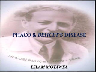
  • 1. PHACO & BEHCET’S DISEASE ESLAM MOTAWEA
  • 2. INTRODUCTION First described in 1937 by Hulusi Behcet, • As classic trisymptom complex of hypopyon, iritis, and orogenital aphthosis. • Turkey has the highest prevalence: 80 to 370 cases per 100,000 population.
  • 3. • Chronic, relapsing-remitting, occlusive vasculitis affecting multiple organ systems • Characterized by oral and genital ulcerations (sores), uveitis, skin rashes, arthritis, thrombophlebitis, colitis, and neurologic symptoms. • Histologically, there is a combination of a perivascular neutrophilic or lymphocytic infiltration, endothelial cell damage or swelling, fibrinoid necrosis coupled with a pro-thrombotic tendency BEHCET’S DISEASE
  • 4. • Predominant age: 3rd to 4th decades • Predominant sex: M > F and with more severe disease in some Mediterranean areas. • In Iran, M:F ratio was 24:1 among 1712 patients. • In Turkey, ratio was 16:1 among 427 patients.
  • 5. Mortality/Morbidity • Chronic morbidity is typical; leading cause is ophthalmic involvement, which can result in blindness. • Effects of disease may be cumulative, especially with neurologic, vascular, and ocular involvement. • Mortality rate is low, but death can occur from neurologic involvement, vascular disease, bowel perforation, cardiopulmonary disease, or as a complication of immunosuppressive therapy.
  • 6. PATHOPHYSIOLOGY • Cause is not known • Immunogenetics, immune regulation, vascular abnormalities, or bacterial and viral infection may have a role
  • 7. History • Signs and symptoms may be recurrent, may precede onset of mucosal membrane ulcerations by 6 months to 5 years. • Prior to onset of disease, patients may experience a variety of symptoms. • Malaise • Anorexia • Weight loss • Generalized weakness • Headache • Perspiration • Decreased or elevated temperature • Lymphadenopathy • Pain of the substernal and temporal regions • A history of repeated sore throats, tonsillitis, myalgias, and migratory erythralgias without overt arthritis is common.
  • 8. spectrum of organ involvement in behçet’s disease.
  • 9. Diagnostic criteria from the Behçet syndrome research committee of Japan (1987 revision) Diagnosis a) Complete – Four major features b) Incomplete – (1) 3 major features, (2) 2 major and 2 minor features, (3) Typical ocular symptom and 1 major or 2 minor features c) Possible – (1) 2 major features (2) 1 major and 2 minor features • Major features • Recurrent aphthous ulceration of oral mucous membrane • Skin lesions -Erythema nodosum – like lesions, subcutaneous thrombophlebitis, folliculitis (acnelike lesions), cutaneous hypersensitivity • Eye lesions - Iridocyclitis, chorioretinitis, retinouveitis, definite history of chorioretinitis or retinouveitis • Genital ulcers • Minor features • Arthritis without deformity and ankylosis • Gastrointestinal lesions characterized by ileocecal ulcers • Epididymitis • Vascular lesions • Central nervous system symptoms
  • 10. • Recurrent oral ulceration - Minor aphthous or major aphthous or herpetiform ulceration recurs at least 3 times in one 12-month period plus 2 of the following: • Recurrent genital ulceration – Recurrent genital aphthous ulceration or scarring, especially males, observed by a physician or reliably reported by a patient • Eye lesions – (1) Anterior uveitis, posterior uveitis, and cells in vitreous upon slit-lamp examination or (2) retinal vasculitis observed by physician (ophthalmologist) • Skin lesions – (1) Erythema nodosum–like lesions observed by physician or reliably reported by a patient, pseudofolliculitis, and papulopustular lesions or (2) acneiform nodules consistent with Behçet disease, observed by a physician, and in postadolescent patients not receiving corticosteroids • Positive pathergy test – An erythematous papule larger than 2 mm at prick site 48 hours after application of a 20- to 22-gauge sterile needle, which obliquely penetrated avascular skin to a depth of 5 mm as read by a physician at 48 hours Findings are applicable if no other clinical explanation is present. International criteria for the classification of Behçet disease (1990)
  • 11. Physical • Oral ulcers (100%) • Indistinguishable from common aphthae (canker sores). • Aphthae may be more extensive, more painful, more frequent, and evolve quickly from a pinpoint flat ulcer to a large sore. • Lesions can be shallow or deep (2-30 mm in diameter) and usually have a central, yellowish, necrotic base and a punched- out, clean margin. • Appear singly or in crops, located anywhere in the oral cavity, persist for 1-2 weeks, and subside without leaving scars. • M/c sites - tongue, lips, buccal mucosa, and gingiva; • Tonsils, palate, and pharynx are less common sites. • Interval b/w recurrences ranges from weeks to months. • 3 types:
  • 12. Minor ulcer Consists of 1-5 small, moderately painful ulcers persisting for 4- 14 days Major ulcer 1-10 very painful ulcers, measuring 10-30 mm, persisting up to 6 weeks, and possibly leaving a scar upon healing Herpetiform ulcer Recurrent crop of as many as 1000 small and painful ulcers
  • 13. Genital manifestations • Genital ulcers (90%, M>F) resemble their oral counterparts but cause greater scarring. • In males, usually occur on scrotum, penis, and groin. • In females, occur on vulva, vagina, groin, and cervix • Ulcers may found in urethral orifice and perianal area. • Epididymitis may arise in men • Orchiepididymitis,10.8% of men.
  • 14. Cutaneous manifestations(58.6-97%)  A variety of skin lesions may appear,  Eg.- Sweet syndrome–like lesion. Papulopustular eruptions (55-83%, M>F) Erythema nodosum–like lesions (44-62%, F>M)
  • 15. Cutaneous manifestations • Erythema multiforme–like lesions • Thrombophlebitis • Ulcers • Bullous necrotizing vasculitis • Pyoderma gangrenosum • Pathergy reaction • Erythematous papule larger than 2 mm develops at prick site after 48 hours • Higher positivity (84-98%) is found in Mediterranean areas and Middle East than in Far East (40-70%), • Western countries having significantly lower positivity Typical positive pathergy reaction at injection site
  • 16. Ocular manifestations(70/90%) The frequency of ocular involvement in patients with BD is approximately 70%-90% The characteristic ocular involvement is a relapsing remitting panuveitis The ocular disease manifests approximately in 2-3 years after the initial symptoms noted Morever, eye involvement may be the first presenting manifestation of BD in approximately 10% to 20 % of the patients. The mean age at onset of uveitis is around 30 years. Males are more frequently involved, had an earlier disease onset, and had a more severe disease. The frequency of bilateral involvement ranges between 78% and 95% in in most of the series Bilateral but asymmetric ocular inflammation is a characteristic feature Behçet panuveitis is a medical emergency, which must be treated immediately. Ocular involvement in patients with BD, especially posterior uveitis presence, is the primary indication of immunosuppressive treatment.
  • 17. Decreased visual acuity is a result of secondary glaucoma, cataracts, or vitreous hemorrhage. Blindness has been reported to occur within 4-5 years from onset of ocular symptoms. Posterior segment involvement is a poor prognostic sign of ocular BD and is seen in up to 93% of patients with ocular disease The most common and universal posterior segment finding are vitritis and retinal perivasculitis involving both the arteries (periarteritis) and veins (periphlebitis). The most common complication of ocular BD is cystoid macular edema (CME), which observed in approximately in half of the patients with uveitis. It may resolve with appropriate treatment or if untreated progress to persistent chronic macular damage and sequel of CME, with structural changes after recurrences resulting in permanent visual loss. In some cases, it may lead to form a partial or full-thickness macular hole formation
  • 18. .
  • 19. Vascular involvement • Occurs in 30-50%, mostly men. • H/P- media thickening, elastic fiber splitting, and perivascular round cell infiltration. • 4 types of vascular lesions – • Aterial and venous occlusions, aneurysms, and varices. • Venous inv. M/C occlusion, rarely varices. • M/C sites - SVC,IVC, deep femoral vein, and subclavian vein.
  • 20. • Gastrointestinal involvement (>10%) • Clinical spectrum is enormously varied • Anorexia, vomiting, dyspepsia, diarrhea, abdominal distention, and abdominal pain • Cardiac manifestations (5-17%) • Coronary vasculitis and thrombosis, pericarditis, myocarditis, endocarditis with granulomatous changes or fibrosis, regurgitation, and diastolic dysfunction • Lung involvement (up to 18%) • Pulmonary vasculitis, hypertension, pleural effusions and Aneurysms
  • 21. Joint manifestations (30 - 40%) • More than half patients develop signs or symptoms of synovitis, arthritis, and/or arthralgia during course of disease. • Most frequent minor feature in childhood-onset BD is reported to be arthritis, occurring in 11 of 40 patients. • Multiple-joint involvement is common. • Clinical features have been reported as pain, tenderness, swelling, limitation of joint movement, warmth, and morning stiffness.
  • 22. Neurologic manifestations (3.2-49%) • May present as meningoencephalitis, a multiple sclerosis–like illness, acute myelitis, stroke, or pseudotumor cerebri. • Most serious, leading to severe disability and a high fatality rate. • Usually occur within 5 years of disease onset. • Severe headache is most frequent initial symptom. • Three categories (Pallis and Fudge (1956) and Wadia and Williams (1957) (1) Brain stem syndrome, A/w fever, arthralgia and skin eruption; brainstem signs developes subacutely with evolving cranial neuropathies, ocular motor dysfunction, nystagmus and gaze palsies, dysarthria and ataxia, bulbar muscle weakness, Headache with meningism, CSF pleocytosis and high protein content (2) Meningomyelitis syndrome, Meningitis with spinal cord and hemisphere signs (Silfverskiold, 1951) (3) Organic confusional syndrome, Meningoencephalitis without focal neurological signs, some times chronic and progressive, resulting ultimately in dementia, Parkinsonism, pseudobulbar palsy and quadriparesis
  • 23. Pregnancy-associated manifestations • Pregnant women may experience more severe symptoms during first trimester. • Overall, pregnancy does not seem to markedly affect the course of BD. • Close follow-up is necessary to monitor the health of mother and baby.
  • 24. Differential Diagnoses • Acute Febrile Neutrophilic Dermatosis • Pyoderma Gangrenosum • Cyclic neutropenia • Aphthous Stomatitis • Erythema Multiforme • SJS-TEN • Erythema Nodosum • Viral infections • Herpes Simplex, coxsackie, echo • Syphilis and other STIs • Lupus Erythematosus, Acute • Reiter disease • VKH syndrome • Bullous autoimmune diseases • JRA • Sarcoidosis • Multiple sclerosis • Ulcerative colitis, crohn disease
  • 25. Laboratory Studies • Mild anemia and leukocytosis in chronic disease. • ESR, C-reactive protein value, and other acute phase reactants may be elevated during the active didease, but they do not correlate well with clinical activity. • IgA, IgG, alpha-2 globulin, IgM, and immune complexes are occasionally elevated. • Rheumatoid factor and antinuclear antibodies are absent. • Antineutrophil cytoplasmic antibody and antiphospholipid antibody test results are usually negative.
  • 26. BD needs multidisciplinary approach • Dermatologist - For evaluation of mucocutaneous lesions (ie, oral ulcer, genital ulcer, skin lesions) • Ophthalmologist - For evaluation of eye involvement • Rheumatologist or orthopedic surgeon - For evaluation of joint involvement • Neurologist or psychiatrist - For evaluation of CNS involvement • Internal medicine specialist - For evaluation of gastrointestinal, pulmonary, renal, or endocrine involvement • General surgeon - For evaluation of gastrointestinal involvement • Chest surgeon or cardiologist - For evaluation of cardiovascular involvement • Ear, nose, and throat specialist or dentist - For evaluation of oral cavity
  • 27. Medical Care • Treatment of BD symptomatic and empiric. • Choice of treatment depends on site and severity of clinical manifestations
  • 28.
  • 29. Local therapy  Recurrent aphthae • mild diet, avoidance of irritating agents, • Caustic solutions (silver nitrate, 1%-2%; tinctura myrrha, 5%-10% weight/volume; H202,0.5%; methylviolet,0.5%) 1-2xld • Antiseptic and anti-inflammatory preparations (amlexanox, 5% in oral paste; triclosan, 0.1% mouthwash solution and in toothpaste ), 3% diclofenac in 2.5% hyaluronic acid, tetracycline mouthwash • Corticosteroids (triamcinolone mucosal ointment, dexamethasone mucosal paste, betamethasone pastilles) 4 x/day or during night (ointment / paste) or intrafocal infiltrations with triamcinolone suspension 0.1-0.5 mL per lesion • Anaesthetics (lidocaine - 2%-5%; mepivacaine - 1.5%, tetracaine - 0.5%-1% gels or mucosal ointments) 2-3 x/day • 5-Aminosalicylic acid (5% cream) 3 x/day (reduces the duration of lesions and the pain • Sucralfate suspension, 5 mL x 4/day (for oral aphthous and genital ulcers) • Topical tacrolimus, nicotine patches, and topical G-CSF
  • 30.  Genital ulcers and skin lesions, • Corticosteroid and antiseptic creams for 7 days. • Painful genital ulcerations topical anaesthetics in cream. • Corticosteroid injections (triamcinolone, 0.1 to 0.5 ml/lesion) in recalcitrant ulcerations.
  • 31. Systemic therapy • Corticosteroids • Mainstay for all clinical manifestations. • Have a beneficial effect on acute manifestations, no definite evidence effectiveness in controlling progression. • Adverse effects of long-term therapy must be considered. • Mucocutaneous lesions and arthritis:- • NSAIDs, zinc sulfate, levamisole, colchicine, dapsone, or sulfapyridine and thalidomide. Immunosuppressive therapy with azathioprine, chlorambucil, or cyclophosphamide • Uveitis and CNS involvement:- • Systemic corticosteroids, azathioprine, or cyclosporine, MTx • In CNS involvement, risks and benefits of cyclosporine need to be considered b/c of neurological side effects.
  • 32. • Anticoagulants are given to patients with thromboses. • Other therapeutic approaches • IFN alfa, IFN gamma, acyclovir, high-dose corticosteroids or cyclophosphamide pulse therapy, and tacrolimus • Japanese tacrolimus study group reported that tacrolimus was effective in treating refractory uveitis in a dosage-dependent manner. • Adverse effects were renal impairment (28.3%), neurologic symptoms (20.8%), gastrointestinal symptoms (18.9%), and hyperglycemia (13.2%).
  • 33. • Subcutaneous IFN alfa-2a therapy • resulted in reduced ulcers and eye disease. • Flulike symptoms were most common adverse effect. • Leukopenia, hair loss, and development of antinuclear and antithyroid antibodies were reported less commonly. • A patient presenting with oral ulcers resistant to prednisone, azathioprine, colchicine, dapsone, and cyclosporin responded well to lenalidomide. • A patient with Behçet disease with ocular involvement, dependant on corticosteroids and refractory to azathioprine, showed improvement with addition of pentoxifylline
  • 34. • A case of Behçet disease resistant to prednisolone, cyclosporin, azathioprine, infliximab with methotrexate, and colchicine has been successfully treated with anakinra. • infliximab, and etanercept, have decreased frequency of attacks • Etanercept has been used at 25 mg twice a week. • Pediatric case responding to infliximab has been reported. • Infliximab has resulted in responses after etanercept failed. • Infliximab infusions of 5-10 mg/kg have been used with variable dosing schedules. • Several patients not responding to infliximab have been treated with adalimumab.
  • 35. drug doses indication Methylprednisolone 40 mg/every 3 wk lM Erythema nodosum (but not orogenital ulcers) Rebamipide 300 mg/day PO Oral ulcers Colchicine 1-2 mg/day PO Erythema nodosum, arthritis, genital Ulcer s (oral ulcer in female) Dapsone 100 mg/day PO Orogenital ulcers, skin lesions, pathergy Azathioprine 2.5 mg/kg/day Recent onset ocular disease lnterferon-alfa-2a 6x10⁶ lU/3x/wk SC Orogenital ulcers, papulopustular lesions Thalidomide 100 mg/day Orogenital ulcers, Papulopustular lesions CyclosporinA 10 mg/kg/day PO Ocular manifestations, skin lesions, orogenital ulcers Etanercept 25 mg/2 x/wk P0 Oral ulcers, papulopustular lesions nodular lesions, (not pathergy test)
  • 36. Behcet’s Disease: Prognosis • Behcet's disease has an undulating course of exacerbations and remissions, and may become less severe after approximately 20 years. • Appears to be more severe in young, male, and Middle Eastern or Far Eastern patients.
  • 37. Cataract in uveitis as a result of the intraocular inflammation per se, from chronic corticosteroid usage or more often from both [1]. The incidence of cataract in uveitis varies from 57% in pars planitis [2] to 78% in Fuchs heterochromic iridocyclitis (FHI) [3].
  • 38. Cataract Surgery in Behcet’s Disease. Cataract forma-tion is the most common anterior segment complication after recurrent inflammation, occurring in up to 36% of cases [1 It was reported that the postoperative visual acuity was found to be significantly lower in eyes with BD than in those with idiopathic uveitis because of the severe posterior segment complications, mainly optic atrophy [48]. Operating on eyes with cataract and uveitis has been previously reviewed by Foster and associates [8]. Their recommendations for a successful cataract surgery and for minimizing the postoperative uveitis are as follows. Uveitis should be inactive for at least 3 months pre-operatively, systemic and topical steroids should be used prophylactically for 1 week preoperatively and continued postoperatively, immunosuppressive drugs should be con-tinued, complete removal of cortical material should take place, and one-piece PMMA posterior chamber intraocular lens should be used if the patient and the surgeon understand the special nature of this surgery, its risks, and the prognosis for success. In another paper by Berker et al., the authors reported results of phacoemulsification and intraocular lensimplan-tation in patients with Behcet’s disease [49]. They reported 72.5% of eyes had improvement in vision after surgery. However, the vision got worse in 17.5% of the eyes. Most frequent complication reported by them was posterior capsular opacification in 37.5% of eyes. Other complications were posterior seynchiae and severe inflammation. Posterior segment complications such as epiretinal membrane forma-tion, cystoid macular edema, and optic atrophy were also reported by the authors.
  • 39.
  • 40. Current Guidelines for the Management of Uveitic Cataract 4.1. Clinical Evaluation of Complicated Cataract and Asso-ciated Uveitis. The eye in which visual loss is mainly attributable to cataract formation is most likely to benefit from cataract surgery. The outcome of surgery depends upon several factors, namely the uveitic diagnosis, proper perioperative management and meticulous surgery. The specific uveitic diagnosis is of paramount importance also when planning the surgical strategy [4], such as determining whether an intraocular lens should be implanted or not.
  • 41.  The visual potential of the eye, determined by. The state of the macula and optic nerve should be thoroughly examined for during the preoperative assessment. Macular ischemia, atrophy, chronic macular edema, or scar, such as that resulting from a choroidal neovascular membrane, are poor prognostic factors. Similarly, optic atrophy and severe cupping of the optic disc are bad prognostic signs
  • 42.  . In presence of dense lens opacity, B scan ultrasonography should be done to rule out retinal detachment which may complicate the eye with uveitis  as pre-existing corneal scars and severe iris atrophy may also compromise the visual outcome.   Regardless of the guarded prognosis, a definite indication for cataract removal in an eye that is not blind, is phacoanti-genic uveitis. This may result from the hypermature state of a cataract, whereby lens proteins leak out of the capsular bag through an intact capsule, or in cases of trauma, where the lens capsule has been breached, leading to persistence of intraocular inflammation. Cataract surgery may also be indicated to permit better visualization of the posterior segment for appropriate medical or surgical management of the eye
  • 43. Control of Pre Operative Inflammation  The risk of reactivation of uveitis must be assessed. Jancevski and Foster recommended the use of supplementary perioperative anti-inflammatory therapy to prevent damage to ocular structures
  • 44.  to reduce the risk of postoperative CME [59], steroid prophylaxis should be given perioperatively to protect against recurrence of macular swelling. Similarly, steroid prophylaxis should be administered in eyes at risk of developing recurrence of uveitis following cataract surgery,  for example, Vogt-Koyanagi Harada disease, Behcet’s disease and birdshot choroidopathy, to name a few. This may take the form of oral steroids 1 mg per kg/day starting 3 days preoperatively, tapering the steroid dose according to the amount of inflammation postoperatively  . Alternatively, if there are no contraindications to periocular steroid injections, such as documented steroid response or infectious uveitis, an orbital floor or sub-tenon’s injection of depot steroid, such as triamcinolone acetonide 40 mg/1 mL may be given,  especially in patients where high doses of oral steroid are contraindicated, for example in poorly controlled diabetics.  In addition, guttae prednisolone acetate 1% 2 hourly administered 2 days prior to surgery, together with an oral and topical non-steroidal anti-inflammatory agent, can be given. Supported by encouraging results in the recent literature,  the authors favour an intravitreal injection of preservative-free triamcinolone acetonide 4 mg in 0.1 mL at the conclusion of cataract surgery [60–62].
  • 45. Complications of Uveitis Adversely Affecting Surgical Outcome: High-Risk Surgical Cases. Determining the surgical risk is a very important aspect of preoperative assessment. Eyes which have generally low intraocular pressures, espe-cially readings of 6 mmHg or less even when quiescent, are at high risk of developing postoperative hypotony or even phthisis bulbi. Other warning signs such as seclusio papillae with normal intraocular pressure reading and apparent phacodonesis without evident zonulysis are important poor prognostic signs for postoperative hypotony. Eyes in which the uveitis is difficult to control are also at high risk of severe postoperative inflammation and hypotony or phthisis bulbi. The presence of choroidal effusion on B scan ultrasonogra-phy or diffusely thickened choroid is a poor prognostic sign. Conducting a careful ultrasonic biomicroscopy is essential in eyes with relative hypotony [65] to assess the state of the ciliary body and its processes. If the ciliary body has undergone atrophy, the risk of hypotony is high. If the ciliary body is found to be detached and processes appear under traction from a ciliary (cyclitic) membrane, cataract surgery should be combined with vitrectomy and trimming of the ciliary membrane aided by indentation of sclera to relieve ciliary body traction and to restore normal IOP.
  • 46. Diagnostic Aids  . Apart from the standard means of assessing macular, optic nerve and retinal function, one may apply additional methods such as pupillary response, light projection, colour perception and B scan ultrasonography, or an attempted OCT in looking for macular atrophy, edema, or hole [66].
  • 47. Optimal Time for Cataract Surgery. Before scheduling surgery, the ophthalmologist should attempt to ensure that the eye has been quiescent for at least 3 months [11]. This has been shown to reduce the risk of postoperative CME [59]. In cases where, despite heavy immunosuppression, the intraocular inflammation is still not completely abolished and surgery is urgently required, such as in an intumes-cent cataract, the patient may be administered intravenous methylprednisolone 1 g one day before surgery. A study from Japan suggests that in patients with Behcet’s disease the eye should be inactive for a minimum of 6 months and that the risk is higher if attacks have occurred within 12 months of cataract surgery [70]. Counselling of the Patient for Uveitic Cataract Surgery. The most important aspect of counselling when planning to perform cataract surgery for the uveitic eye is explaining the visual prognosis. The general risks involved in surgery, such as infection and other intraoperative complications will also need to be thoroughly explained, especially if there is phacodonesis, hypotony, or glaucoma.
  • 48. 5. Surgical Technique 5.1. Choice of Surgery. The choice of cataract surgery tech-nique is best left to the surgeon Cataract removal by phacoemulsification is safer for the uveitic cataract as less inflammation is induced than that by a man-ual extracapsular cataract extraction. During the surgery, the anatomy of the anterior segment should be restored to a state as close to normal as possible. Some uveitic cataract eyes are complicated by glaucoma or retinal problems that may also benefit from surgery. For eyes with concomitant uveitic glaucoma, surgery is preferably not combined with the cataract surgery as the risk of bleb failure is increased with drainage of post-cataract surgery inflammatory exudate through a healing bleb. Where possible, cataract surgery should be done first.
  • 49.  Regarding retinal complications, such as epiretinal mem-branes or coexisting retinal detachment, cataract surgery may be combined with vitreoretinal surgery.  In cases with major retinal problems, the eye may be safely rendered aphakic until the retinal problem has been dealt with. In eyes with intermediate uveitis, or FHI, cataract surgery may be combined with vitrectomy, performed to clear the vitreous gel, thereby reducing vitreous clouding. In intermediate uveitis, this often not only improves vision but also controls intraocular inflammation and helps resolve the cystoid macular edema.  At the end of surgery, especially with combined surgical procedures, having excluded steroid responders and eyes with infectious uveitis, an intravitreal injection of triamcinolone acetonide, is often adequate to control the postoperative inflammation and prevent CME.
  • 50. .2. Intraoperative Surgical Techniques and Skills Posture. Patients with ankylosing spondylitis with a fixed flexion deformity of the axial spine, especially when the cervical spine is involved Surgical Challenges. The uveitic eye poses numerous surgical challenges. These include the small pupil, shallow anterior chamber, posterior synechiae, peripheral ante-rior synechiae, pupillary membranes and even zonulolysis
  • 51. Anaesthesia. Whilst phacoemulsification surgery may be done under topical anesthesia, manipulation of the iris may induce ocular discomfort or pain. Either regional anesthesia or an intracameral injection of preservative- free lignocaine 1% can provide adequate analgesia. For children and in patients for whom prolonged surgical time is anticipated, as in severe zonulolysis requiring modified capsular tension rings that need suturing, general anesthesia may be preferred.
  • 52. Incision. Either a scleral or temporal clear corneal incision may be used. However, the incision should be of adequate length in order to prevent iris prolapse in eyes with small or stretched pupils. Pupil Enlargement. balanced salt solution with adrenaline (1 : 1000 0.5 mL adrenaline in 500 mL) into eyes with pupils that are not bound by synechiae or membranes. Preservative-free intracameral lignocaine 1% may also be used to help dilate the pupil only if it is not bound. Healon 5 is useful can physically roll open the pupil and keep it dilated as long as the aspiration flow rate is kept low.
  • 53.  (f) Synechiolysis and Removal of Pupillary Membrane.  When both are present, the PAS should be released before the PS. Release of PAS may be done by injecting viscoelastic, such as Healon 5 to physically separate the iris from the cornea  tip of the viscoelastic cannula may be used to sweep the iris away from the peripheral cornea as the viscoelastic material is being injected into the angle of the anterior chamber. This should be done very gently and carefully, taking care not to detach Descemet’s membrane in the process.
  • 54. sweeping the pupil free from the lens capsule with a cannula. In cases where a narrow strip of membrane is present at the site of PS formation, a 27-gauge needle may be used to simultaneously nick the membrane to segment and release it from the anterior capsule and release the PS, thus stretching the pupil. Pupil Expansion. The most user-friendly instrument for extensive synechiae once an edge of the iris has been viscodissected off the anterior capsule is a bent Kuglen hook Alternative means of opening the pupil include the use of a Beehler pupil dilator (2 or 3 pronged) to mechanically stretch the pupil in a single injector system
  • 55.
  • 56.
  • 57.
  • 58. Continuous Circular Capsulorhexis. It is generally prefer-able to keep the size of the capsulorhexis slightly smaller than the pupil so that iris chaffing does not occur and results in progressive intraoperative miosis as nuclear fragments are being moved out of the capsular bag (Figure 8). This also contributes to increased postoperative inflammation. When the pupil size is small, the capsulorhexis inevitably needs to be larger than the pupil. The anterior chamber must be kept deep and the anterior capsule flattened using adequate viscoelastic material in order to control the tearing of a capsulorhexis. The capsulotomy can be initiated by a 26- or 27-gauge bent cystitome
  • 59.  Creating the ideal capsulorhexis is also very important in preventing posterior capsule opacification. The capsulorhexis should be centred, overlapping the edge of the optic at all times, but not so small as to prevent capsular phimosis.  Nucleus Management. In small pupils, the safest tech- nique is the vertical chop employed in the in situ chop technique. Chopping of fragments is done within the pupillary aperture with the phaco tip kept in view at all times with minimal risk of engaging and traumatizing the iris
  • 60. Irrigation and Aspiration. This step must be done thor-oughly so as not to leave cortical material behind. Intraocular Lens Implantation Contraindications and Type of IOL. the most biocompatible IOL for the anterior chamber and the capsular bag is a single-piece, square-edged acrylic (either hydrophilic or hydrophobic) IOL. They also found that the posterior capsular opaci-fication rate was highest (34.2%) in eyes with silicone IOLsof postoperative cystoid macular edema and PS formation, and pupillary membranes
  • 61.  hydrophilic acrylic material has good uveal but worse capsular biocompatibility, but hydrophobic acrylic material had lower uveal but better capsular biocompatibility [78]. In eyes with chronic uncontrolled uveitis, IOL implant should be deferred.   Removal of viscoelastic from under the IOL is an important step in reducing the space behind the IOL into which lens epithelial cells tend to migrate, thus causing PCO. Pressing the optic against the posterior capsule when using a single-piece hydrophobic IOL also encourages its adhesion to the posterior capsule, thereby reducing the risk of PCO
  • 62. Complications and Postoperative Management 5.3.1. Intraoperative Complication An infrequent intraoperative complication is Zonulolysis (CTR) is often necessary in order to prevent IOL decentration Failure to use a CTR in the presence of weak zonules may result in capsular phimosis, due to unopposed capsular bag fibrosis and shrinkage.
  • 63. Retained Lens or Nuclear Fragments.  Due to the small pupil size, small hard nuclear  These small fragments can cause recurrent anterior uveitis ;may also cause localized corneal edema localized corneal decompensation in the long term. Hence, at the end of phaco, the eye should be given a gentle shake whilst aspirating to ensure no nuclear fragments are inadvertently left behind. Any retained soft lens material or nuclear fragment should be removed surgically as soon as possible [80].
  • 64. . Early Postoperative Complications Excessive Postoperative Inflammation. One of the most com-mon postoperative complications is cystoid macular edema phacoemulsification, the incidence has been reported to range from 12% to 59 Generally, if preoperative prophylactic oral steroids have been given and maximal topical steroids and cycloplegics have proven ineffective in controlling the uveitis, the dose of oral steroids may be sharply increased. If no prophylactic oral steroids had been given the patient should be given an oral pulse of steroids or injection of periocular steroids. An alternative means would be to give an intravitreal injection of triamcinolone acetonide
  • 65. Recurrence of Uveitis. Increased frequency of recurrence following cataract surgery may occur. This is thought to be triggered by the intraocular procedure. The recurrence rate has been reported to be as high as 51% [15]. Hence, stepping up the immunosuppression for the long term may be necessary to prevent further recurrences
  • 66.  Intraocular Pressure (IOP) Abnormalities.  The IOP may be raised transiently during the early postoperative period in eyes with compromised trabecular meshwork or angles. This can often be managed with topical and systemic anti-glaucoma medications.   However, the surgeon’s greatest fear is hypotony.  Once wound leakage has been ruled out, the next step is to increase the anti- inflammatory therapy topically and systemically. This is often effective in raising the IOP, but the topical steroids may be difficult to taper or withdraw and patients may require long-term topical steroids to maintain the IOP.  In severe cases, vitrectomy and trimming of ciliary body traction membranes and silicone oil filling may be needed if UBM shows the presence of ciliary body detachment secondary to tractional membranes not addressed during the cataract surgery [83].
  • 67. 5.3.3. Late Postoperative Complications  Posterior Capsular Opacification. the most common complication following any type of cataract surgery.  reported an incidence of 48.0%, Rauz et al. [84] 81.7% and Kuc¸¨ukerd¨onmez¨ et al. [82] 34.2% at 1 year Their corresponding Nd:YAG capsulotomy rates were 32.2%, 8.3%, and 3.6%.  Preventive measures include creating a circular well-centred capsulorhexis which is smaller than the optic size, using an acrylic IOL with a square-edged optic design, meticulous removal of viscoelastic from within the capsular bag and ensuring the optic is stuck on to the posterior capsule at the conclusion of surgery. Control of postoperative inflammation also plays an important role in preventing PCO.
  • 68.  Explanting an IOL. Removal of intraocular lens in uveitic  eyes is rarely necessary. Foster et al. reported that their indications include the formation of perilental membrane, chronic low-grade inflammation not responding to anti-inflammatory treatment and cyclitic membrane resulting in hypotony and maculopathy
  • 69.
  • 70. 6. Conclusion and Summary  Preoperative factors include proper patient selection and counseling and preoperative control of inflam-mation  therefore control of inflammation, both pre-and postoperatively, is vital. The use of immunosuppressive agents other than steroids also helps control inflammation  Management of postopera-tive complications, especially inflammation and glaucoma, earlier rather than later, has also contributed to improved outcomes  conclusion, management of the uveitic cataract requires careful case selection, proper timing of surgery, meticulous surgery and close monitoring with appropriate handling of the postoperative complications that may occur
  • 71.
  • 72.
  • 73. Take home msg 1.Revise the diagnosis of Behcet. I have seen many cases where the previous diagnosis is not true.  2- If it is Behcet, giving steroids only without an immune modulator is forbidden.  Clinical Evaluation of Complicated Cataract and Asso-ciated Uveitis.  Control of Pre Operative Inflammation  Complications of Uveitis Adversely Affecting Surgical Outcome  Diagnostic Aids  Optimal Time for Cataract Surgery  Counselling of the Patient for Uveitic Cataract Surgery  Surgical Technique  Choice of Surgery.  Intraoperative Surgical Techniques and Skills  Posture.  Surgical Challenges.  Anaesthesia.  Incision.  Pupil Enlargement.  Synechiolysis and Removal of Pupillary Membrane.  Pupil Expansion.  Continuous Circular Capsulorhexis.  Nucleus Management  Irrigation and Aspiration.  Intraocular Lens Implantation Contraindications and Type of IOL.  complication intrea ,,,early ///late after starting the proper treatment, we wait for three months of documented systemic as well as ocular inactivity.  Since there is no ocular activity now, assess for systemic activity. If the disease is systemically active, ask for CBC and liver enzymes, and if they are normal, add lmmuran 50 mg, one tablet twice daily for two weeks then three times daily for two weeksand inform me of the response of systemic activity to this teatment four weeks from now.