
Recommended
More Related Content
What's hot
What's hot (20)
Similar to Toxoplasma gondii
Similar to Toxoplasma gondii (20)
Recently uploaded
Recently uploaded (20)
Toxoplasma gondii
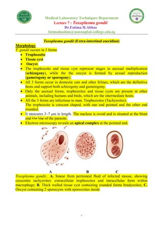
- 1. 1 Medical Laboratory Techniques Department Lecture 7 : Toxoplasma gondii Dr.Fatima H.Abbas fatimahashim@mustaqbal-college.edu.iq Toxoplasma gondii (Extra-intestinal coccidian) Morphology T. gondii occurs in 3 forms Trophozoite Tissue cyst Oocyst. The trophozoite and tissue cyst represent stages in asexual multiplication (schizogony), while the the oocyst is formed by sexual reproduction (gametogony or sporogony). All 3 forms occur in domestic cats and other felines, which are the definitive hosts and support both schizogony and gametogony. Only the asexual forms, trophozoites and tissue cysts are present in other animals, including humans and birds, which are the intermediate hosts. All the 3 forms are infectious to man. Trophozoites (Tachyzoites). The trophozoite is crescent shaped, with one end pointed and the other end rounded. It measures 3–7 μm in length. The nucleus is ovoid and is situated at the blunt end حادة نهاية of the parasite. Electron microscopy reveals an apical complex at the pointed end. A B C Toxoplasma gondii:. A. Smear from peritoneal fluid of infected mouse, showing crescentic tachyzoites: extracellular trophozoites and intracellular form within macrophage; B. Thick walled tissue cyst containing rounded forms bradyzoites; C. Oocyst containing 2 sporocysts with sporozoites inside
- 2. 2 Toxoplasma gondii: Trophozoite (tachyzoite), fine structure seen by electron microscopy The trophzoite stains well with Giemsa stain, the cytoplasm appearing azure blue and the nucleus, red. The actively multiplying trophozoite is seen intracellularly in various tissues during early acute phase of infection. Extracellular trophozoites can also be seen in impression smears. It can invade any nucleated cell and replicate within cytoplasmic vacuoles by a process called endogony (internal budding), wherein 2 daughter trophozites are formed, each surrounded by a membrane, while still within the parent cell. When the host cell becomes distended with the parasite, it disintegrates, releasing the trophozoites that infect other cells. During acute infection, the proliferating trophozoites within host cell may appear rounded and enclosed by host cell membrane. This is, called pseudocyst or colony and can be differentiated from tissue cysts by staining reactions. The rapidly proliferating trophozoites in acute infection are called tachyzoites The trophozoites are susceptible to drying, freeze thawing, and gastric digestion. Tissue cyst Tissue cysts are the resting form of the parasite. They are found during chronic stage of the infection and can be found in the brain (most common site), skeletal muscles, and various other organs. The cyst wall is eosionophilic and stains with silver, in contrast to the pseudocyst. With periodic acid Schiff (PAS) stain, the cyst wall stains weakly, and the parasites inside are stained deeply. The slowly multiplying parasites within the cyst are called bradyzoites.
- 3. 3 The cyst is round or oval, 10–20 μm in size and contains numerous bradyzoites. Cysts remain viable in tissue for several years. In immunologically normal hosts, the cysts remain silent, but in the immunodefcient subjects, they may get reactivated, leading to clinical disease. It is relatively resistant and when the raw or undercooked meat containing the cysts is eaten, infection occurs. The cyst wall is disrupted by peptic or tryptic digestion and the released parasites initiate infection by invading intestinal epithelial cells. They reach various tissues and organs through blood and lymphatic dissemination. Cysts are susceptible to desiccation, freezing, and thawing, and heat above 60°C. Oocyst Oocysts develop only in intestine of the definitive hosts . It is oval in shape and measures 10–12 μm in diameter. Each cyst is surrounded by a thick resistant wall. The oocystis formed by sexual reproduction (gametogony). Cats shed millions of oocysts per day in feces for about 2 weeks during the primary infection. The freshly passed oocyst is not infectious. They undergo sporulation in the soil with formation of 2 sporocysts, each containing 4 sporozoites. The sporulated oocyst is infective. Oocyst is very resistant to environmental conditions and can remain infective in soil for about a year. When the infective oocyst is ingested, it releases sporozoites in the intestine, which initiates infection. Life Cycle T. gondii completes its life cycle in 2 hosts. Definitive host: Cats and other felines, in which both sexual and asexual cycle takes place. Intermediate hosts: Man and other mammals, in which only the asexual cycle takes place. T. gondii has 2 types of life cycles: 1. Enteric cycle 2. Exoentric cycle.
- 4. 4 Life cycle of Toxoplasma gondii. Enteric cycle Enteric cycle occurs in cat and other definitive hosts. Both sexual reproduction (gametogony) and asexual reproduction (schizogony) occur within the mucoscal epithelial cells of the small intestine of the cat. Cat acquires infection by ingestion of tissue cysts in the meat of rats and other animals or by ingestion of oocysts passed in its feces. The bradyzoites are released in the small intestine and they undergo asexual multiplication (schizogony) leading to formation of merozoites. Some merozoites enter extraintestinal tissues resulting in the formation of tissue cysts in other organs of the body. Other merozoites transform into male and female gametocytes and sexual cycle (gametogony) begins, with the formation of microgamete and macrogamete.
- 5. 5 A macrogamete is fertilized by motile microgamete resulting in the formation of an oocyst, which passes through maturation stages (sporulation) in the soil after being excreted from host through feces. A mature oocyst containing 8 sporozoites is the infective form which may be ingested by rats or other mammals to repeat the cycle Exoenteric cycle Exoenteric cycle occurs in humans, mice, rats, sheep, cattle, pigs and bird , which are the intermediate hosts. Humans acquire infection after: Eating uncooked or undercooked infected meat, particularly lamb and pork containing tissue cysts. Ingestion of mature oocysts through food, water, or fingers contaminated with cat feces directly or indirectly. Intrauterine infection from mother to fetus (congenital toxoplasmosis) Blood transfusion or transplantation from infected donors. Sporozoites from the oocysts and bradyzoites from the tissue cysts enter into the intestinal mucosa and multiply asexually and tachyzoites are formed (endodyogeny). Tachyzoites continue to multiply and spread locally by lymphatic system and blood. Some tachyzoites also spread to distant extraintestinal organs like brain, eye, liver, spleen, lung, and skeletal muscles and form tissue cysts. The slowly multiplying forms inside the tissue cysts are known as bradyzoites, which remain viable for years. The dormant bradyzoites inside the cyst may be reactived in immune suppression causing renewed infection in the host. Human infection is a dead end for the parasite. Human toxoplasmosis is a zoonosis. The full natural cycle is maintained predominantly by cats and mice. Mice eat materials contaminated with oocysts shed in cats feces. Tissue cysts develop in mice. When such mice are eaten by cats, they get infected and again shed oocysts in feces. Pathogenicity and Clinical Features The outcome of Toxoplasma infection depends on the immune status of the infected person. Active progression of infection is more likely in immunocompromised individuals. Toxoplasmosis has acquired great importance as one of the major fatal complications in acquired immunodeficiency syndrome (AIDS). Most human infections are asymptomatic. Clinical toxoplamosis may be congenital or acquired. Congenital toxoplasmosis Congenital toxoplasmosis results when T. gondii is transmitted transplacentally from mother to fetus.
- 6. 6 Parasite which can be transmitted from mother to fetus: Toxoplasma gondii Plasmodium spp. Trypanosoma cruzi This occurs when the mother gets primary Toxoplasma infection, whether clinical or asymptomatic, during the pregnancy. The risk of fetal infection rises with progress of gestation; from 25%, when the mother acquires primary infection in first trimester to 65% in the third trimester. Conversely, the severity of fetal damage is highest, when infection is transmitted in early pregnancy. Mothers with chronic or latent Toxoplasma infection, acquired earlier, do not ordinarily infect their babies. But in some women with latent or chronic infection, the tissue cyst may be reactivated during pregnancy and liberate trophozoites, which may infect the fetus in utero Most infected newborns are asymptomatic at birth and may remain so throughout. Some develop clinical manifestations of toxoplasmosis weeks, months, and even years after birth. The manifestations of congenital toxoplasmosis include chorioretinitis التهاب المشيمة, cerebral calcifications الدماعية التكلسات, convulsions تشنجات, strabismus الحول, deafness الصم, blindness العمى, mental retardation العقلي التخلف, microcephaly الرأس صغر, and hydrocephalus الرأس استسقاء. A few children are born with manifestations of acute toxoplasmosis, which may include fever, jaundice, petechial rashes, microphthalmia, cataract, glaucoma, chorioretintis, lymphadenopathy, hepatosplenomegaly, myocarditis, cerebral calcifications, and chorioretinitis. Acquired Toxoplasmosis Infection acquired postnatally الوالدة بعد is mostly asymptomatic. The most common manifestation of acute acquired toxoplasmosis is lymphadenopathy اللمفاوية العقد اعتالل ; the cervical lymph nodes being most frequently affected. Fever, headache, myalgia العضلي االلم, and splenomegaly are often present. The illness may resemble mild flu and is self-limited, although the lymphadenopathy may persist. In some cases, there may be a typhus-like exanthema التيفوس يشبة طفح with pneumonitis رئوي التهاب, myocarditis القلب عضلة التهاب, and meninogoencephalitis والدماغ السحايا التهاب, which may be fatal. Ocular Toxoplasmosis Another type of toxoplasmosis is ocular. It may present as uveitis, choroiditis, or chorioretinitis. Some cases may be so severe that they require enucleation. Toxoplamosis in Immunocompromised Patients. Toxoplasmosis is most serious and often fatal in immunocompromised patients, particularly in AIDS, whether it may be due to reactivation of latent infection or new acquisition of infections.
- 7. 7 In these patients, involvement of brain is most common. Clinical manifestation include encephalitis, altered mental state, seizures, cerebellar signs, meningismus, and neuropsychiatric manifestations. Besides central nervous system involvement, other organs involved are lungs, pancreas, gastrointestinal tract, eyes, heart, and liver. Toxoplasma pneumonia can be confused with pneumocystis pneumonia. Host Immunity Host defense against Toxoplasma infection involves both humoral (antibody- mediated) and cellular responses. Specific IgG antibody can lyse extracellular trophozoites, but activated T cells and natural killer cells appear to be more important in containing the infection and preventing clinical disease. Laboratory Diagnosis -Presence of only tissue cysts does not differentiate between active and chronic infection. -The presence of cysts in placenta or tissues of newborn establishes congenital Toxoplasma infection. Antibody detection Diagnosis of acute infection with T. gondii can be made by detection of the simultaneous presence of IgM and IgG antibodies. Tests for detecting IgG antibody include: 1. Enzyme-linked immunosorbent assay (ELISA) 2. Indirect fluroscent antibody test (IFAT) 3. Latex agglutination test 4. Sabin-Feldman dye test.
- 8. 8 -Positive IgG titer (>1:10) can be detected as early as 2–3 weeks after infection. Peak level of antibody is observed in blood 4–8 weeks after infection. -A positive IgM antibody titer indicates an early primary infection. The serum IgM titer can be measured by double sandwich IgM ELISA or IgM immunosorbent assay (IgM-ISAGA). Both assays are equally specific and sensitive. Negative IgM titer and postive IgG titer indicate distant infection. -The double sandwich IgA-ELISA test is used for detecting congenital infection in newborns. Antigen detection Detection of antigen by ELISA indicates recent Toxoplasma infection. -In AIDS and other immonocompromised patients, antigen detection is very useful. -Detection of antigen in amniotic fluid is helpful to diagnose congenital toxoplasmosis. Immunocompromised Patients AIDS patients who are seropositive for T. gondii and have a CD4+ Tlymphocyte count below <100/μL, should receive primary prophylaxis against Toxoplasma encephalitis. *Trimethoprimsulfamethoxazole is the drug of choice. If this drug cannot be tolerated by patients, dapsonepyrimethamine is the recommended alternative drug of choice. * Prophylaxis against toxoplasma encephalitis should be discontionued in patients who have responded to antiretroviral therapy (ART) and whose CD4+ T lymphocyte count has been above 200/μL for 3 months. Notes Individuals at risk, particularly pregnant women, children, and immunocompromised person's should avoid contact with cat and its feces. Proper cooking of meal. Proper washing of hands and washing of vegetables and fruits before eating. Blood or blood products from seropositive persons should not be given and screening for T. gondii antibody should be done in all blood banks. Control It is difficult to control toxoplasmosis because of wide range of animal reservoirs. Currently, there is no effective vaccine available for humans. A genetically engineered vaccine is under development for use in cats. Key points of Toxoplasma