
Recommended
More Related Content
What's hot
What's hot (20)
Similar to Anatomy of Paranasal Sinuses
Similar to Anatomy of Paranasal Sinuses (20)
Recently uploaded
Recently uploaded (20)
Anatomy of Paranasal Sinuses
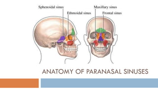
- 1. ANATOMY OF PARANASAL SINUSES
- 2. There are 4 Paired Paranasal Sinuses; Air Containing spaces 1. Anterior Group of Sinuses Frontal Anterior Ethmoidal Maxillary All of these open into the middle meatus anterior to the middle turbinate. 2. Posterior Group of Sinuses- Posterior Ethmoidal (opens in the superior meatus) Sphenoid (opens in sphenoethmoidal recess)
- 3. FUNCTION Lighten skull bones Add resonance to speech Vital role in conditioning the inspired air Thermal insulators Extra surface for olfaction Local immunological defence Buffers against trauma to brain
- 5. MAXILLARY SINUS (Antrum Of Highmore) Largest sinus, average 15 ml capacity Pyramidal in shape Boundaries- 1)Roof- Floor of the orbit 2) Medial wall- middle and inferior meatuses 3) Floor- maxilla ( palatine and alveolar processes) 4) Posterior wall- infratemporal and pterygopalatine fossae 5) Anterior wall - facial surface of maxilla 6) Lateral wall- Zygomatic process of maxilla or Zygomatic bone itself
- 8. ETHMOIDAL SINUS • Group of 3-18 thin walled air cavities in the lateral masses of ethmoid bone • Openings- • 1) Anterior ethmoid- Middle meatus • 2) Posterior ethmoid- Superior meatus and into supreme meatus • Roof - Medial extension of the orbital plate of the frontal bone • Lateral - Lamina papyracea • Anterior Group: 1. Agger nasi cells 2. Ethmoid bulla 3. Supraorbital cells 4. Frontoethmoid cells 5. Haller cells • Posterior Group: Onodi cell (sphenoethmoidal cell)
- 11. SPHENOID SINUS Occupies the body of the Sphenoid. Right & left sinus separated by thin bony septum. Ostium of the sinus opens within sphenoethmoidal recess, medial to the superior turbinate. Volume of an adult spenoid sinus: 2 cm x 2 cm x 2 cm Radiologically, sphenoid sinuses can be identified at 4 years of age Associated with the least common sinusitis. Mucocilliary clearnace is towards the ostium into the sphenoethmoidal recess Blood supply - sphenopalatine artery and posterior ethmoidal artery. Nerve supply - first and second divisions of the trigeminal nerve. Relations: 1) Laterally: Optic nerve, Maxillary nerve, Internal carotid artery 2) Inferiorly: Vidian nerve. 3) Superiorly: Pituitary, Olfactory nerve. 4) Posteriorly it forms Clivus
- 14. FRONTAL SINUS Situated between inner and outer tables of frontal bone, above and deep to supra orbital margin Relations: Anterior wall - skin over the forehead Inferior wall - orbit and its contents Posterior wall - meninges and frontal lobe of the brain Drains through its ostium into the frontal recess Radiologically, identifiable at 6 years of age. Anterior ethmoidal cells may obstruct drainage into the frontal recess and cause sinusitis Mucocilliary clearnace is anticlockwise in the right and clockwise in the left sinuses and though its natural ostium. Frontal sinus is supplied by the supraorbital and supratrochlear arteries of the ophthalmic artery It is innervated by the supraorbital and supratrochlear nerves of the first division of the trigeminal nerve
- 16. Lymphatic Drainage of PNS Lymphatics of maxillary, ethmoid, frontal and sphenoid sinuses form a capillary network in their lining mucosa and collect with lymphatics of nasal cavity They drain into lateral retropharyngeal and/or jugulodigastric nodes
- 18. THANK YOU