2. E3␣ (22, 27–32).
Despite these extensive findings, the physiological signifi-
cance of the N-end rule pathway in vivo has long been unclear,
and its importance has even been questioned (33). Yeast mu-
tants that lack this pathway (i.e. mutants in Ubc2, which
encodes the yeast homologue of E214k, or in Ubr1, which en-
codes the yeast homologue of E3␣) have only minor phenotypes
(34), and efforts to identify substrates with destabilizing N
termini or ones that undergo tRNA-dependent proteolysis have
not succeeded thus far. We report here the unexpected finding
that this ubiquitination system, involving E214k and E3␣, as
well as tRNA-dependent substrate modification, is responsible
for up to 60% of the ATP-dependent degradation of soluble
proteins in extracts of normal skeletal muscles.
EXPERIMENTAL PROCEDURES
Protein Reagents—Ubiquitin, actin, myosin, bovine serum albumin,
and human ␣-lactalbumin were from Sigma. Lysozyme was from Boeh-
ringer Mannheim. Radioiodination of protein substrates was performed
by the chloramine T method as described previously (8). The dominant
negative inhibitor mutants of the E2s were kindly provided by Jackie
Pierce, Margaret Read, and Vincent Chau (Proscript, Inc.). Escherichia
coli strains engineered to express E214k, E2-F1 (UbcH7), and UbcH5b
were kindly provided by S. Wing, M. Scheffner, and A. Weissman,
respectively.
Muscle Extract Preparation—Male New Zealand White rabbits (3–4
kg) were killed by lethal injection of sodium pentobarbital, and extracts
from psoas muscles were prepared as described earlier (8). Homoge-
nates were centrifuged at 30,000 ϫ g for 30 min to remove myofibrils.
“Crude extracts” were prepared by centrifuging the supernatants at
100,000 ϫ g for 1 h and were either studied directly or fractionated on
DEAE-cellulose (8) into fraction II, the resin-bound material, which
contains the proteasomes and most of the enzymes required for Ub
conjugation, and fraction I, the flow-through, which contains Ub and
70% of cell proteins. Both crude extracts and fraction II were then
dialyzed against buffer containing 20 mM Tris-HCl, pH 7.6, 2 mM DTT,
5 mM MgCl2 and 10% glycerol and stored at Ϫ70 °C until use.
Purification of E1, E2s, and E3␣—E1 was prepared from rabbit
muscle fraction II extract using Ub-Sepharose affinity chromatography
by elution with AMP and PPi as described previously (35). The E1 was
further purified by MonoQ FPLC (Amersham Pharmacia Biotech).
E214k was purified from E. coli fraction II by MonoQ FPLC and size
exclusion chromatography on Sephacryl S-100 HiPrep FPLC (Amer-
sham Pharmacia Biotech). UbcH5b and E2-F1 were prepared directly
from crude E. coli lysates using HiTrapS FPLC (Amersham Pharmacia
Biotech) followed by Sephacryl S-100 HiPrep FPLC.
Partially purified E3␣ was prepared from crude muscle extract using
a protocol kindly provided by A. Haas and co-workers.2
Crude muscle
extract (35 mg) was twice passed over a 1.0-ml E214k-Affi-Gel 10 (Bio-
Rad) affinity column (prepared according to the manufacturer’s instruc-
tions using 10 M E214k and 5.0 ml of resin). The column was washed
exhaustively with 20 mM Tris-HCl, pH 7.6, 50 mM NaCl, 1 mM DTT, and
then E3␣ was eluted with 20 mM Tris-HCl, pH 7.6, 1 M NaCl, 0.5 mM
DTT into tubes containing bovine serum albumin to a final concentra-
tion of 0.1 mg/ml. The material was dialyzed against 20 mM Tris-HCl,
pH 7.6, 5 mM MgCl2, 0.5 mM DTT, 10% glycerol and stored at Ϫ70 °C.
Protein Degradation Assays in Skeletal Muscle Extracts—Degrada-
tion of endogenous proteins in crude extracts and fraction II was meas-
ured by assaying the generation of free tyrosine in the trichloroacetic
acid-soluble supernatant (8). Reaction mixtures contained the following
in a volume of 100 l: 20 mM Tris-HCl, pH 7.6, 5 mM MgCl2, 2 mM DTT,
ATP regenerating system (10 g of creatine phosphokinase and 10 mM
creatine phosphate), 1 mM ATP, 25 g of Ub, and approximately 1 mg
of dialyzed crude extract or fraction II. Following incubation at 37 °C for
2 h, the reactions were terminated by the addition of an equal volume
of 20% trichloroacetic acid. The reaction mixtures were centrifuged, and
the amount of tyrosine generated in the supernatant was measured by
fluorescence spectroscopy as described previously (8). When degrada-
tion of 125
I-myosin, 125
I-actin, or 125
I-lysozyme in the crude extracts was
studied, the reaction mixture contained 3 g of labeled proteins, and
their degradation was measured by following the appearance of trichlo-
roacetic acid-soluble radioactivity using a ␥-counter. The results shown
are typical of those obtained in three independent experiments. The
absolute amount of protein degraded in the absence or presence of ATP
varied from extract to extract.
Measurement of 125
I-Ub Conjugation to Muscle Proteins—To measure
Ub conjugation in the crude muscle extracts, the dialyzed extracts (50
g protein) were incubated in a 20-l volume containing 20 mM Tris-
HCl, pH 7.6, 20 mM KCl, 5 mM MgCl2, 1 mM DTT, 10% glycerol, 2 mM
AMP-PNP, 125
I-Ub (ϳ150,000 cpm, 5–10 M), 20 g/ml bestatin (to
block aminopeptidases), 20 M MG132 (to block proteasomal activities),
and 2 M ubiquitin aldehyde (to inhibit the hydrolysis of ubiquitin
conjugates by deubiquitinating isopeptidases (36)).
For ubiquitination assays in fraction II, which lack endogenous ubiq-
uitin, preparations were centrifuged for 6–8 h at 100,000 ϫ g to remove
most of the proteasomes (37). The supernatant (60 g in a volume of 25
l) was then incubated with 125
I-Ub (ϳ150,000 cpm, 5–10 M) at 37 °C
in a buffer containing 20 mM Tris-HCl, pH 7.4, 1 mM DTT, 5 mM MgCl2,
2 mM ATP␥S, 20 g/ml bestatin, and 5% glycerol in the absence or
presence of the various inhibitors. All dipeptides and methyl esters
were added at a concentration of 2 mM. All reactions were carried out in
parallel under identical conditions. The reactions were incubated at
37 °C for 60 min and terminated by the addition of Laemmli sample
buffer. Finally, equal amounts of 125
I-labeled proteins were loaded onto
the gel, and SDS-polyacrylamide gel electrophoresis was performed as
described by Laemmli (38) on 12% acrylamide gels. The gels were then
dried and autoradiographed.
RESULTS
Inhibitors of E3␣ Reduce Breakdown of Endogenous Proteins
in Muscle Extracts—We reported previously that crude ex-
tracts of rabbit psoas muscle degrade endogenous proteins all
the way to amino acids via the Ub-proteasome system as shown
by the ATP-dependent generation of free tyrosine (8). Because
this amino acid cannot be synthesized or degraded by muscle,
its appearance indicates the degradation of cell proteins (8).
This process occurred at a linear rate for 2 h at 37 °C, was
stimulated 3–6-fold by ATP, and was blocked by removal or
inhibition of proteasomes (8). To test whether the N-end rule
pathway contributes to this degradation of endogenous pro-
teins, various competitive inhibitors of E3␣ were added to these
muscle extracts. The addition of arginine methyl ester reduced
the ATP-dependent degradation of soluble muscle proteins by
about 50% (Table I). The inhibitor of the hydrophobic site of
E3␣, leucine methyl ester, also reduced proteolysis but to a
lesser extent (30%). In contrast, alanine methyl ester, which
does not inhibit E3␣ (24), caused little reduction (Ͻ10%) in the
breakdown of muscle proteins. Similarly, the dipeptide inhibi-
tor of E3␣, Phe-Ala, also reduced ATP-stimulated degradation
of endogenous proteins by 30%, but its isomer, Ala-Phe, had
little effect on this process (Ͻ5% inhibition). There was also a
low amount of ATP-independent proteolysis in these prepara-
tions, but it was not affected by these dipeptides or amino acid
esters (data not shown). These observations suggest that a
large fraction (perhaps 60–80%) of the ATP-dependent degra-
dation of soluble proteins in crude muscle extracts involves
E3␣, the Ub-protein ligase of the N-end rule pathway.
A very similar effect of these inhibitors on proteolysis was
seen after the extracts were fractionated by DEAE chromatog-
raphy. Most cell proteins, including Ub, flow through the col-
umn (fraction I), but about 30% of cell proteins were bound and
eluted with high salt. This material (fraction II) contains E1,
E214k, E3␣, and proteasomes (26, 39, 40). When ATP and Ub
were added to fraction II, they stimulated the breakdown of
endogenous proteins 3–5-fold (8), and as shown in Table I, most
of the degradation of proteins in fraction II (like those in crude
extracts) appears to involve E3␣. This ATP- and Ub-stimulated
degradation was inhibited when arginine methyl ester or Phe-
Ala was added, while alanine methyl ester or Ala-Phe, which do
not inhibit E3␣, had very little effect (Ͻ5%) on proteolysis. In
fact, the pattern of inhibition in fraction II with the dipeptide
and methyl ester inhibitors of E3␣ was almost identical to that
2
R. Crinelli, O. V. Baboshina, and A. L. Haas, personal
communication.
N-end Rule Pathway in Muscle Proteolysis 25217
byguestonMay15,2015http://www.jbc.org/Downloadedfrom
3. seen in crude extracts (Table I).
Evidence for a tRNA Requirement in Muscle Proteolysis—
Another type of substrate for the N-end rule pathway is pro-
teins with acidic N-terminal residues. Genetic studies by Var-
shavsky (27) and biochemical studies by Ferber and
Ciechanover (28) have shown that such proteins are modified
before degradation by covalent linkage of an arginine residue to
their N terminus. This modification requires arginyl-tRNA and
leads to rapid Ub conjugation by E3␣ (27–32). To test if this
system also contributes to muscle proteolysis, the muscle ex-
tracts were treated in ways that inhibit this tRNA-dependent
modification of the acidic N termini in reticulocyte extracts (27,
28). Treatment of the extract with RNase A caused a 25%
inhibition of the ATP-dependent degradation of endogenous
proteins (Table I), but RNase A had no effect on the breakdown
of 125
I-lysozyme, which does not require tRNA-mediated argi-
nylation (data not shown) (28). This inhibitory effect appeared
to be specifically due to RNA hydrolysis, since DNase I even at
high concentrations did not affect proteolysis. Also, when
RNase A was preincubated with the specific RNase inhibitor
from human placenta before the addition to muscle extract, it
did not reduce muscle proteolysis (Table I). Finally, to verify
that this inhibition by RNase A was due to destruction of a
tRNA species, we initially treated the extract with RNase A,
and then added the ribonuclease inhibitor to inactivate the
enzyme; when a mixture of tRNAs was then added to this
preparation, it partially restored protein breakdown toward
control levels. Furthermore, if the RNase A inhibitor was not
added before the addition of tRNA, no restoration of proteolysis
was observed. These findings strongly suggest that a signifi-
cant fraction (up to 25%) of the soluble proteins in muscle
extracts undergo a tRNA-dependent modification prior to ubiq-
uitination by E3␣ and subsequent degradation.
Specificity of Dipeptide and Amino Acid Ester Inhibitors of
E3␣—Since the above inhibitors of the N-end rule pathway are
weak substrate analogs and must therefore be utilized at rel-
ative high concentrations (24), it was important to establish
that the reduction in ATP-dependent proteolysis shown in Ta-
bles I and II was actually due to inhibition of E3␣ and that
these agents do not nonspecifically inhibit other ubiquitinating
enzymes or the proteasome. We therefore compared in rabbit
muscle extracts the effects of these inhibitors on the breakdown
of exogenously added 125
I-labeled lysozyme (a classic substrate
of the N-end rule pathway that contains an N-terminal lysine)
as well as 125
I-myosin and actin that have N-␣-acetylated N
termini and are, therefore, not N-end rule substrates. The
dipeptide and amino acid ester with basic N termini, which
reduced breakdown of endogenous muscle proteins by 60%,
inhibited similarly the rapid degradation of 125
I-lysozyme. In
muscle fraction II (Table II), as found previously in reticulocyte
extracts (24), Lys-Ala and arginine methyl ester selectively
inhibited the ATP-dependent degradation of 125
I-lysozyme
(60–70%), while Phe-Ala, alanine methyl ester, leucine methyl
ester, or RNase A caused very little (0–15%) inhibition of
125
I-lysozyme breakdown (see Table II). The Ub-mediated deg-
radation of proteins with N-␣-acetylated termini involves a
different E3 and is not sensitive to inhibitors of E3␣ (41, 42).
Exogenously added 125
I-labeled myosin and actin are degraded
in rabbit crude muscle extracts by the Ub-proteasome system
(8). At concentrations that inhibited degradation of endogenous
muscle proteins by 60% and 125
I-lysozyme by 60%, arginine
methyl ester or Lys-Ala reduced the degradation of 125
I-actin
and 125
I-myosin only slightly (5–15%) (Table II). These obser-
vations indicate that the large inhibition of breakdown of en-
dogenous proteins in crude extracts and fraction II is not a
nonspecific effect on proteolysis by the Ub-proteasome system.
Exogenous E2s and E3␣ Stimulate Proteolysis in Rabbit
Muscle Extracts—To further evaluate the selectivity of the
TABLE I
Inhibitors of the N-end rule pathway reduce the ATP-stimulated
degradation of endogenous proteins in rabbit muscle extracts
As indicated, the reaction mixtures included various L-amino acid
dipeptide or methyl ester inhibitors of E3␣ at 2 mM. To prevent hydrol-
ysis of the added dipeptide, bestatin (20 g/ml), an inhibitor of amin-
opeptidases, was added to all reaction mixtures, including controls.
Bestatin alone reduced tyrosine production slightly (less than 20%).
100% proteolytic activity is the value measured in the absence of any
inhibitors. Typically, crude extracts produced 150 pmol of tyrosine in
the absence of ATP, 550 pmol in its presence, and for fraction II, 120
pmol of tyrosine was generated in the absence of ATP and 700 pmol in
the presence of ATP and Ub in 2 h. The ATP-independent proteolysis
was subtracted from all samples, and the results were normalized to
100%. The data on this ATP-independent process are not shown, since
the addition of inhibitors had no effect on degradation in the absence of
ATP and/or Ub. For the RNase A inhibition assay, the reaction mixtures
were preincubated for 30 min with 0.02 units of RNase A (from bovine
pancreas) prior to the addition of ATP and Ub. The concentration of
bovine pancreas DNase I used was 5 units. The data shown in this and
subsequent tables were obtained in a single experiment and are the
averages of triplicate determinations, which agreed within 10%. All
experiments were repeated at least three times with similar results.
Additions (2 mM)
Inhibition of
degradation
Crude Fraction II
%
None 0 0
Arginine methyl ester 50 45
Leucine methyl ester 30 25
Alanine methyl ester 10 5
Phe-Ala 30 25
Ala-Phe 5 5
RNase A 25 NAa
RNase A pretreated with RNase inhibitor 5 NA
RNase A ϩ RNase inhibitor ϩ tRNAb
15 NA
DNase I 0 NA
a
NA, not assayed.
b
The crude extract was preincubated with RNase A (0.02 units), and
then excess human placental RNase inhibitor (0.05 units) was added to
inactivate the nuclease. Subsequently, tRNA from fetal calf liver (0.13
units) was added.
TABLE II
Inhibitors of the N-end rule pathway reduce the ATP-stimulated
degradation of endogenous proteins and 125
I-lysozyme but not of 125
I-
myosin and actin
Extracts were preincubated for 5 min at 37 °C with the various
inhibitors. 125
I-Labeled substrates, 1 mM ATP, and the ATP regenerat-
ing system were then added, and the incubation was continued for 2 h.
Degradation of endogenous proteins and 125
I-labeled myosin and actin
was measured in the crude lysate as discussed in Table I. Also shown
for comparison is the degradation of 125
I-lysozyme measured in fraction
II. Degradation of the 125
I-labeled substrates was then measured with
and without inhibitors. Since the various inhibitors had negligible effect
on the ATP-independent degradation, these data are not shown. With-
out ATP, 0.5% of 125
I-lysozyme, 1% of 125
I-actin, and 1.5% of 125
I-myosin
were degraded, and with ATP, 4% of 125
I-lysozyme, 4% of 125
I-actin, and
4.5% of 125
I-myosin were degraded.
Additions (2 mM)
Inhibition of degradation
Endogenous
proteins
125
I-Lysozyme 125
I-Myosin 125
I-Actin
%
None 0 0 0 0
Arginine methyl
ester
50 60 10 15
Leucine methyl
ester
30 15 7 5
Alanine methyl
ester
10 10 5 10
Lys-Ala 60 70 15 10
Phe-Ala 30 10 0 12
Ala-Phe 5 10 0 5
RNase A 25 0 0 0
N-end Rule Pathway in Muscle Proteolysis25218
byguestonMay15,2015http://www.jbc.org/Downloadedfrom
4. dipeptide inhibitors and the involvement of E3␣ and E214k in
the degradation of muscle proteins generally, we studied the
effects of the addition of both of these enzymes as well as of two
other E2s, E2-F1 and UbcH5b, on ATP-dependent breakdown
of endogenous proteins. These experiments also allowed us to
investigate whether the components of the N-end rule pathway
might actually be rate-limiting for proteolysis in these muscle
extracts. The addition of all of these Ub-conjugating enzymes
stimulated the ATP-dependent degradation of endogenous pro-
teins in crude muscle extracts without affecting ATP-independ-
ent proteolysis (Table III). When recombinant E214k (a major
E2 in fraction II), E2-F1, or UbcH5b (both in fraction I) were
added to crude muscle extracts at 1 M, total ATP-dependent
proteolysis was increased by more than 50% (Table III). These
E2s had approximately equal activity/mol when assayed by
thioester bond formation (data not shown). These findings sug-
gest that the supply of E2s limits overall proteolysis under
these conditions. Furthermore, the addition of E3␣ (partially
purified by affinity chromatography on E214k-Affi-Gel 10) also
stimulated the ATP-dependent degradation of endogenous
muscle proteins by about 90% but had no effect on the ATP-
independent degradation of muscle proteins. To determine to
what extent E214k and E3␣ may be functioning together ac-
cording to the N-end rule and whether UbcH5b and E2-F1 act
through a distinct Ub-protein ligase, these agents were added
in the presence or absence of dipeptide inhibitors of E3␣ (Lys-
Ala and Phe-Ala). As expected, most of the E214k- and E3␣-
stimulated proteolysis (70–80%) was sensitive to these dipep-
tides. By contrast, the E2-F1- and UbcH5b-stimulated protein
degradation was unchanged upon the addition of these inhibi-
tors. These experiments argue strongly that the dipeptide in-
hibitors reduce muscle protein breakdown by selectively block-
ing the N-end rule pathway and do not nonspecifically reduce
Ub-dependent proteolysis in general.
Effect of Inhibitors of E3␣ on Ub Conjugation—If these in-
hibitors are in fact reducing ATP-dependent proteolysis in the
muscle extracts by acting on E3␣, then they should also gen-
erally suppress Ub conjugation to endogenous proteins in these
extracts. To test this, rates of 125
I-Ub conjugation to proteins
were measured in crude extracts and in fraction II from rabbit
and rat muscle. ATP␥S or AMP-PNP was utilized in the reac-
tions, because these nucleotides, unlike ATP, can support Ub
conjugation but not the degradation of Ub conjugates by the 26
S proteasome (43). The amount of 125
I-Ub conjugation to pro-
teins was assayed by measuring the appearance of new high
molecular weight 125
I-labeled bands (larger then 8 kDa) in the
SDS-polyacrylamide gel electrophoresis. Formation of 125
I-Ub
conjugates required the addition of a nucleotide and was linear
for up to 60 min at 37 °C (8). In the crude muscle extracts, the
inhibitor of E3␣, Lys-Ala, greatly reduced Ub conjugation,
while the isomeric dipeptide, Ala-Lys, had no significant effect
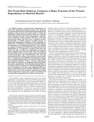
(Fig. 1, lane 7 versus lane 6).
FIG. 1. Effect of E3␣ inhibitors on ubiquitination of endoge-
nous proteins in rabbit muscle extract. Crude muscle extract as the
source for both ubiquitination enzymes and endogenous protein sub-
strates was mixed with 125
I-Ub as described under “Experimental Pro-
cedures.” Lane 1 contained hexokinase (1.5 units, 5 g) and 2-deoxy-
glucose (10 mM) to deplete endogenous ATP. The remaining lanes
contained 2 mM AMP-PNP. Lanes 3–5 contained 2 mM E214k, C88S E
214k and C114S E 2-C, respectively. Lanes 6 and 7 contained 2 mM
Ala-Lys and Lys-Ala, respectively. The intense bands just above the
125
I-Ub in lanes 4 and 5 reflect the stable Ub-E2 ester linkage formed by
the dominant negative E214k and E2-C, respectively (white arrow-
heads). A, SDS-polyacrylamide gel electrophoresis. B, densitometry
analysis of A. Control level of ubiquitination (100%) was taken to be
lane 2, which contains AMP-PNP but no other addition.
TABLE III
Effects of different E2s and E3␣ on ATP-dependent proteolysis of
endogenous soluble proteins in rabbit muscle extracts
by E3␣-dependent and -independent pathways
These experiments utilized soluble crude extracts of rabbit skeletal
muscles. Shown in the column “ATP-dependent proteolysis” are values
for ATP-stimulated degradation of endogenous soluble proteins after
subtracting the ATP-independent activity from total proteolysis. Typi-
cally, crude extracts in this specific experiment produced 230 pmol of
tyrosine in the absence of ATP, 580 pmol in its presence (total proteol-
ysis). “Not sensitive to dipeptide inhibitors” is the amount of tyrosine
generated due to protein degradation in the presence of dipeptide in-
hibitors (1 mM Lys-Ala and 1 mM Phe-Ala). “Sensitive to dipeptide
inhibitors” proteolysis is what remains after subtracting “Not sensitive
to dipeptide inhibitors” from total “ATP-dependent proteolysis.” The
addition of 1 mM Ala-Lys and 1 mM Ala-Phe had no effect on degrada-
tion. Also, the addition of various ubiquitinating enzymes or any inhib-
itors of E3␣ had no effect on ATP-independent degradation; therefore,
these data are not shown. Concentration of various E2s was 1 M. 2.5 l
of E3␣ preparation (described under “Experimental Procedures”) was
added to a total assay volume of 100 l in appropriate reactions. For all
other conditions, see the legend to Table I.
Additions
ATP-dependent
proteolysis
Sensitive to dipeptide
inhibitors
Not sensitive to
dipeptide inhibitors
pmol tyrosine released/2 h
None 350 230 120
Increase upon addition
pmol tyrosine
E1 NSa
E214k 130 100 30
E2-F1 180 30 150
UbcH5b 160 0 160
E3␣ 310 260 50
a
No significant stimulation of degradation was observed upon addi-
tion of E1.
N-end Rule Pathway in Muscle Proteolysis 25219
byguestonMay15,2015http://www.jbc.org/Downloadedfrom
5. In accord with our findings on protein breakdown, these
experiments indicated that the various inhibitors that reduced
overall ATP-dependent proteolysis in muscle fraction II did so
by reducing Ub conjugation. Upon the addition of arginine
methyl ester, Ub conjugation was inhibited by up to 50% and to
a lesser extent by leucine methyl ester or Phe-Ala (Table IV). In
contrast, alanine methyl ester or Ala-Phe, which do not inhibit
E3␣, reduced the ubiquitination of muscle proteins only
slightly (Ͻ10%). In addition, preincubation of fraction II with
RNase A markedly reduced conjugate formation, but not if the
RNase inhibitor was also present (Table IV). Thus, these find-
ings are in full agreement with the measurements of overall
protein degradation (Table I) and indicate that most of the Ub
conjugation to proteins in rabbit and rat muscle extracts in-
volves E3␣. Related experiments by us and others (42) in a
reconstituted ubiquitination system with the model N-end rule
substrate, 125
I-␣-lactalbumin, or with a substrate, 125
I-tropo-
nin, that is ubiquitinated by a distinct Ub-protein ligase, E3L
(42), also indicated that these inhibitors are highly selective in
their effects. When the purified Ub carrier protein, E214k, E2-
F1, or UbcH5b was incubated in the presence of Ub, E1, par-
tially purified E3␣, and 125
I-␣-lactalbumin, only E214k was able
to support Ub conjugation, and this process was inhibited al-
most completely by Lys-Ala but not by Ala-Lys (data not
shown). By contrast, only the addition of E2-F1 (and not E214k)
could support the ubiquitination of 125
I-troponin in fraction II,
and this E2-F1-dependent ubiquitination of troponin was not
sensitive to the dipeptide inhibitors of E3␣ (data not shown).
A Dominant Negative Form of E214k Inhibits Ub Conjugation
in Muscle Extracts—The addition of recombinant E214k, at
concentrations that stimulated overall proteolysis (Table III),
enhanced Ub conjugation to endogenous proteins in the crude
muscle extracts (Fig. 1, lane 3 versus lane 2). These findings
suggest that E214k is rate-limiting in overall Ub conjugation in
these extracts. By contrast, when a similar concentration of a
mutated form of E214k, in which the active site cysteine at
residue 88 (C88S) was mutated to serine (44), was added, it
inhibited Ub conjugate formation (Fig. 1, lane 4). This Cys 3
Ser mutant forms a stable ester linkage with Ub, which cannot
be transferred by E3␣ to a substrate (44). The effect of the
dominant negative E214k was even more pronounced in rat
muscle fraction II, where it inhibited Ub conjugation by over
75% (data not shown). As a control, a dominant negative form
of another E2, UbcH10 (active site Cys114
mutated to Ser),
which is involved in cyclin B ubiquitination at the end of
mitosis (21, 45), had no effect on Ub conjugation in these
extracts (Fig. 1, lane 5). At this concentration of the dominant
negative E214k, Ub-thioester formation to another exogenously
added E2 was not reduced; therefore, the dominant negative
E214k did not deplete the system of E1 or ubiquitin. This
inhibition by the dominant negative E214k, but not by the
dominant negative UbcH10, further demonstrates that the
E214k/E3␣ pathway is a major contributor to the total amount
of Ub conjugation in the muscle extracts.
The N-end Rule Pathway Is More Active in Skeletal Muscle
than in HeLa Cells—Because these findings indicated a major
role for the N-end rule pathway in protein breakdown in skel-
etal muscle, we tested whether this system is of equal impor-
tance in all mammalian cells. The conjugation of 125
I-Ub to
endogenous proteins could be readily measured in crude ex-
tracts from HeLa cells, where ubiquitination has been fre-
quently studied (Fig. 2). Although overall Ub conjugation was
FIG. 2. The E3␣-dependent ubiquitination pathway plays a
larger role in ubiquitination of endogenous proteins in rabbit
muscle extract than in HeLa extract. HeLa cell extract was kindly
provided by O. Coux and prepared according to (47). Upper panel,
125
I-Ubiquitin conjugation to endogenous extract proteins. Lower panel,
ubiquitin conjugation to 125
I-␣-lactalbumin in HeLa extract. Ubiquiti-
nation reactions were as described in Fig. 1 and under “Experimental
Procedures.” Lane 1, hexokinase (1.5 units, 5 g) and 2-deoxyglucose
(10 mM) were added instead of AMP-PNP. Lanes 2–4, 2 mM AMP-PNP
added. Lane 3 included 2 mM Lys-Ala, and lane 4 included 2 mM
Ala-Lys. Control level of ubiquitination (100%) was taken to be lane 2,
which contains AMP-PNP but no other addition. Solid bars, HeLa
extract. Shaded bars, Rabbit muscle extract.
TABLE IV
Effect of various inhibitors of the N-end rule pathway on the 125
I-Ub
conjugation to soluble proteins in rabbit and rat muscle extracts
Values shown are relative amounts of ubiquitination of endogenous
proteins in the absence or presence of various inhibitors of E3␣. 100% is
the amount of 125
I radioactivity incorporated, in the absence of any
inhibitor, into higher molecular weight forms (defined arbitrarily as
125
I-Ub migration with molecular mass greater than 20 kDa). For all
other conditions see the legend to Fig. 1.
Additions
Inhibition of
ubiquitination
Rabbit Rat
%
None 0 0
Arginine methyl ester 40 45
Leucine methyl ester 30 25
Alanine methyl ester 5 0
Phe-Ala 25 30
Ala-Phe 10 10
RNase A 20 25
RNase A pretreated with RNase inhibitor 0 5
DNase I 5 0
N-end Rule Pathway in Muscle Proteolysis25220
byguestonMay15,2015http://www.jbc.org/Downloadedfrom
6. markedly inhibited by Lys-Ala in the rabbit muscle extract,
this dipeptide had little or no effect on Ub conjugation in HeLa
cell lysate (compare lanes 3 and 4, Fig. 2, upper panel). How-
ever, HeLa cell lysates do contain the N-end rule pathway.
Specifically, human 125
I-␣-lactalbumin was ubiquitinated in
the HeLa cell lysate, and this process was highly sensitive to
the inhibitor Lys-Ala (Fig. 2, lower panel). Thus, the N-end rule
pathway appears to play a much more important role in overall
proteolysis in skeletal muscle than in rapidly dividing HeLa
cells. Moreover, since Lys-Ala markedly affected ␣-lactalbumin
ubiquitination but not ubiquitination of endogenous proteins in
the HeLa cells (even at 2 mM), these data are further evidence
that this dipeptide selectively inhibited E3␣.
DISCUSSION
The various observations presented here demonstrate that
the ubiquitination system involving E3␣ and E214k catalyzes
the degradation of a large fraction of soluble proteins in skel-
etal muscle. Studies utilizing a variety of inhibitors of the
N-end rule pathway (the dipeptides, amino acid esters, and the
dominant negative inhibitor of E214k) as well as experiments in
which we supplemented extracts with purified ubiquitination
enzymes all clearly implicate E3␣ in protein degradation gen-
erally. The latter experiments also indicate that levels of E3␣
and E214k enzymes are rate-limiting for overall protein break-
down in extracts of normal muscles. Therefore, protein sub-
strates bearing destabilizing N-terminal residues appear to be
present in these muscles, since a significant inhibition of pro-
tein degradation and ubiquitination was seen with competitive
inhibitors that block selectively the binding of either type of
substrate to E3␣ (24, 25). In fact, when two types of inhibitors,
e.g. arginine methyl ester and Phe-Ala, were added together,
they had additive effects on 125
I-Ub conjugation and ATP-de-
pendent proteolysis (data not shown). However, in these exper-
iments, dipeptides or amino acid esters with basic N-terminal
residues were consistently more effective inhibitors of overall
proteolysis and ubiquitination than those with hydrophobic N
termini, presumably because proteins with basic N termini are
more abundant in muscle. However, some substrates of the
N-end rule pathway (e.g. bovine ␣-lactalbumin and soybean
trypsin inhibitor) actually begin with acidic or amide-contain-
ing N-terminal residues and undergo a tRNA-dependent mod-
ification that attaches N-terminal arginine residues (27, 31).
The partial inhibition of overall proteolysis and Ub conjugation
by RNase A and restoration of proteolysis by tRNA addition
further suggests that some of the endogenous substrates in
muscles have acidic N termini and undergo a tRNA-mediated
arginylation reaction to enter this degradative pathway.
Although these dipeptide and amino acid esters are weak
inhibitors that had to be utilized in the millimolar range, a
variety of findings have confirmed that the large inhibition
seen in these experiments is due to specific inhibition of E3␣.
For example, 1) the dipeptide and amino acid ester inhibitors of
E3␣ suppressed Ub conjugation and the ATP-dependent, but
not ATP-independent, degradation of endogenous proteins in
these muscle extracts. 2) The isomers of these inhibitors Ala-
Lys, Ala-Phe, or the analogous alanine methyl ester at similar
concentrations had no significant effect on these processes. 3)
These agents specifically inhibited Ub conjugation and ATP-
dependent degradation of those model substrates, lysozyme
and ␣-lactalbumin, known to require E214k and E3␣ but did not
affect Ub-dependent breakdown of 125
I-myosin or actin. 4)
These inhibitors reduced the stimulation of proteolysis upon
the addition of E214k and E3␣ but not the stimulation induced
by addition of other E2s (E2-F1 and UbcH5b). 5) Although
overall Ub conjugation was markedly inhibited by Lys-Ala in
the muscle extract, this dipeptide had no general effect on Ub
conjugation to protein in HeLa cell lysate, where it did block
ubiquitination of exogenously added ␣-lactalbumin. 6) Further-
more, Howley and co-workers (46) have previously shown that
these inhibitors of E3␣ do not affect p53 ubiquitination in other
cell extracts, and Ciechanover et al. (42) have noted that they
have no effect on E3L, another major E3. 7) Perhaps the strong-
est independent evidence for the importance of the N-end rule
pathway in muscles comes from the experiments with the dom-
inant negative form of E214k. This mutant E2, which can form
an ester with ubiquitin but cannot transfer the Ub to a sub-
strate, inhibited overall ubiquitination of muscle proteins. By
contrast, the addition of an analogous dominant negative form
of another E2, UbcH10, had no effect on Ub conjugation in
these extracts. Thus these experiments, as well as those involv-
ing supplementation with E214k and E3␣, completely support
the conclusion obtained with the low molecular weight
inhibitors.
The presence in normal tissues of substrates for the N-end
rule pathway was certainly not expected, since all cell proteins
when synthesized begin with methionine, and the N termini of
most cellular proteins are blocked by N-acetylation in muscles
as in other eukaryotic cells. One possible trivial explanation for
our findings could have been that in the course of muscle
homogenization or extract preparation, there was some proteo-
lytic cleavage of cell proteins leading to the generation of
polypeptides with unnatural hydrophobic or basic N termini.
To minimize this possibility, a variety of special procedures
were employed: 1) extracts were prepared at 4 °C in the pres-
ence of 1% glycerol, immediately after dissection, or after quick
freezing of the muscles in liquid nitrogen; 2) several different
homogenization procedures were tried; 3) a number of protease
inhibitors, including chymostatin and E-64 (20 g/ml), EDTA
(1 mM), and EGTA (1 mM) were included in the extraction buffer
to block the activity of lysosomal, Ca2ϩ
-activated and mast cell
(chymase and tryptase) proteases. None of these procedures
significantly altered the results.
The existence of large quantities of protein substrates for the
N-end rule pathway in skeletal muscle (but not in HeLa cells)
raises the possibility that a rate-limiting step in the degrada-
tion of the long lived muscle proteins is a slow exo- or endopro-
teolytic cleavage that exposes a destabilizing N-terminal resi-
due. Such a modification in vivo could even be a regulated
mechanism that triggers the acceleration of muscle proteolysis
in fasting or other catabolic states (6). Our studies involving
readdition of different ubiquitination enzymes strongly argue
that substrates for E214k and E3␣ (as well as for the other
ubiquitinating enzymes E2-F1 and UbcH5b) are present in
excess. Therefore, their degradation may be regulated simply
by changes in the levels of these various ubiquitinating en-
zymes. It is noteworthy that in muscle, E214k is among the
most abundant Ub carrier proteins.3
Moreover, its expression
appears to rise together with expression of poly-Ub and protea-
some subunits in atrophying muscles, when proteolysis (6) and
Ub conjugation are enhanced.4
In fact, in related studies, we
have found that the activity of the N-end rule pathway in-
creases in physiological and pathological states where muscle
protein breakdown rises.4
Thus, this ubiquitination system not
only accounts for most of the breakdown of soluble proteins in
extracts of normal muscle, but its activity is precisely regu-
lated, and changes in its activity appear to account for much of
the enhancement in Ub-dependent proteolysis in atrophying
muscles.
3
S. Lecker, unpublished observations.
4
Solomon, V., Baracos, V., Sarrat, P., and Goldberg, A. L. (1998) Proc.
Natl. Acad. Sci U. S. A., in press.
N-end Rule Pathway in Muscle Proteolysis 25221
byguestonMay15,2015http://www.jbc.org/Downloadedfrom
7. Acknowledgments—We are especially grateful to Jackie Pierce, Mar-
garet Read, and Vincent Chau (Proscript, Inc.) for providing the domi-
nant negative inhibitor mutants of the E2s and to Art Haas for the
method of purification of E3␣. We are grateful to Aurora Scott for
assistance in preparing the manuscript.
REFERENCES
1. Finley, D., and Chau, V. (1991) Annu. Rev. Cell Biol. 7, 25–69
2. Hershko, A., and Ciechanover, A. (1992) Annu. Rev. Biochem. 61, 761–807
3. Goldberg, A. L. (1992) Eur. J. Biochem. 203, 9–23
4. Jentsch, S. (1992) Annu. Rev. Genet. 26, 179–207
5. Rock, K. L., Gramn, C., Rothstein, L., Clark, K., Stein, R., Dick, L., Hwang, D.,
and Goldberg, A. L. (1994) Cell 78, 761–771
6. Mitch, W. E., and Goldberg, A. L. (1996) N. Engl. J. Med. 335, 1897–1905
7. Tawa, N. E., Jr., Odessey, R., and Goldberg, A. L. (1997) J. Clin. Invest. 100,
197–203
8. Solomon, V., and Goldberg, A. L. (1996) J. Biol. Chem. 271, 26690–26697
9. Tawa, N. E., and Goldberg, A. L. (1994) in Myology: Basic and Clinical (Engle,
A. G., Franzini-Armstrong, C., eds) 2nd Ed., pp. 683–707, Field & Wood,
New York
10. Temparis, S., Asensi, M., Taillandier, D., Aurousseau, E., Larbaud, D., Obled,
A., Bechet, D., Ferrara, M., Estrela, J. M., and Attaix, D. (1994) Cancer Res.
54, 5568–5573
11. Tiao, G., Fagan, J. M., Samuels, N., James, J. H., Hudson, K., Lieberman, M.,
Fischer, J. E., and Hasselgreen, P. O. (1994) J. Clin. Invest. 94, 2255–2264
12. Baracos, V. E., De Vivo, C., Hoyle, D. H., and Goldberg, A. L. (1995) Am. J.
Physiol. 268, E996–E1006
13. Medina, R., Wing, S. S., and Goldberg, A. L. (1995) Biochem. J. 307, 631–637
14. Wing, S. S., Hass, A. L., and Goldberg, A. L. (1995) Biochem. J. 307, 639–645
15. Rechsteiner, M., Hoffman, L., and Dubiel, W. (1993) J. Biol. Chem. 268,
6065–6068
16. Coux, O., Tanaka, K., and Goldberg, A. L. (1996) Annu. Rev. Biochem. 65,
801–847
17. Shkedy, D., Gonen, H., Bercovich, B., and Ciechanover, A. (1994) FEBS Lett.
348, 126–130
18. Orian, A., Whiteside, S., Israel, A., Stancovski, I., Schwartz, A. L., and
Ciechanover, A. (1995) J. Biol. Chem. 270, 21707–21714
19. Stancovski, I., Gonen, H., Orian, A., Schwartz, A. L., and Ciechanover, A.
(1995) Mol. Cell. Biol. 15, 7106–7116
20. Sudakin, V., Ganoth, D., Dahan, A., Heller, H., Hershko, J., Luca, F. C.,
Ruderman, J. V., and Hershko, A. (1995) Mol. Biol. Cell 6, 185–197
21. Aristarkhov, A., Eytan, E., Moghe, A., Admon, A., Hershko, A., and Ruderman,
J. V. (1996) Proc. Natl. Acad. Sci. U. S. A. 93, 4294–4299
22. Bachmair, A., Finley, D., and Varshavsky, A. (1986) Science 234, 179–186
23. Gonda, D. K., Bachmair, A., Wunning, I., Tobias, J. W., Lane, W. S., and
Varshavsky, A. (1989) J. Biol. Chem. 264, 16700–16712
24. Reiss, Y., Kaim, D., and Hershko, A. (1988) J. Biol. Chem. 263, 2693–2698
25. Reiss, Y., and Hershko, A. (1990) J. Biol. Chem. 265, 3685–3690
26. Wing, S. S., Dumas, F., and Banville, D. (1992) J. Biol. Chem. 267, 6495–6501
27. Varshavsky, A. (1996) Proc. Natl. Acad. Sci. U. S. A. 93, 12142–12149
28. Ferber, S., and Ciechanover, A. (1986) J. Biol. Chem. 261, 3128–3134
29. Ciechanover, A. (1987) J. Cell. Biochem. 34, 81–100
30. Ferber, S., and Ciechanover, A. (1987) Nature 326, 808–811
31. Ciechanover, A., Ferber, S., Ganoth, D., Elias, S., Hershko, A., and Arfin, S.
(1988) J. Biol. Chem. 263, 11155–11167
32. Elias, S., and Ciechanover, A. (1990) J. Biol. Chem. 265, 15511–15517
33. Ciechanover, A. (1994) Cell 79, 13–21
34. Bartel, B., Wunning, I., and Varshavsky, A. (1990) EMBO J. 9, 3179–3189
35. Hershko, A., Heller, H., Elias, S., and Ciechanover, A. (1983) J. Biol. Chem.
258, 8206–8214
36. Hershko, A., and Rose, I. A. (1987) Proc. Natl. Acad. Sci. U. S. A. 87,
1829–1833
37. Hegde, A. N., Goldberg, A. L., and Schwartz, J. H. (1993) Proc. Natl. Acad. Sci.
U. S. A. 90, 7436–7440
38. Laemmli, U. K. (1970) Nature 227 680–685
39. Fagan, J. M., Waxman, L., and Goldberg, A. L. (1987) Biochem. J. 243,
335–343
40. Ciechanover, A., Elias, S., Heller, H. and Hershko, A. (1982) J. Biol. Chem.
257, 2537–2542
41. Blumenfeld, N., Gonen, H., Mayer, A., Smith, C. E., Siegel, N. R., Schwartz,
A. L., and Ciechanover, A. (1994) J. Biol. Chem. 269, 9574–9581
42. Gonen, H., Stancovski, I., Shkedy, D., Hadari, T., Bercovich, B., Bengal, E.,
Mesilati, S., Abu-Hatoum, O., Schwartz, A. L., and Ciechanover, A. (1996)
J. Biol. Chem. 271, 302–310
43. Johnston, N. L., and Cohen, R. E. (1991) Biochemistry 30, 7514–7522
44. Sung, P., Prakash, S., and Prakash, L. (1991) J. Mol. Biol. 221, 745–749
45. Townsley, F. M., Aristarkhov, A., Beck, S., Hershko, A., and Ruderman, J. V.
(1997) Proc. Natl. Acad. Sci. U. S. A. 94, 2362–2367
46. Scheffner, M., Takahashi, T., Huibregtse, J. M., Minna, J. D., and Howley,
P. M. (1992) J. Virol. 66, 5100–5105
47. Coux, O., and Goldberg, A. L. (1998) J. Biol. Chem. 273, 8820–8828
N-end Rule Pathway in Muscle Proteolysis25222
byguestonMay15,2015http://www.jbc.org/Downloadedfrom
8. L. Goldberg
Vered Solomon, Stewart H. Lecker and Alfred
in Skeletal Muscle
Major Fraction of the Protein Degradation
The N-end Rule Pathway Catalyzes a
CELL BIOLOGY AND METABOLISM:
doi: 10.1074/jbc.273.39.25216
1998, 273:25216-25222.J. Biol. Chem.
http://www.jbc.org/content/273/39/25216Access the most updated version of this article at
.JBC Affinity SitesFind articles, minireviews, Reflections and Classics on similar topics on the
Alerts:
When a correction for this article is posted•
When this article is cited•
to choose from all of JBC's e-mail alertsClick here
http://www.jbc.org/content/273/39/25216.full.html#ref-list-1
This article cites 46 references, 28 of which can be accessed free at
byguestonMay15,2015http://www.jbc.org/Downloadedfrom