Wound Healing
•
0 likes•263 views

This topic is mainly for MBBS Studnts. It is under the General Principles of Surgery. Students shoud know the phases of wound healing so as to treat them appropriately and select the correct method of dressing material....

Recommended
More Related Content
What's hot
What's hot (20)
Similar to Wound Healing
Similar to Wound Healing (20)
More from Uthamalingam Murali
More from Uthamalingam Murali (20)
Recently uploaded
Recently uploaded (20)
Wound Healing
- 1. Prof. U. Murali. Wound Healing
- 2. Learning Objectives •Introduction •Phases of wound healing •Types of wound healing •Factors of wound healing •Complications
- 3. • Wound healing is a complex and dynamic biological process. • It is related to tissue reconstitution which is the process by which the body replenishes cells that are being lost. • All wounds heal following a specific sequence of phases which may overlap. • The process of wound healing depends on the type of tissue which has been damaged and the nature of tissue disruption. Introduction
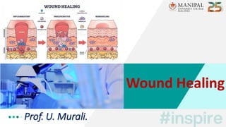
- 4. 4 • Classically, wound healing has been arbitrarily described in 3 overlapping but distinct stages / phases and include: • Inflammatory phase [ Lag / Substrate / Exudative phase ] • Proliferative phase [ Collagen / Fibroblastic phase ] • Remodeling phase [ Maturation phase ] Phases of Wound Healing
- 5. 5 • It begins immediately after wounding & and lasts 2–3 days. • Hemostasis, is often described as the immediate phase occurring before inflammation. • Hemostasis is achieved by vasoconstriction with formation of platelet plug [PT adhere, activate and aggregate] & activation of clotting pathway, resulting in formation of fibrin matrix. • The fibrin clot helps to stabilize the platelet plug and form a scaffold for migration of inflammatory cells [PMLs] into the wound. I. Phase – H & I
- 6. • In the early inflammatory phase (days 1–2), platelet activation causes an influx of inflammatory cells led by PMLs, particularly neutrophils. • Neutrophils – the first infiltrating cells to enter the wound site. • They are important for minimizing bacterial contamination of the wound, by phagocytosis. • Platelets and the local injured tissue release vasoactive amines such as histamine & serotonin, which increase vascular permeability, thereby aiding infiltration of further inflammatory cells. I. Inflammatory Phase - Early
- 7. • During the late inflammatory phase (days 2–3) monocytes appear in the wound and differentiate into macrophages. • Macrophages play a vital role in wound healing. • They function as phagocytic cells and release proteolytic enzymes to help debride the wound. • They are also the primary producer of cytokines and growth factors promoting fibroblast proliferation & angiogenesis. • Historically, this phase has been described by rubor (redness), tumor (swelling), calor (heat) and dolor (pain) – RTCD. I. Inflammatory Phase - Late
- 8. 8 • The proliferative phase starts around day 3 and lasts for 2–4 weeks. • It is during this phase that the wound continuity is re-established. • It consist mainly of fibroblast activity with the production of collagen & ground substance (glycosaminoglycans and proteoglycans). • Also, the growth of new blood vessels as capillary loops (angio-neogenesis) & the re-epithelialization of the wound surface. II. Proliferative Phase
- 9. 9 • The wound tissue formed in the early part of this phase is called granulation tissue (contains fibroblasts, macrophages & endothelial cells). • It has a pink and granular appearance. • Some fibroblasts differentiate into myofibroblasts, which are contractile cells. • These play an important role in contraction to bring the edges of the wound together. II. Proliferative Phase
- 10. • In the latter part of this phase, there is an increase in the tensile strength of the wound due to increased collagen, which is at first deposited in a random fashion and consists of type III collagen. • Initial angiogenesis occurs following release of factors from keratinocytes & macrophages. • Later re-epithelialization of the wound surface occurs by migration of basal layer of the retained epidermis which proliferates, differentiates and stratifies to form wound closure. II. Proliferative Phase
- 11. 11 • The remodeling phase begins 2–3 weeks after injury and lasts for a year (or) more. This phase is characterized by maturation of collagen. • Type III collagen, which is prevalent during proliferation, is replaced by stronger type I collagen until the normal skin ratio of 4:1 type I to type III collagen is re-established. III. Remodeling Phase
- 12. • The collagen becomes more cross-linked and uniformly aligned. • This maturation of collagen leads to increased tensile strength in the wound/scar, which is maximal 12 weeks post injury and represents approximately 80% of the uninjured skin strength. • The final matured scar is acellular and avascular. III. Remodeling Phase
- 16. 16 • Wound healing is accomplished in one of the following 3 ways – • Healing by primary intention [wounds with opposed edges] • Healing by secondary intention [wounds with separated edges] • Healing by tertiary intention [tertiary wound healing] Types of Wound Healing
- 17. • It occurs in a clean incised wound (or) surgical wound with good apposition of the edges. • The incision causes only focal disruption of epithelial BM continuity & death of a relatively few epithelial connective tissue cells. • As a result, there is more epithelial regeneration than fibrosis. • Wound heals rapidly with complete closure and leaving best scar. Scar will be linear, smooth, and supple. Primary Intention
- 18. 18 • This occurs in open wounds, particularly when there has been significant loss of tissue, or wounds with irregular margins. • Regeneration of parenchymal cells cannot completely reconstitute the original architecture. • It heals slowly with fibrosis. It leads into a wide scar, often hypertrophied and contracted. It may lead into disability. Secondary Intention
- 19. • Delayed primary healing occurs when the wound edges are not opposed immediately, which may be necessary in contaminated or untidy wounds. • After debridement of non-viable tissue and when the wound is clean, the wound edges may be surgically approximated. This is also called healing by tertiary intention. • Primary contaminated or mixed tissue wounds heal by tertiary intention. Tertiary Intention
- 22. 22
- 24. • Various factors can adversely affect wound healing and include – •Local Factors •Systemic / General Factors Factors - Wound Healing
- 55. Complications – W H • Excessive Scar Formation: Hypertrophic scar, Keloid. • Deficient Scar Formation: Result in wound dehiscence [or] rupture of the wound due to inadequate formation of granulation tissue. • Exuberant Granulation (Proud Flesh): Excessive GT that protrudes above the skin level. • Deficient contraction – Skin grafts Excessive contraction – In Burns • Others: Pigmentary changes, Incisional hernia, Dystrophic calcification, Neoplastic changes.
- 56. 56 • Phases of wound healing. • The changes in each phases - pathophysiology. • Types of wound healing. • Factors affecting wound healing – Local & Systemic factors. • Complications of wound healing. To Summarize
- 57. References
- 58. • Explain the first phase of wound healing. • List 4 differences between 1 & 2 union of wound healing. • Enumerate 5 local factors affecting wound healing. • Mention the complications of wound healing. • Write about tertiary intention. • Illustrate with flow-chart the phases of wound healing. • Describe the nutritional deficiencies affecting wound healing. • Write the stages of wound healing. Question Time
- 59. Factors impairing wound healing include all the following, except – ◼ a) Excessive tension. ◼ b) Lack of hemostasis. ◼ c) Inversion of wound edges. ◼ d) Drains. ◼
- 60. The tensile strength of the wound starts and increases after – ◼ a) Immediate suture of the wound. ◼ b) 3 – 4 days. ◼ c) 7 – 10 days. ◼ d) 6 months. ◼
- 61. Which one of the following cells plays an important role in bringing the edges of the wound together? – ◼ a) Myofibroblasts. ◼ b) Macrophages. ◼ c) Polymorphonuclear leukocytes. ◼ d) Fibroblasts. ◼
- 62. Factors that may adversely affect the healing of wounds include all the following, except – ◼ a) Exposure to radiation. ◼ b) Advanced neoplasia. ◼ c) Exposure to UV light. ◼ d) Obstructive jaundice. ◼
- 63. When is the maximum collagen content of wound tissue? – ◼ a) 2 – 5 days. ◼ b) 6 – 10 days. ◼ c) 11 – 16 days. ◼ d) 17 – 21 days. ◼
- 64. 64