SlideShare a Scribd company logo
1 of 70
CORNEAL ECTASIAS
CORNEAL ECTASIAS
• It is a group of diseases affecting the corneal
shape due to CORNEAL THINNING.
– Anterior Keratoconus
– Keratoglobus
– Pellucid Marginal Degeneration
– Posterior Keratoconus
• Key Features:
– Usually Bilateral, often asymmetrical
– Isolated to cornea
– Non inflammatory
KERATOCONUS
• Progressive corneal steepening, typically inferior
to the centre of the cornea.
• Non inflammatory, usually bilateral.
• Incidence: Between 50 and 230 per 100 0001.
• Unilateral involvement is 2–4%2.
• Onset starts at the age of puberty.
1. Krachmer JH, Feder RS, Belin MW. Keratoconus and related noninflammatory corneal thinning disorders. Surv Ophthalmol. 1984;28:293–322.
2. Holland DR, Maeda N, Hannush SB, et al. Unilateral keratoconus. Incidence and quantitative topographic analysis.
Ophthalmology.1997;104:1409–1413.
ASSOCIATED DISEASES
Systemic
 Atopic Diseases (35%)1.
 Down’s Syndrome (5.5-15%)2.
 Ehler-Danlos and osteogenesis imperfecta.
 Mitral valve prolapse.
 Marfan Syndrome.
 Ocular
 Vernal Conjunctivitis.
 Retinitis Pigmentosa.
 Leber’s congenital amaurosis
 Retinopathy of prematurity.
 Fuchs’ Dystrophy.
1. Rahi A, Davies P, Ruben M, et al. Keratoconus and coexisting atopic disease. Br J Ophthalmol. 1977;61:761–764.
2. Cullen JF, Butler HG. Mongolism (Down’s syndrome) and keratoconus. Br J Ophthalmol. 1963;47:321–330.
ETIOLOGY
BIOCHEMICAL:
• Up-regulation of degradative enzymes (acid
esterase and acid phosphatase, cathepsins B and
G, and some matrix metalloproteinases) and the
down-regulation of proteinase inhibitors.
• There is a four fold increase in the receptors of
INTERLEUKIN 1 causing apoptosis of stromal
keratocytes.
• Microtrauma Release of IL-1 from epithelial
epithelium.
HEREDITY
• Autosomal dominant with incomplete penetrance.
• Frequency of inheritance is 6%.
• Keratoconus prevalence in first-degree relatives to be
3.34%, about 68 times that found in the general
population.
PATHOLOGY
• Keratoconus can involve every layer and tissue of
cornea.
• Superficial cell enlargement and prominence of
elongated cells in the corneal epithelium.
Early degeneration of basal epithelial cells.
Disruption of epithelial basement membrane.
Epithelium grows posteriorly into the Bowman’s Layer,
with collagen growing anteriorly into the epithelium.
Z shaped interruptions at the level of the basement
membrane.
Fragmentation of the Bowman’s layer. (Early change)
Fleischer ring found at the base of the cone is a
hallmark of keratoconus.
Breaks in Bowman’s layer fill with scar tissue.
Fine scars consolidate leading to formation of reticular
branched opacities at this level.
Collagen lamellae are released from their interlamellar
attachments or from their attachments to Bowman’s
layer and become free to slide Thinning without
collagenolysis.
Alteration of the normal orthogonal arrangement of
the collagen fibrils leads to stromal tissue
instability.
Most common location for the apex of the cone, in
the central or inferior cornea, isrelated to this.
Endothelial cell pleomorphism and polymegathism
DIAGNOSIS
• Onset of the disease is at puberty.
• Usually appears in the 2nd or 3rd decade.
SYMPTOMS:
• Progressive visual blurring & disortion due to
irregular astigmatism.
• Frequent change of glasses.
• Pain, photophobia.
• Eye rubbing.
Scissoring of red reflex on
retinoscopy
One of the earliest signs
SIGNS:
Munson Sign
Buldging of
the lower
eye lid in
downward
gaze.
Papillae, trantas dots, limbal nodule, pigmentation, ropy
discharge.
Corneal thinning, scarring.
Corneal ectasia:
Apex is inferior to the
horizontal line to the
pupillary axis. Corneal
thinning of about 1/2-1/5 is
observed at the apex.
2 types of cones:
Round/ Nipple shaped
Oval shaped
Fleischer Ring
Iron depositions in the
corneal epithelium.
The ring configuration is
due to irregular
distribution of tears at
the base of the cone and
resultant deposition.
Vogt’s Striae
Fine vertical lines
formed due to
compression of
descemet’s
membrane.
Parallel to the
axis of the cone
Prominent corneal
nerves
Rizzuti
phenomenon
• Breaks in Descemet Membrane
• Stromal edema
Charleux Sign
Dark reflex in the area of
the cone on observation
of the cornea with pupil
dilated using a direct
ophthalmoscope set on
plano.
DIFFERENTIAL DIAGNOSIS
• Pellucid marginal degeneration.
• Keratoglobus.
• Post traumatic corneal ectasia.
• Protrusion of cornea subsequent of corneal
thinning from ulceration.
• Corneal ectasia following keratorefractive
surgery.
CLASSIFICATION
AMSLER KRUMEICH CLASSIFICATION
INVESTIGATIONS
• KERATOMETRY:
Mires are commonly steep, highly astigmatic, often
egg shaped.
Inability to superimpose the central keratometric
rings is due to irregular astigmatism. (hallmark)
LIMITATIONS:
Provides information only of the central 3mm of
cornea.
Not useful in irregular astigmatism.
• VIDEOKERATOMETRY
Based on Placoid disc principle.
Early disease: Isolated area of smaller ring spacing
and distortion.
Advanced disease: Decrease in overall ring spacing
and becomes increasingly irregular
LIMITATIONS:
Does not give accurate information about the
posterior curvature and corneal thickness.
Cannot detect early keratoconus.
Astigmatism progression:
Symmetric Bow tie pattern
Asymmetric appearance
Inferotemporally displaced steep sided cone
• ORBSCAN
Uses the principle of scanning slit combined with a
Placoid system.
Provides reliable information about the anterior and
posterior elevation and best fit sphere and a
corneal pachymetry map.
LIMITATIONS
The posterior curvature map might not be 100%
accurate.
Requires patient fixation for accurate data.
Data pertaining to the central cornea is not
accurate.
• PENTACAM
USES a rotating Scheimpflug camera.
Provides reliable data of anterior and posterior
corneal elevation and accurate measurement of
corneal thickness.
The images taken by a pentacam maintain the
central point of each meridian hence the software
can re register these central points and eliminate
the eye movement.
Data is 10 times more accurate than the that
obtained from Orbscan.
• PACHYMETRY
Ultrasonic and optical based devices can be used.
Measurement of corneal thickness is useful for
diagnosis, documenting progression, and planning
treatment.
• OCULAR RESPONSE ANALYSER
Helps in making a diagnosis and classifying by
assessing corneal hysteresis and resistance.
TREATMENT
• NON SURGICAL
– Spectacles: Only in early cases.
• Once irregular astigmatism develops this type of visual
correction cannot be used.
– Contact lens: Soft toric lenses, standard bicurved
hard lenses, custom back toric lenses, piggyback
systems, hybrid lens, scleral and mini scleral lens.
RIGID GAS PERMEABLE LENS
• First choice in correcting irregular astigmatism.
• Fitting strategies:
Apical clearance: There is no touch in the apical area.
Advantage: 1. Reduced risk of scarring
2. Whorled keratopathy
3. Corenal erosion
Limitation: Hampers tear exchange, edge can come in
visual axis.
Rarely used now days.
Apical touch : Aims at providing primary lens support at
the apex of the cornea.
The central optic zone of the lens touches the central
cornea.
ADVANTAGE: Better quality of vision
LIMITATION: Corneal scarring and intolerance over
long term use.
 Three point touch: Most popular technique
The lens bearing is shared between apex and mid
peripheral cornea. Minimizes apical scarring, better
comfort and longer wearing time.
PIGGYBACK LENS SYSTEM
• Consists of the fitting a rigid gas permeable on
top of a soft contact lens.
• The soft lens is used to improve wearing
comfort and provide a more regular area for
the gas permeable contact lens to sit.
• The gas permeable contact lens is used for
providing adequate visual acuity.
HYBRID CONTACT LENS
• Contain a rigid gas permeable center with a
soft skirt.
• They are fitted with no or minimal apical
touch in the central cornea.
ADVANTAGE: Can be fit on cones of any severity.
LIMITATION: Can cause hypoxia related changes
like vascularisation and central corneal
clouding.
ROSE K LENSES
• Multicurve lenses with small optical zone
which fits snugly over the cone.
• Rose K contact lens provides greater comfort,
better quality of vision and requires lesser
chair time.
• Rose K2 IC is a larger diameter, intralimbal lens
that can be used for oval cones.
SCLERAL LENSES
• They rest on the sclera without touching the
cornea or the limbus leaving a clear area
between the contact lens and the cornea.
• They are of two types:
– Prosthetic Replacement Of the ocular Surface
Ecosystem (PROSE)
– Boston Ocular Surface Prosthesis (BOSP)
ADVANTAGE
Good centration, stability and improved Visual
acuity.
Extermely useful in patients with advanced
ectasias who are intolerant to contact lenses
or when immediate surgery is not possible
LIMITATION
High cost, reduced tear exchange and difficult
insertion and removal.
• SURGICAL MANAGEMENT
– INTRACORNEAL RING SEGMENT INSERTS
– CORNEAL CROSS LINKAGE
– LENTICULAR REFRACTIVE SURGERY
– CORNEAL TRANSPLANT
INTRACORNEAL RING SEGMENT INSERTS
• Ferrara rings:
Fixed inner diameter of 5.00 mm with a triangular
anterior contour.
• Intacs:
Inner diameter of 6.8 mm and a flat anterior contour.
Hexagonal in cross section.
Intacs SK: Inner diameter of 6.00 mm and elliptical cross
section.
The ring segments should be inserted upto 2/3rd depth of
the cornea.
Minimum corneal thickness should be 450 µ.
They do not prevent the progression of the disease.
The technique involves
the implantation of
one of two
polymethy metha
acrylate segments in
the stroma to flatten
the central cornea.
Intralamellar pockets
are created with the
help of a vaccum
lamellar dissector or
femtosecond laser.
CORNEAL COLLAGEN CROSSLINKING
• The most recent addition to surgical management.
• It may slow or halt the progression with photo-
oxidative treatment to increase the rigidity of the
corneal stroma.
• 60-70% patients show some stabilization following
this treatment.
• The therapeutic effects are limited upto 300 µ of the
anterior stroma.
• This technique should not be performed for corneas
less than 400µ in thickness.
• It carries a slight risk of development of keratitis.
STEPS:
Under topical anaesthesia 6-7 mm of central corneal
epithelium is removed.
Riboflavin 0.1% solution is applied topically (10-30
mins) and cornea is irradiated with UVA light of 370 nm
for 30 mins.
Production of reactive oxygen species helps create
covalent bonds between collagen fibrils.
CORNEAL TRANSPLANTATION
Penetrating Keratoplasty:
Success rate in 90-95%.
Visual recovery takes
several weeks to months.
Full stabilisation might
even take a year.
Hydrops that does not
clear by 3-4 months is
best treated by
Penetrating keratoplasty.
DEEP ANTERIOR LAMELLAR
KERATOPLASTY
• It is the procedure of choice as there is good
endothelial count even after hydrops.
• Advantages over Penetrating keratoplasty:
Reduced risk of Rejection
Reduced risk of Endopthalmitis
Greater wound stability
Reduced need for topical steroids Faster healing
LAYER BY LAYER MANUAL DISSECTION
Partial trephination of variable depth ranging from
50-70% of corneal thickness, stroma is removed with
the help of a crescent.
Layer by layer removal of the stroma to reach as
close to the Descemet’s membrane.
LIMITATION:
Poor visual outcome due to residual stroma and
interface haze.
AIR ASSISTED DALK
Archila first described this technique involving air
injection to achieve dissection as close to DM as
possible.
The Big bubble technique was described by Anwar
and Teichmann and is the most widely used
technique.
Steps:
Air injection into the stroma through a groove created by
trephining 60-80% of the stromal thickness.
Air infiltrates the potential space between deep stroma
and DM and creates a Dome shaped detachment of DM.
Once the plane of separation is created the stroma can be
easily excised.
FEMTO LASER ASSISTED DALK
It can be used to create corneal incisions with
customised graft edges and lamellar planes for both
donor and recipient corneas.
ADVANTAGE:
Accuracy of forming the bubble at desired
corneal depth
Positive refractive outcome
LIMITATIONS:
Cost and availability
KERATOGLOBUS
• Bilateral ectatic disorder which is non
progressive or minimally progressive.
• Generalised thinning with marked peripheral
thinning.
• Types:
– Congeital: Ehler’s-Danlos type IV and brittle
cornea syndrome.
– Acquired: Initially keratoconus keratoglobus.
Clinical Features
Blurring of vision.
Corneal perforation spontaneous or following
minimal trauma.
Rarely might present as photohobia, glare, pain,
conjunctival injection following acute hydrops.
Limbus- limbus corneal thinning
Blue sclera due to thinning
Differential Diagnosis
Keratoconus
Pellucid Marginal Degeneration
Megalocornea
Congenital glaucoma
Investigations
Diagnosis is essentially clinical.
Ultrasonic Pachymetry shows reducude corneal
thinckness.
Corneal Topography shows generalised thinning and
irregular astigmatism is difficult to perform in
advaced cases .
Management
Conservative:
Observation is the rule in early cases.
Patient is advised to use polycarbonate glasses.
Visual rehab is done using refractive correction
for high myopia.
Scleral lens, rigid gas permeable lens, reverse
geometry hydrogel lens, large diameter inverse
geometry rigid gas permeable lenses can be
used.
Surgical Management
It is difficult to manage these patients by surgery:
Large sized grafts are required increasing the risk
of rejection.
Peripheral sutures often cut through.
Proximity of the graft to the limbus increases risk
of rejection.
Higher chances of perforation.
PELLUCID MARGINAL DEGENERATION
• Bilateral corneal ectatic
disorder.
• Band of thinning 1-2mm
in diameter extending
from 4 to 8 o’clock.
• Area of thinning is
separated from the
limbus by 1-2mm of
normal cornea.
• Onset: 2nd- 5th decade of
life.
Beer Belly
appearance
• Blurring of vision due to against the rule
astigmatism.
• Frequent chances of glasses.
• Ocular examination reveals thinning of inferior
cornea with 1-2 mm separation from the
limbus.
• Ectasia is just superior to the thinning.
• Protruding cornea is of normal thickness.
• Occasional concentric Descemet folds seen
inferiorly.
Investigations
• Corneal topography:
Inferior peripheral steepening
extending into the mid
periphery.
Crab- claw appearance.
Against the rule: Superiorly.
With the rule: Inferiorly.
Management
• Spectacles in early cases.
• PROSE, BOSP and hybrid contact lens can be
used.
• Large diameter Penetrating keratoplasty.
• Combined Lamellar with penetrating
keratoplasty.
• Crescentric lamellar keratoplasty.
• Crescentric or wedge excision if ectasia in
small sector.
POSTERIOR KERATOCONUS
• It is a congenital corneal anomaly
in which the posterior corneal
surface protrudes into the stroma.
• Sporadic, unilateral, non
progressive.
• Variant of corneal mesenchymal
dysgenesis.
• Scarring is seen anterior to the
descemet bulge.
• Pathologic Examination: Scarring
at Bowman’s membrane and
thinning of descmet membrane
with excrescences.
Corneal ectasias
Corneal ectasias

More Related Content

What's hot

Refrective surgery ppt
Refrective surgery pptRefrective surgery ppt
Refrective surgery pptsubhadri manna
 
Central Retinal Vein Occlsion (CRVO)
Central Retinal Vein Occlsion (CRVO)Central Retinal Vein Occlsion (CRVO)
Central Retinal Vein Occlsion (CRVO)Yousaf Jamal Mahsood
 
Corneal dystrophies
Corneal dystrophiesCorneal dystrophies
Corneal dystrophiesNajara Thapa
 
Peripheral ulcerative keratitis (puk)
Peripheral ulcerative keratitis (puk)Peripheral ulcerative keratitis (puk)
Peripheral ulcerative keratitis (puk)Desta Genete
 
Gonioscopy presentation
Gonioscopy presentationGonioscopy presentation
Gonioscopy presentationHira Dahal
 
Intermitent exotropia
Intermitent exotropiaIntermitent exotropia
Intermitent exotropiaSSSIHMS-PG
 
Congenital optic disc anomalies
Congenital optic disc anomaliesCongenital optic disc anomalies
Congenital optic disc anomaliesJagdish Dukre
 
Ocular Manifestations of Leprosy - EYE
Ocular Manifestations of Leprosy - EYEOcular Manifestations of Leprosy - EYE
Ocular Manifestations of Leprosy - EYEChetan Ganteppanavar
 
Assessment of corneal endothelium
Assessment of corneal endotheliumAssessment of corneal endothelium
Assessment of corneal endotheliumdrvasant162
 
Fuchs dystrophy and pseudophakic bullous keratopathy
Fuchs dystrophy and pseudophakic bullous keratopathyFuchs dystrophy and pseudophakic bullous keratopathy
Fuchs dystrophy and pseudophakic bullous keratopathyunleng
 

What's hot (20)

Refrective surgery ppt
Refrective surgery pptRefrective surgery ppt
Refrective surgery ppt
 
Central Retinal Vein Occlsion (CRVO)
Central Retinal Vein Occlsion (CRVO)Central Retinal Vein Occlsion (CRVO)
Central Retinal Vein Occlsion (CRVO)
 
Corneal dystrophies
Corneal dystrophiesCorneal dystrophies
Corneal dystrophies
 
Intermittent exotropia
Intermittent exotropiaIntermittent exotropia
Intermittent exotropia
 
Corneal Ectasias
Corneal Ectasias Corneal Ectasias
Corneal Ectasias
 
Peripheral ulcerative keratitis (puk)
Peripheral ulcerative keratitis (puk)Peripheral ulcerative keratitis (puk)
Peripheral ulcerative keratitis (puk)
 
Gonioscopy presentation
Gonioscopy presentationGonioscopy presentation
Gonioscopy presentation
 
Intermitent exotropia
Intermitent exotropiaIntermitent exotropia
Intermitent exotropia
 
Corneal Dystrophies
Corneal DystrophiesCorneal Dystrophies
Corneal Dystrophies
 
Congenital optic disc anomalies
Congenital optic disc anomaliesCongenital optic disc anomalies
Congenital optic disc anomalies
 
Aphakia
AphakiaAphakia
Aphakia
 
Mooren’s ulcer
Mooren’s ulcerMooren’s ulcer
Mooren’s ulcer
 
Keratoconus
KeratoconusKeratoconus
Keratoconus
 
Macular hole
Macular holeMacular hole
Macular hole
 
Ocular Manifestations of Leprosy - EYE
Ocular Manifestations of Leprosy - EYEOcular Manifestations of Leprosy - EYE
Ocular Manifestations of Leprosy - EYE
 
Assessment of corneal endothelium
Assessment of corneal endotheliumAssessment of corneal endothelium
Assessment of corneal endothelium
 
Corneal edema
Corneal edemaCorneal edema
Corneal edema
 
Keratoconus
KeratoconusKeratoconus
Keratoconus
 
Keratoconus
Keratoconus Keratoconus
Keratoconus
 
Fuchs dystrophy and pseudophakic bullous keratopathy
Fuchs dystrophy and pseudophakic bullous keratopathyFuchs dystrophy and pseudophakic bullous keratopathy
Fuchs dystrophy and pseudophakic bullous keratopathy
 

Similar to Corneal ectasias

CORNEAL DYSTROPHY and degen.pptx
CORNEAL DYSTROPHY and degen.pptxCORNEAL DYSTROPHY and degen.pptx
CORNEAL DYSTROPHY and degen.pptxManoAarthi1
 
Contact lens options in keratoconus hira
Contact lens options in keratoconus hiraContact lens options in keratoconus hira
Contact lens options in keratoconus hiraHira Dahal
 
Contact Lens Options In Keratoconus Patients
Contact Lens Options In Keratoconus Patients Contact Lens Options In Keratoconus Patients
Contact Lens Options In Keratoconus Patients Simran Pahuja
 
Cornea- Anatomy & Physiology.pptx
Cornea- Anatomy & Physiology.pptxCornea- Anatomy & Physiology.pptx
Cornea- Anatomy & Physiology.pptxAyurgyan2077
 
KERATOCONUS-1.pptx
KERATOCONUS-1.pptxKERATOCONUS-1.pptx
KERATOCONUS-1.pptxSujit Shah
 
Available options for keratoconus management
Available options for keratoconus managementAvailable options for keratoconus management
Available options for keratoconus managementAmr Mounir
 
Abnormalities of Shape of Cornea and Corneal Opacity.pptx
Abnormalities of Shape of Cornea and Corneal Opacity.pptxAbnormalities of Shape of Cornea and Corneal Opacity.pptx
Abnormalities of Shape of Cornea and Corneal Opacity.pptxKAJAYKIRAN41
 
RGP Complications
RGP ComplicationsRGP Complications
RGP ComplicationsHira Dahal
 

Similar to Corneal ectasias (20)

Rosek2 part1
Rosek2 part1Rosek2 part1
Rosek2 part1
 
Keratoconus
KeratoconusKeratoconus
Keratoconus
 
keratoconus-161126084010.pdf
keratoconus-161126084010.pdfkeratoconus-161126084010.pdf
keratoconus-161126084010.pdf
 
Ophthalmology 5th year, 4th lecture (Dr. Bakhtyar)
Ophthalmology 5th year, 4th lecture (Dr. Bakhtyar)Ophthalmology 5th year, 4th lecture (Dr. Bakhtyar)
Ophthalmology 5th year, 4th lecture (Dr. Bakhtyar)
 
CORNEAL DYSTROPHY and degen.pptx
CORNEAL DYSTROPHY and degen.pptxCORNEAL DYSTROPHY and degen.pptx
CORNEAL DYSTROPHY and degen.pptx
 
Keratoconus 2016
Keratoconus 2016Keratoconus 2016
Keratoconus 2016
 
Keratoconus
KeratoconusKeratoconus
Keratoconus
 
Keratoconus.pptx
Keratoconus.pptxKeratoconus.pptx
Keratoconus.pptx
 
Contact lens options in keratoconus hira
Contact lens options in keratoconus hiraContact lens options in keratoconus hira
Contact lens options in keratoconus hira
 
Contact Lens Options In Keratoconus Patients
Contact Lens Options In Keratoconus Patients Contact Lens Options In Keratoconus Patients
Contact Lens Options In Keratoconus Patients
 
KERATOCONUS
KERATOCONUSKERATOCONUS
KERATOCONUS
 
Cornea- Anatomy & Physiology.pptx
Cornea- Anatomy & Physiology.pptxCornea- Anatomy & Physiology.pptx
Cornea- Anatomy & Physiology.pptx
 
Kc divz
Kc divzKc divz
Kc divz
 
KERATOCONUS-1.pptx
KERATOCONUS-1.pptxKERATOCONUS-1.pptx
KERATOCONUS-1.pptx
 
Keratoconus
KeratoconusKeratoconus
Keratoconus
 
Available options for keratoconus management
Available options for keratoconus managementAvailable options for keratoconus management
Available options for keratoconus management
 
Abnormalities of Shape of Cornea and Corneal Opacity.pptx
Abnormalities of Shape of Cornea and Corneal Opacity.pptxAbnormalities of Shape of Cornea and Corneal Opacity.pptx
Abnormalities of Shape of Cornea and Corneal Opacity.pptx
 
RGP Complications
RGP ComplicationsRGP Complications
RGP Complications
 
Anatomy of Cornea
Anatomy of CorneaAnatomy of Cornea
Anatomy of Cornea
 
Corneal_dystrophies_&_misc.pptx
Corneal_dystrophies_&_misc.pptxCorneal_dystrophies_&_misc.pptx
Corneal_dystrophies_&_misc.pptx
 

Recently uploaded

Call Girls in Lucknow Just Call 👉👉8630512678 Top Class Call Girl Service Avai...
Call Girls in Lucknow Just Call 👉👉8630512678 Top Class Call Girl Service Avai...Call Girls in Lucknow Just Call 👉👉8630512678 Top Class Call Girl Service Avai...
Call Girls in Lucknow Just Call 👉👉8630512678 Top Class Call Girl Service Avai...soniyagrag336
 
Bhawanipatna Call Girls 📞9332606886 Call Girls in Bhawanipatna Escorts servic...
Bhawanipatna Call Girls 📞9332606886 Call Girls in Bhawanipatna Escorts servic...Bhawanipatna Call Girls 📞9332606886 Call Girls in Bhawanipatna Escorts servic...
Bhawanipatna Call Girls 📞9332606886 Call Girls in Bhawanipatna Escorts servic...Dipal Arora
 
Goa Call Girl Service 📞9xx000xx09📞Just Call Divya📲 Call Girl In Goa No💰Advanc...
Goa Call Girl Service 📞9xx000xx09📞Just Call Divya📲 Call Girl In Goa No💰Advanc...Goa Call Girl Service 📞9xx000xx09📞Just Call Divya📲 Call Girl In Goa No💰Advanc...
Goa Call Girl Service 📞9xx000xx09📞Just Call Divya📲 Call Girl In Goa No💰Advanc...Sheetaleventcompany
 
Pune Call Girl Service 📞9xx000xx09📞Just Call Divya📲 Call Girl In Pune No💰Adva...
Pune Call Girl Service 📞9xx000xx09📞Just Call Divya📲 Call Girl In Pune No💰Adva...Pune Call Girl Service 📞9xx000xx09📞Just Call Divya📲 Call Girl In Pune No💰Adva...
Pune Call Girl Service 📞9xx000xx09📞Just Call Divya📲 Call Girl In Pune No💰Adva...Sheetaleventcompany
 
Call 8250092165 Patna Call Girls ₹4.5k Cash Payment With Room Delivery
Call 8250092165 Patna Call Girls ₹4.5k Cash Payment With Room DeliveryCall 8250092165 Patna Call Girls ₹4.5k Cash Payment With Room Delivery
Call 8250092165 Patna Call Girls ₹4.5k Cash Payment With Room DeliveryJyoti singh
 
Cheap Rate Call Girls Bangalore {9179660964} ❤️VVIP BEBO Call Girls in Bangal...
Cheap Rate Call Girls Bangalore {9179660964} ❤️VVIP BEBO Call Girls in Bangal...Cheap Rate Call Girls Bangalore {9179660964} ❤️VVIP BEBO Call Girls in Bangal...
Cheap Rate Call Girls Bangalore {9179660964} ❤️VVIP BEBO Call Girls in Bangal...Sheetaleventcompany
 
Call Girl in Chennai | Whatsapp No 📞 7427069034 📞 VIP Escorts Service Availab...
Call Girl in Chennai | Whatsapp No 📞 7427069034 📞 VIP Escorts Service Availab...Call Girl in Chennai | Whatsapp No 📞 7427069034 📞 VIP Escorts Service Availab...
Call Girl in Chennai | Whatsapp No 📞 7427069034 📞 VIP Escorts Service Availab...amritaverma53
 
❤️Amritsar Escorts Service☎️9815674956☎️ Call Girl service in Amritsar☎️ Amri...
❤️Amritsar Escorts Service☎️9815674956☎️ Call Girl service in Amritsar☎️ Amri...❤️Amritsar Escorts Service☎️9815674956☎️ Call Girl service in Amritsar☎️ Amri...
❤️Amritsar Escorts Service☎️9815674956☎️ Call Girl service in Amritsar☎️ Amri...Sheetaleventcompany
 
Gastric Cancer: Сlinical Implementation of Artificial Intelligence, Synergeti...
Gastric Cancer: Сlinical Implementation of Artificial Intelligence, Synergeti...Gastric Cancer: Сlinical Implementation of Artificial Intelligence, Synergeti...
Gastric Cancer: Сlinical Implementation of Artificial Intelligence, Synergeti...Oleg Kshivets
 
Call Girls Rishikesh Just Call 9667172968 Top Class Call Girl Service Available
Call Girls Rishikesh Just Call 9667172968 Top Class Call Girl Service AvailableCall Girls Rishikesh Just Call 9667172968 Top Class Call Girl Service Available
Call Girls Rishikesh Just Call 9667172968 Top Class Call Girl Service Availableperfect solution
 
Gorgeous Call Girls Dehradun {8854095900} ❤️VVIP ROCKY Call Girls in Dehradun...
Gorgeous Call Girls Dehradun {8854095900} ❤️VVIP ROCKY Call Girls in Dehradun...Gorgeous Call Girls Dehradun {8854095900} ❤️VVIP ROCKY Call Girls in Dehradun...
Gorgeous Call Girls Dehradun {8854095900} ❤️VVIP ROCKY Call Girls in Dehradun...Sheetaleventcompany
 
💚Chandigarh Call Girls Service 💯Piya 📲🔝8868886958🔝Call Girls In Chandigarh No...
💚Chandigarh Call Girls Service 💯Piya 📲🔝8868886958🔝Call Girls In Chandigarh No...💚Chandigarh Call Girls Service 💯Piya 📲🔝8868886958🔝Call Girls In Chandigarh No...
💚Chandigarh Call Girls Service 💯Piya 📲🔝8868886958🔝Call Girls In Chandigarh No...Sheetaleventcompany
 
7 steps How to prevent Thalassemia : Dr Sharda Jain & Vandana Gupta
7 steps How to prevent Thalassemia : Dr Sharda Jain & Vandana Gupta7 steps How to prevent Thalassemia : Dr Sharda Jain & Vandana Gupta
7 steps How to prevent Thalassemia : Dr Sharda Jain & Vandana GuptaLifecare Centre
 
ANATOMY AND PHYSIOLOGY OF REPRODUCTIVE SYSTEM.pptx
ANATOMY AND PHYSIOLOGY OF REPRODUCTIVE SYSTEM.pptxANATOMY AND PHYSIOLOGY OF REPRODUCTIVE SYSTEM.pptx
ANATOMY AND PHYSIOLOGY OF REPRODUCTIVE SYSTEM.pptxSwetaba Besh
 
Call Girl In Chandigarh 📞9809698092📞 Just📲 Call Inaaya Chandigarh Call Girls ...
Call Girl In Chandigarh 📞9809698092📞 Just📲 Call Inaaya Chandigarh Call Girls ...Call Girl In Chandigarh 📞9809698092📞 Just📲 Call Inaaya Chandigarh Call Girls ...
Call Girl In Chandigarh 📞9809698092📞 Just📲 Call Inaaya Chandigarh Call Girls ...Sheetaleventcompany
 
Low Cost Call Girls Bangalore {9179660964} ❤️VVIP NISHA Call Girls in Bangalo...
Low Cost Call Girls Bangalore {9179660964} ❤️VVIP NISHA Call Girls in Bangalo...Low Cost Call Girls Bangalore {9179660964} ❤️VVIP NISHA Call Girls in Bangalo...
Low Cost Call Girls Bangalore {9179660964} ❤️VVIP NISHA Call Girls in Bangalo...Sheetaleventcompany
 
Call Girls Mussoorie Just Call 8854095900 Top Class Call Girl Service Available
Call Girls Mussoorie Just Call 8854095900 Top Class Call Girl Service AvailableCall Girls Mussoorie Just Call 8854095900 Top Class Call Girl Service Available
Call Girls Mussoorie Just Call 8854095900 Top Class Call Girl Service AvailableJanvi Singh
 
ANATOMY AND PHYSIOLOGY OF RESPIRATORY SYSTEM.pptx
ANATOMY AND PHYSIOLOGY OF RESPIRATORY SYSTEM.pptxANATOMY AND PHYSIOLOGY OF RESPIRATORY SYSTEM.pptx
ANATOMY AND PHYSIOLOGY OF RESPIRATORY SYSTEM.pptxSwetaba Besh
 
👉 Chennai Sexy Aunty’s WhatsApp Number 👉📞 7427069034 👉📞 Just📲 Call Ruhi Colle...
👉 Chennai Sexy Aunty’s WhatsApp Number 👉📞 7427069034 👉📞 Just📲 Call Ruhi Colle...👉 Chennai Sexy Aunty’s WhatsApp Number 👉📞 7427069034 👉📞 Just📲 Call Ruhi Colle...
👉 Chennai Sexy Aunty’s WhatsApp Number 👉📞 7427069034 👉📞 Just📲 Call Ruhi Colle...rajnisinghkjn
 
Difference Between Skeletal Smooth and Cardiac Muscles
Difference Between Skeletal Smooth and Cardiac MusclesDifference Between Skeletal Smooth and Cardiac Muscles
Difference Between Skeletal Smooth and Cardiac MusclesMedicoseAcademics
 

Recently uploaded (20)

Call Girls in Lucknow Just Call 👉👉8630512678 Top Class Call Girl Service Avai...
Call Girls in Lucknow Just Call 👉👉8630512678 Top Class Call Girl Service Avai...Call Girls in Lucknow Just Call 👉👉8630512678 Top Class Call Girl Service Avai...
Call Girls in Lucknow Just Call 👉👉8630512678 Top Class Call Girl Service Avai...
 
Bhawanipatna Call Girls 📞9332606886 Call Girls in Bhawanipatna Escorts servic...
Bhawanipatna Call Girls 📞9332606886 Call Girls in Bhawanipatna Escorts servic...Bhawanipatna Call Girls 📞9332606886 Call Girls in Bhawanipatna Escorts servic...
Bhawanipatna Call Girls 📞9332606886 Call Girls in Bhawanipatna Escorts servic...
 
Goa Call Girl Service 📞9xx000xx09📞Just Call Divya📲 Call Girl In Goa No💰Advanc...
Goa Call Girl Service 📞9xx000xx09📞Just Call Divya📲 Call Girl In Goa No💰Advanc...Goa Call Girl Service 📞9xx000xx09📞Just Call Divya📲 Call Girl In Goa No💰Advanc...
Goa Call Girl Service 📞9xx000xx09📞Just Call Divya📲 Call Girl In Goa No💰Advanc...
 
Pune Call Girl Service 📞9xx000xx09📞Just Call Divya📲 Call Girl In Pune No💰Adva...
Pune Call Girl Service 📞9xx000xx09📞Just Call Divya📲 Call Girl In Pune No💰Adva...Pune Call Girl Service 📞9xx000xx09📞Just Call Divya📲 Call Girl In Pune No💰Adva...
Pune Call Girl Service 📞9xx000xx09📞Just Call Divya📲 Call Girl In Pune No💰Adva...
 
Call 8250092165 Patna Call Girls ₹4.5k Cash Payment With Room Delivery
Call 8250092165 Patna Call Girls ₹4.5k Cash Payment With Room DeliveryCall 8250092165 Patna Call Girls ₹4.5k Cash Payment With Room Delivery
Call 8250092165 Patna Call Girls ₹4.5k Cash Payment With Room Delivery
 
Cheap Rate Call Girls Bangalore {9179660964} ❤️VVIP BEBO Call Girls in Bangal...
Cheap Rate Call Girls Bangalore {9179660964} ❤️VVIP BEBO Call Girls in Bangal...Cheap Rate Call Girls Bangalore {9179660964} ❤️VVIP BEBO Call Girls in Bangal...
Cheap Rate Call Girls Bangalore {9179660964} ❤️VVIP BEBO Call Girls in Bangal...
 
Call Girl in Chennai | Whatsapp No 📞 7427069034 📞 VIP Escorts Service Availab...
Call Girl in Chennai | Whatsapp No 📞 7427069034 📞 VIP Escorts Service Availab...Call Girl in Chennai | Whatsapp No 📞 7427069034 📞 VIP Escorts Service Availab...
Call Girl in Chennai | Whatsapp No 📞 7427069034 📞 VIP Escorts Service Availab...
 
❤️Amritsar Escorts Service☎️9815674956☎️ Call Girl service in Amritsar☎️ Amri...
❤️Amritsar Escorts Service☎️9815674956☎️ Call Girl service in Amritsar☎️ Amri...❤️Amritsar Escorts Service☎️9815674956☎️ Call Girl service in Amritsar☎️ Amri...
❤️Amritsar Escorts Service☎️9815674956☎️ Call Girl service in Amritsar☎️ Amri...
 
Gastric Cancer: Сlinical Implementation of Artificial Intelligence, Synergeti...
Gastric Cancer: Сlinical Implementation of Artificial Intelligence, Synergeti...Gastric Cancer: Сlinical Implementation of Artificial Intelligence, Synergeti...
Gastric Cancer: Сlinical Implementation of Artificial Intelligence, Synergeti...
 
Call Girls Rishikesh Just Call 9667172968 Top Class Call Girl Service Available
Call Girls Rishikesh Just Call 9667172968 Top Class Call Girl Service AvailableCall Girls Rishikesh Just Call 9667172968 Top Class Call Girl Service Available
Call Girls Rishikesh Just Call 9667172968 Top Class Call Girl Service Available
 
Gorgeous Call Girls Dehradun {8854095900} ❤️VVIP ROCKY Call Girls in Dehradun...
Gorgeous Call Girls Dehradun {8854095900} ❤️VVIP ROCKY Call Girls in Dehradun...Gorgeous Call Girls Dehradun {8854095900} ❤️VVIP ROCKY Call Girls in Dehradun...
Gorgeous Call Girls Dehradun {8854095900} ❤️VVIP ROCKY Call Girls in Dehradun...
 
💚Chandigarh Call Girls Service 💯Piya 📲🔝8868886958🔝Call Girls In Chandigarh No...
💚Chandigarh Call Girls Service 💯Piya 📲🔝8868886958🔝Call Girls In Chandigarh No...💚Chandigarh Call Girls Service 💯Piya 📲🔝8868886958🔝Call Girls In Chandigarh No...
💚Chandigarh Call Girls Service 💯Piya 📲🔝8868886958🔝Call Girls In Chandigarh No...
 
7 steps How to prevent Thalassemia : Dr Sharda Jain & Vandana Gupta
7 steps How to prevent Thalassemia : Dr Sharda Jain & Vandana Gupta7 steps How to prevent Thalassemia : Dr Sharda Jain & Vandana Gupta
7 steps How to prevent Thalassemia : Dr Sharda Jain & Vandana Gupta
 
ANATOMY AND PHYSIOLOGY OF REPRODUCTIVE SYSTEM.pptx
ANATOMY AND PHYSIOLOGY OF REPRODUCTIVE SYSTEM.pptxANATOMY AND PHYSIOLOGY OF REPRODUCTIVE SYSTEM.pptx
ANATOMY AND PHYSIOLOGY OF REPRODUCTIVE SYSTEM.pptx
 
Call Girl In Chandigarh 📞9809698092📞 Just📲 Call Inaaya Chandigarh Call Girls ...
Call Girl In Chandigarh 📞9809698092📞 Just📲 Call Inaaya Chandigarh Call Girls ...Call Girl In Chandigarh 📞9809698092📞 Just📲 Call Inaaya Chandigarh Call Girls ...
Call Girl In Chandigarh 📞9809698092📞 Just📲 Call Inaaya Chandigarh Call Girls ...
 
Low Cost Call Girls Bangalore {9179660964} ❤️VVIP NISHA Call Girls in Bangalo...
Low Cost Call Girls Bangalore {9179660964} ❤️VVIP NISHA Call Girls in Bangalo...Low Cost Call Girls Bangalore {9179660964} ❤️VVIP NISHA Call Girls in Bangalo...
Low Cost Call Girls Bangalore {9179660964} ❤️VVIP NISHA Call Girls in Bangalo...
 
Call Girls Mussoorie Just Call 8854095900 Top Class Call Girl Service Available
Call Girls Mussoorie Just Call 8854095900 Top Class Call Girl Service AvailableCall Girls Mussoorie Just Call 8854095900 Top Class Call Girl Service Available
Call Girls Mussoorie Just Call 8854095900 Top Class Call Girl Service Available
 
ANATOMY AND PHYSIOLOGY OF RESPIRATORY SYSTEM.pptx
ANATOMY AND PHYSIOLOGY OF RESPIRATORY SYSTEM.pptxANATOMY AND PHYSIOLOGY OF RESPIRATORY SYSTEM.pptx
ANATOMY AND PHYSIOLOGY OF RESPIRATORY SYSTEM.pptx
 
👉 Chennai Sexy Aunty’s WhatsApp Number 👉📞 7427069034 👉📞 Just📲 Call Ruhi Colle...
👉 Chennai Sexy Aunty’s WhatsApp Number 👉📞 7427069034 👉📞 Just📲 Call Ruhi Colle...👉 Chennai Sexy Aunty’s WhatsApp Number 👉📞 7427069034 👉📞 Just📲 Call Ruhi Colle...
👉 Chennai Sexy Aunty’s WhatsApp Number 👉📞 7427069034 👉📞 Just📲 Call Ruhi Colle...
 
Difference Between Skeletal Smooth and Cardiac Muscles
Difference Between Skeletal Smooth and Cardiac MusclesDifference Between Skeletal Smooth and Cardiac Muscles
Difference Between Skeletal Smooth and Cardiac Muscles
 

Corneal ectasias

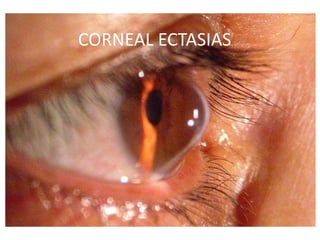
  • 2. CORNEAL ECTASIAS • It is a group of diseases affecting the corneal shape due to CORNEAL THINNING. – Anterior Keratoconus – Keratoglobus – Pellucid Marginal Degeneration – Posterior Keratoconus • Key Features: – Usually Bilateral, often asymmetrical – Isolated to cornea – Non inflammatory
  • 3. KERATOCONUS • Progressive corneal steepening, typically inferior to the centre of the cornea. • Non inflammatory, usually bilateral. • Incidence: Between 50 and 230 per 100 0001. • Unilateral involvement is 2–4%2. • Onset starts at the age of puberty. 1. Krachmer JH, Feder RS, Belin MW. Keratoconus and related noninflammatory corneal thinning disorders. Surv Ophthalmol. 1984;28:293–322. 2. Holland DR, Maeda N, Hannush SB, et al. Unilateral keratoconus. Incidence and quantitative topographic analysis. Ophthalmology.1997;104:1409–1413.
  • 4. ASSOCIATED DISEASES Systemic  Atopic Diseases (35%)1.  Down’s Syndrome (5.5-15%)2.  Ehler-Danlos and osteogenesis imperfecta.  Mitral valve prolapse.  Marfan Syndrome.  Ocular  Vernal Conjunctivitis.  Retinitis Pigmentosa.  Leber’s congenital amaurosis  Retinopathy of prematurity.  Fuchs’ Dystrophy. 1. Rahi A, Davies P, Ruben M, et al. Keratoconus and coexisting atopic disease. Br J Ophthalmol. 1977;61:761–764. 2. Cullen JF, Butler HG. Mongolism (Down’s syndrome) and keratoconus. Br J Ophthalmol. 1963;47:321–330.
  • 5. ETIOLOGY BIOCHEMICAL: • Up-regulation of degradative enzymes (acid esterase and acid phosphatase, cathepsins B and G, and some matrix metalloproteinases) and the down-regulation of proteinase inhibitors. • There is a four fold increase in the receptors of INTERLEUKIN 1 causing apoptosis of stromal keratocytes. • Microtrauma Release of IL-1 from epithelial epithelium.
  • 6. HEREDITY • Autosomal dominant with incomplete penetrance. • Frequency of inheritance is 6%. • Keratoconus prevalence in first-degree relatives to be 3.34%, about 68 times that found in the general population. PATHOLOGY • Keratoconus can involve every layer and tissue of cornea. • Superficial cell enlargement and prominence of elongated cells in the corneal epithelium.
  • 7. Early degeneration of basal epithelial cells. Disruption of epithelial basement membrane. Epithelium grows posteriorly into the Bowman’s Layer, with collagen growing anteriorly into the epithelium. Z shaped interruptions at the level of the basement membrane. Fragmentation of the Bowman’s layer. (Early change)
  • 8. Fleischer ring found at the base of the cone is a hallmark of keratoconus. Breaks in Bowman’s layer fill with scar tissue. Fine scars consolidate leading to formation of reticular branched opacities at this level. Collagen lamellae are released from their interlamellar attachments or from their attachments to Bowman’s layer and become free to slide Thinning without collagenolysis.
  • 9. Alteration of the normal orthogonal arrangement of the collagen fibrils leads to stromal tissue instability. Most common location for the apex of the cone, in the central or inferior cornea, isrelated to this. Endothelial cell pleomorphism and polymegathism
  • 10. DIAGNOSIS • Onset of the disease is at puberty. • Usually appears in the 2nd or 3rd decade. SYMPTOMS: • Progressive visual blurring & disortion due to irregular astigmatism. • Frequent change of glasses. • Pain, photophobia. • Eye rubbing.
  • 11. Scissoring of red reflex on retinoscopy One of the earliest signs SIGNS:
  • 12. Munson Sign Buldging of the lower eye lid in downward gaze.
  • 13. Papillae, trantas dots, limbal nodule, pigmentation, ropy discharge.
  • 15. Corneal ectasia: Apex is inferior to the horizontal line to the pupillary axis. Corneal thinning of about 1/2-1/5 is observed at the apex. 2 types of cones: Round/ Nipple shaped Oval shaped
  • 16. Fleischer Ring Iron depositions in the corneal epithelium. The ring configuration is due to irregular distribution of tears at the base of the cone and resultant deposition.
  • 17. Vogt’s Striae Fine vertical lines formed due to compression of descemet’s membrane. Parallel to the axis of the cone
  • 20. • Breaks in Descemet Membrane • Stromal edema
  • 21. Charleux Sign Dark reflex in the area of the cone on observation of the cornea with pupil dilated using a direct ophthalmoscope set on plano.
  • 22. DIFFERENTIAL DIAGNOSIS • Pellucid marginal degeneration. • Keratoglobus. • Post traumatic corneal ectasia. • Protrusion of cornea subsequent of corneal thinning from ulceration. • Corneal ectasia following keratorefractive surgery.
  • 25. INVESTIGATIONS • KERATOMETRY: Mires are commonly steep, highly astigmatic, often egg shaped. Inability to superimpose the central keratometric rings is due to irregular astigmatism. (hallmark) LIMITATIONS: Provides information only of the central 3mm of cornea. Not useful in irregular astigmatism.
  • 26. • VIDEOKERATOMETRY Based on Placoid disc principle. Early disease: Isolated area of smaller ring spacing and distortion. Advanced disease: Decrease in overall ring spacing and becomes increasingly irregular LIMITATIONS: Does not give accurate information about the posterior curvature and corneal thickness. Cannot detect early keratoconus.
  • 27. Astigmatism progression: Symmetric Bow tie pattern Asymmetric appearance Inferotemporally displaced steep sided cone
  • 28. • ORBSCAN Uses the principle of scanning slit combined with a Placoid system. Provides reliable information about the anterior and posterior elevation and best fit sphere and a corneal pachymetry map. LIMITATIONS The posterior curvature map might not be 100% accurate. Requires patient fixation for accurate data. Data pertaining to the central cornea is not accurate.
  • 29.
  • 30. • PENTACAM USES a rotating Scheimpflug camera. Provides reliable data of anterior and posterior corneal elevation and accurate measurement of corneal thickness. The images taken by a pentacam maintain the central point of each meridian hence the software can re register these central points and eliminate the eye movement. Data is 10 times more accurate than the that obtained from Orbscan.
  • 31.
  • 32.
  • 33. • PACHYMETRY Ultrasonic and optical based devices can be used. Measurement of corneal thickness is useful for diagnosis, documenting progression, and planning treatment. • OCULAR RESPONSE ANALYSER Helps in making a diagnosis and classifying by assessing corneal hysteresis and resistance.
  • 34.
  • 35. TREATMENT • NON SURGICAL – Spectacles: Only in early cases. • Once irregular astigmatism develops this type of visual correction cannot be used. – Contact lens: Soft toric lenses, standard bicurved hard lenses, custom back toric lenses, piggyback systems, hybrid lens, scleral and mini scleral lens.
  • 36. RIGID GAS PERMEABLE LENS • First choice in correcting irregular astigmatism. • Fitting strategies: Apical clearance: There is no touch in the apical area. Advantage: 1. Reduced risk of scarring 2. Whorled keratopathy 3. Corenal erosion Limitation: Hampers tear exchange, edge can come in visual axis. Rarely used now days.
  • 37. Apical touch : Aims at providing primary lens support at the apex of the cornea. The central optic zone of the lens touches the central cornea. ADVANTAGE: Better quality of vision LIMITATION: Corneal scarring and intolerance over long term use.  Three point touch: Most popular technique The lens bearing is shared between apex and mid peripheral cornea. Minimizes apical scarring, better comfort and longer wearing time.
  • 38. PIGGYBACK LENS SYSTEM • Consists of the fitting a rigid gas permeable on top of a soft contact lens. • The soft lens is used to improve wearing comfort and provide a more regular area for the gas permeable contact lens to sit. • The gas permeable contact lens is used for providing adequate visual acuity.
  • 39.
  • 40. HYBRID CONTACT LENS • Contain a rigid gas permeable center with a soft skirt. • They are fitted with no or minimal apical touch in the central cornea. ADVANTAGE: Can be fit on cones of any severity. LIMITATION: Can cause hypoxia related changes like vascularisation and central corneal clouding.
  • 41.
  • 42. ROSE K LENSES • Multicurve lenses with small optical zone which fits snugly over the cone. • Rose K contact lens provides greater comfort, better quality of vision and requires lesser chair time. • Rose K2 IC is a larger diameter, intralimbal lens that can be used for oval cones.
  • 43. SCLERAL LENSES • They rest on the sclera without touching the cornea or the limbus leaving a clear area between the contact lens and the cornea. • They are of two types: – Prosthetic Replacement Of the ocular Surface Ecosystem (PROSE) – Boston Ocular Surface Prosthesis (BOSP)
  • 44. ADVANTAGE Good centration, stability and improved Visual acuity. Extermely useful in patients with advanced ectasias who are intolerant to contact lenses or when immediate surgery is not possible LIMITATION High cost, reduced tear exchange and difficult insertion and removal.
  • 45. • SURGICAL MANAGEMENT – INTRACORNEAL RING SEGMENT INSERTS – CORNEAL CROSS LINKAGE – LENTICULAR REFRACTIVE SURGERY – CORNEAL TRANSPLANT
  • 46. INTRACORNEAL RING SEGMENT INSERTS • Ferrara rings: Fixed inner diameter of 5.00 mm with a triangular anterior contour. • Intacs: Inner diameter of 6.8 mm and a flat anterior contour. Hexagonal in cross section. Intacs SK: Inner diameter of 6.00 mm and elliptical cross section. The ring segments should be inserted upto 2/3rd depth of the cornea. Minimum corneal thickness should be 450 µ. They do not prevent the progression of the disease.
  • 47. The technique involves the implantation of one of two polymethy metha acrylate segments in the stroma to flatten the central cornea. Intralamellar pockets are created with the help of a vaccum lamellar dissector or femtosecond laser.
  • 48. CORNEAL COLLAGEN CROSSLINKING • The most recent addition to surgical management. • It may slow or halt the progression with photo- oxidative treatment to increase the rigidity of the corneal stroma. • 60-70% patients show some stabilization following this treatment. • The therapeutic effects are limited upto 300 µ of the anterior stroma. • This technique should not be performed for corneas less than 400µ in thickness. • It carries a slight risk of development of keratitis.
  • 49. STEPS: Under topical anaesthesia 6-7 mm of central corneal epithelium is removed. Riboflavin 0.1% solution is applied topically (10-30 mins) and cornea is irradiated with UVA light of 370 nm for 30 mins. Production of reactive oxygen species helps create covalent bonds between collagen fibrils.
  • 50.
  • 51.
  • 52. CORNEAL TRANSPLANTATION Penetrating Keratoplasty: Success rate in 90-95%. Visual recovery takes several weeks to months. Full stabilisation might even take a year. Hydrops that does not clear by 3-4 months is best treated by Penetrating keratoplasty.
  • 53. DEEP ANTERIOR LAMELLAR KERATOPLASTY • It is the procedure of choice as there is good endothelial count even after hydrops. • Advantages over Penetrating keratoplasty: Reduced risk of Rejection Reduced risk of Endopthalmitis Greater wound stability Reduced need for topical steroids Faster healing
  • 54. LAYER BY LAYER MANUAL DISSECTION Partial trephination of variable depth ranging from 50-70% of corneal thickness, stroma is removed with the help of a crescent. Layer by layer removal of the stroma to reach as close to the Descemet’s membrane. LIMITATION: Poor visual outcome due to residual stroma and interface haze.
  • 55. AIR ASSISTED DALK Archila first described this technique involving air injection to achieve dissection as close to DM as possible. The Big bubble technique was described by Anwar and Teichmann and is the most widely used technique. Steps: Air injection into the stroma through a groove created by trephining 60-80% of the stromal thickness. Air infiltrates the potential space between deep stroma and DM and creates a Dome shaped detachment of DM. Once the plane of separation is created the stroma can be easily excised.
  • 56. FEMTO LASER ASSISTED DALK It can be used to create corneal incisions with customised graft edges and lamellar planes for both donor and recipient corneas. ADVANTAGE: Accuracy of forming the bubble at desired corneal depth Positive refractive outcome LIMITATIONS: Cost and availability
  • 57. KERATOGLOBUS • Bilateral ectatic disorder which is non progressive or minimally progressive. • Generalised thinning with marked peripheral thinning. • Types: – Congeital: Ehler’s-Danlos type IV and brittle cornea syndrome. – Acquired: Initially keratoconus keratoglobus.
  • 58.
  • 59. Clinical Features Blurring of vision. Corneal perforation spontaneous or following minimal trauma. Rarely might present as photohobia, glare, pain, conjunctival injection following acute hydrops. Limbus- limbus corneal thinning Blue sclera due to thinning
  • 60. Differential Diagnosis Keratoconus Pellucid Marginal Degeneration Megalocornea Congenital glaucoma
  • 61. Investigations Diagnosis is essentially clinical. Ultrasonic Pachymetry shows reducude corneal thinckness. Corneal Topography shows generalised thinning and irregular astigmatism is difficult to perform in advaced cases .
  • 62. Management Conservative: Observation is the rule in early cases. Patient is advised to use polycarbonate glasses. Visual rehab is done using refractive correction for high myopia. Scleral lens, rigid gas permeable lens, reverse geometry hydrogel lens, large diameter inverse geometry rigid gas permeable lenses can be used.
  • 63. Surgical Management It is difficult to manage these patients by surgery: Large sized grafts are required increasing the risk of rejection. Peripheral sutures often cut through. Proximity of the graft to the limbus increases risk of rejection. Higher chances of perforation.
  • 64. PELLUCID MARGINAL DEGENERATION • Bilateral corneal ectatic disorder. • Band of thinning 1-2mm in diameter extending from 4 to 8 o’clock. • Area of thinning is separated from the limbus by 1-2mm of normal cornea. • Onset: 2nd- 5th decade of life. Beer Belly appearance
  • 65. • Blurring of vision due to against the rule astigmatism. • Frequent chances of glasses. • Ocular examination reveals thinning of inferior cornea with 1-2 mm separation from the limbus. • Ectasia is just superior to the thinning. • Protruding cornea is of normal thickness. • Occasional concentric Descemet folds seen inferiorly.
  • 66. Investigations • Corneal topography: Inferior peripheral steepening extending into the mid periphery. Crab- claw appearance. Against the rule: Superiorly. With the rule: Inferiorly.
  • 67. Management • Spectacles in early cases. • PROSE, BOSP and hybrid contact lens can be used. • Large diameter Penetrating keratoplasty. • Combined Lamellar with penetrating keratoplasty. • Crescentric lamellar keratoplasty. • Crescentric or wedge excision if ectasia in small sector.
  • 68. POSTERIOR KERATOCONUS • It is a congenital corneal anomaly in which the posterior corneal surface protrudes into the stroma. • Sporadic, unilateral, non progressive. • Variant of corneal mesenchymal dysgenesis. • Scarring is seen anterior to the descemet bulge. • Pathologic Examination: Scarring at Bowman’s membrane and thinning of descmet membrane with excrescences.

Editor's Notes

  1. Scarring of Bowman’s layer and the anterior stroma are common and correlate histopathologically with collagen fragmentation, fibrillation, and fibroblastic activity.
  2. Arrow head pattern
  3. Lysyl hydroxylase activity normal in brittle cornea synd