Oxidative stress response of Daphnia magna exposed to silver nanoparticles - CROSS11-1
1. ReferencesConclusion
Owing to their exceptional catalytic,
antimicrobial, and plasmonic
properties, silver nanoparticles
(AgNPs) are among the most widely
used engineered nanoparticles
(ENPs) in both consumer and medical
applications. Thus, increased
likelihood of AgNPs entering the
environment urges the need for
environmentally relevant toxicity
testing using appropriate test species
in order to minimize and quantify all
risk. In this work, a comprehensive
toxicity assessment of AgNPs was
conducted using D. magna as a
standardized test organism.
Introduction
Methods
Results
Obtained data showed significantly
different toxicity of nano and ionic
form of Ag in the aquatic system,
with Ag+ being more toxic to D.
magna. This study provided strong
evidence of the antioxidation
mechanism and suggested that
introduced nanomaterials can
significantly affect the toxicity of
nanoparticles on aquatic organisms.
Four biomarkers were evaluated:
reduced glutathione (GSH) level,
reactive oxidative species (ROS)
content using fluorescent probes
DCFH-DA and DHE, catalase (CAT),
and superoxide dismutase (SOD)
activities, all of reflect the responses
of chemically induced stress. Citrate-
coated AgNPs were synthesized
following a method described by Li et
al. Stability evaluation of purified
AgNPs was performed in both
ultrapure water (UPW) and standard
culture medium (SCM) using the
dynamic light scattering (DLS)
method and TEM measurements.
Oxidative stress response of Daphnia magna
exposed to silver nanoparticles
Tea Crnkovid¹, Lea Ulm², Adela Krivohlavek², Ivana Vinkovid Vrček³
¹ Faculty of Pharmacy and Biochemistry, University of Zagreb ² Institute of Public Health “Dr. Andrija Štampar”, Zagreb ³ Institute for
Medical Research and Occupational Health, Zagreb
Contact Tea Crnković: tcrnkovic@pharma.hr; tea.crnkovic92@gmail.com
Fabrega J, Luoma SN, Tyler CR, Galloway TS, Lead JR.
Silver nanoparticles: behaviour and effects in the aquatic
environment. Environ Int. 2011; 37:517–531.
Li H, Xia H, Wang D, Tao X. Simple synthesis of
monodisperse, quasi-spherical, citrate-stabilized silver
nanocrystals in water. Langmuir. 2013; 29:5074−5079.
Jemec A, Tišler T, Drobne D, Sepčić K, Jamnik P, Roš M.
Biochemical biomarkers in chronically metal-stressed
daphnids. Comp. Biochem. Physiol. Part C. 2008; 147:61–
68.
Marklund SL, Marklund G. Involvement of the
superoxide anion radical in the autoxidation of pyrogallol
and a convenient assay for superoxide dismutase. Eur. J.
Biochem. 1974; 47(3):469.
Ellman GL. Tissue sulfhydryl groups. Arch. Biochem.
Biophys. 1959; 82(1):70–77.
Barata C, Varo I, Navarro JC, Arun S, Porte C. Antioxidant
enzyme activities and lipid peroxidation in the freshwater
cladoceran Daphnia magna exposed to redox cycling
compounds. Comp. Biochem. Physiol.C Toxicol.
Pharmacol. 2005; 140(2):175–186.
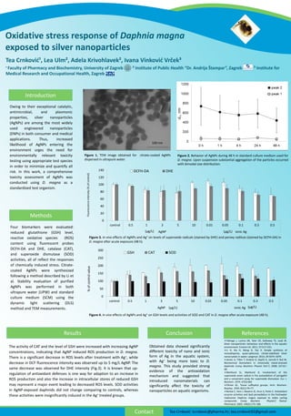
The activity of CAT and the level of GSH were increased with increasing AgNP
concentrations, indicating that AgNP induced ROS production in D. magna.
There is a significant decrease in ROS levels after treatment with Ag+, while
decrease in DCF fluorescence intensity was observed up to 5 mg/L AgNP. The
same decrease was observed for DHE intensity (Fig.3). It is known that up-
regulation of antioxidant defenses is one way for adaption to an increase in
ROS production and also the increase in intracellular stores of reduced GSH
may represent a major event leading to decreased ROS levels. SOD activities
in AgNP-exposed daphnids did not change comparing to controls, whereas
these activities were insignificantly induced in the Ag+ treated groups.
Figure 3. In vivo effects of AgNPs and Ag⁺ on levels of superoxide radicals (stained by DHE) and peroxy radilcas (stained by DCFH-DA) in
D. magna after acute exposure (48 h).
0
50
100
150
200
250
300
control 0.5 1 3 5 10 0.01 0.05 0.1 0.3 0.5
AgNP ionic Ag
%ofcontrolvalue
GSH CAT SOD
(μg/L) (μg/L)
0
20
40
60
80
100
120
140
control 0.5 1 3 5 10 0.01 0.05 0.1 0.3 0.5
AgNP ionic Ag
Fluorescenceintensity(%ofcontrol)
DCFH-DA DHE
(μg/L)(μg/L)
Figure 2. Behavior of AgNPs during 48 h in standard culture medium used for
D. magna. Upon suspension substantial aggregation of the particles occurred
with bimodal size distribution.
Figure 1. TEM image obtained for citrate-coated AgNPs
dispersed in ultrapure water.
100 nm
Figure 4. In vivo effects of AgNPs and Ag⁺ on GSH levels and activities of SOD and CAT in D. magna after acute exposure (48 h).