1. Electrophoresis and
Movement of Molecules
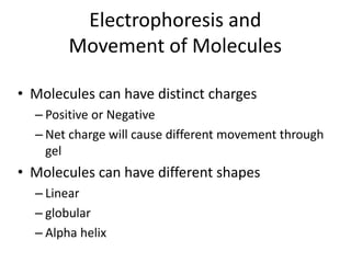
• Molecules can have distinct charges
– Positive or Negative
– Net charge will cause different movement through
gel
• Molecules can have different shapes
– Linear
– globular
– Alpha helix
+
2.
3. Macromolecular charge
• Macromolecules have a
variable net charge that
depends on pH
• pH at which net charge is
zero = pI
• Electrical shielding of charge
occurs when counterions
are solvated
V=
V =
4. Electrophoresis
• Horizontal Agarose Gels
• Agarose forms a gel or molecular sieve
that supports the movement of small
materials in solution used for DNA
• Vertical Polyacrylamide Gels
• Made of Polyacrylamide
• Used for Protein molecular size, shape, charge
• IEF electrophoresis
• Western Blot technique
6. Agarose gels
• Usually used in DNA analysis
• Made up of linear polysaccharide mol wt of
12,000
• Basic repeating unit is agarobiose
• Gels are prepared at 1% to 3% providing
tunnels for molecules to move through
• DNA can be much larger then most proteins
7. Agarose Gel with DNA Bands
• DNA is negatively
charged
• Smaller sized DNA
moves faster than
Larger DNA
• Markers are used to
determine relative
sizes of DNA pieces
markers
8. PAGE
• Native : Protein is prepared with little
disturbance to the cellular material
– Proteins are associated
– Movement of samples through the gel can be
inconsistent
• SDS : Sodium Dodecyl Sulfate Is a detergent
– Protein coated with a negative charge in
proportion to its molecular weight
– Denatures and unfolds protein
– Reducing agents (DTT)break amino acid cross-links
11. Uses for PAGE
• Separates proteins from each other
– Proteins separated by size
– Isoelectric point
• Determines
– Molecular size of protein
– Quantifies the amount present
– Displays Impurities
– Used in western blot assays by antigen interactions
12. Determine Molecular Weight
1. Run standard molecular weight markers
on gel
2. Run unknown protein on the same gel
3. Create a graph of the mol wt versus
distance molecule has moved
4. Using the distance the unknown has moved
determine the molecular weight from graph
14. Western Blot Analysis
• Identifies protein through antibody interaction
• Run proteins on denatured gel (SDS-PAGE)
• Transfer (blot) proteins onto membrane
• Probe the membrane with primary antibody
• Add secondary antibody (this antibody is linked
to an enzyme)
• Substrate is added and color appears
16. SDS Effect on Protein Movement
• Sodium Dodecyl Sulfate denatures protein and
covers it with negative charges : moves to + end
• Vertical gels are designed so the top of the gel
box is attached to the negative power outlet
• The bottom of the gel box is attached to the
positive power outlet
• Movement through the PAGE gel is
proportional to mass not to charge
17. Movement of Proteins on an SDS
Gel
Stacking of
proteins at top of
gel at start
Low weight
molecular dye
-
+
Distribution of
proteins in a
charged field
Protein Migration
Highest
Molecular
Wt. protein
18. % Polyacrylamide in Gel
• Gels can be made at different concentrations of
polyacrylamide
• Example: gels made at 3%,6%,9% and 12% will
produce different openings through which the
molecule will migrate
• The larger the opening allows large molecules
to move through the gel
28. Transfer
• Make proteins accessible to antibody detection
• Gel to membrane (nitrocellulose or PVDF)
• Manual or Electroblotting
• Manual:
-Membrane on gel, stack of filters
papers on both sides
- Entire stack in buffer, proteins move by
capillary action
30. Blocking
• Steps taken to prevent interactions between
the membrane and the antibody
• Placing the membrane in a dilute solution of
protein with a minute percentage of detergent
such as Tween 20
31. Detection
• Antibody against the target protein
• Antibody can be labeled with:
- Enzyme
- Chemiluminescent
- Fluorescence
• Single Step
• Two Step
• Third Alternative is radioactivity
33. Medical Diagnostic Applications
• Confirmatory HIV test
• Definitive test for Bovine spongiform encephalopathy
(Mad Cow Disease)
• Confirmatory test for Hepatitis B infection
• Western blot is sometimes used to confirm FIV status
in cats
34.
35.
36. Interpretations of Results (HIV)
• Negative: No Band Present
• Positive: 2 Env Band Present (WHO)
• Indeterminate: Any bands present but
do not meet criteria for positive
37. Electrophoresis of Samples
Setting Up and Running
Mini-PROTEAN TGX Precast
Gels –
Youhttp://www.youtube.co
m/watch?v=XnEdmk1SqvgT
ube
Samples: boiled 3’ with
loading dye (2x Laemmli
buffer + running dye)
Mini-PROTEAN tetra cell:
Set up according to SOP
given in workbook
Power settings: 75 volts for
45 – 60 minutes
Running dye should not
run off the bottom of gel