1. MYOCARDIAL INFARCT AND MYOCARDITIS
Definition - DISCRETE FOCUS OF ISCHEMIC NECROSIS OF MUSCLE OF
HEART
- *exclude small foci of necrosis caused by drugs | toxins | viruses
- *dev. Of infarct related to :
- DURATION OF ISCHEMIA
- METABOLIC RATE OF ISCHEMIC TISSUE
- *Foci of necrosis form after 20 min of ischemia—become more
extensive as period of ischemia lengthens
Types of MI .
classified according to
:
a) SIZE AND
EXTENT OF
INFARCTION
b) ANATOMIC
LOCATION OF
THE INFARCT
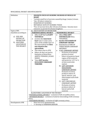
A) SIZE N EXTENT OF INFARCTION
SUBENDOCARDIAL INFARCT TRANSMURAL INFARCT
Affect 1/3rd to ½ of LEFT
VENTRICLE
involves the innermost
layer and, in some cases,
parts of the middle layer of
the myocardium but does
not extend to the
epicardium.
This infarct due to ATH
Others dz that can lead to
limitation of blood flow :
AORTIC STENOSIS | H’AGIC
SHOCK
*does NOT involve
OCCLUSIVE CORONARY
THROMBI
Affect FULL LEFT
VENTRICULAR WALL
THICKNESS
Involve OCCLUSION OF
CORONARY ARTERY
Distribution of ONE OF
THREE MAJOR CORONARY
ARTERIES:
A) RT. CORONARY ARTERY
- Occlusion of proximal
portion of vessel
- Infarct of posterior basal
region of left ventricle
and posterior 1/3rd to ½
of interventricular
septum
B) LEFT ANTERIOR
DESCENDING
CORONARY ARTERY
- Occlusion of artery
produces infarct of
apical, anterior, and
anteroseptal wall of left
ventricle
C) LEFT CIRCUMFLEX
CORONARY ARTERY
- Occlusion of artery
produces infarct of
lateral wall of left
ventricle
B) ANATOMIC LOCATION OF THE INFARCT
- POSTEROLATERAL INFARCT – occlusion of left circumflex artery
- ANTERIOR INFARCT – occlusion of anterior descending of left coronary
artery
- POSTERIOR INFARCT – occlusion of right coronary artery
Development of MI - Does not occur spontaneously
- 1st develop in SUBENDOCARDIUM
2. - Progress to SUBEPICARDIUM OVER COURSE OF SEVERAL HOURS
- Transient occlusion only cause SUBENDOCARDIAL INFARCT
- Persistent occlusion leads to TRANSMURAL INFARCT
- Infarcts MOST occur in LEFT VENTRICLE due to:
a) Greater workload
b) Greater thickness
MACROSCOPIC
CHARACTERISTIC OF
INFARCT
Ischemia for up to 20-30 min result in REVERSIBLE CYANOSIS n
BULGING during systole
By 24hr –INFARCT CAN BE RECOGNISE ON CUT S/F INVOLVED
VENTRICLE BY ITS PALLOR
After 3-5dy – INFARCTED AREA BECOME MOTTED, MORE SHARPLY
OUTLINED, CENTRAL PALE, YELLOWISH, NECROTIC AREA
BORDERED BY HYPEREMIC ZONE
Within 2-3 weeks – INFARCTED AREA IS DEPRESSED N SOFT W
GELATINOUS APPEARANCES
After several months – HEALED INFARCTS ARE FIRM N
CONTRACTED , HV PALE GRAY OF SCAR TISSUE
3. MICROSCOPIC
CHARACTERISTIC OF
INFARCT
24HR First 24 hr : after 30-60min of
ischemia,mitochondria sweollen w
disorganized cristae
: nucleus clumping n margination of
chromatin
:sarcolemma focally distrupted
:loss of sarcolemma integrity leads to
release of intracellular proteins(myoglobin, CK,
and troponin I and T)
After 24 hr : myocytes deeply eosinophilic n
show coagulation necrosis
: take several dys for myocyte nucleus
to disappear totally
2-3 days - PMN attracted to necrotic myocytes
- PMN accumulate at infarct borders
- Interstitial oedema n h’age appear
- Myocytes necrotic, striation less
prominent
- Some PMN begins to undergo karyorrhexis
5-7 days a) PMNs remain
b) The infarcted area shows phagocytosis of
dead muscle by macrophage
c) Fibroblast begins to proliferate
d) New collagen deposited
e) Lymphocytes and pigment-laden
macrophage prominent
f) Replacing necrotic muscle with scar
tissue
1-3 weeks - Collagen deposition proceeds
- Inflammatory infiltrates gradually recedes
More than 4
weeks
o Debris progressively removed
o Scar becomes more solid
After 3 months Infarcted area been replaced by scar tissue
MYOCARDITIS
Definition Inflammation of myocardium Assoc. with myocytes necrosis n degeneration
Excludes ischemic heart dz
Occur at ANY AGE
COMMON IN CHILDREN (a year-10)
Can produce ACUTE HEART FAILURE
Aetiology - Idiopathic
- Infectious : Viral | Rickettsial | Bacterial | Fungi n protozoa | Metazoan
parasites
- Non infectious : Hypersensitivity n immunologic related dz | Radiation |
Sarcoidosis | uremia
4. Microscopic
characteristic
-present of LYMPHOCYTES (called lymphocytes myocarditis)
-Hvneutrophilic component
-infiltrates myocytes – result in myocytes necrosis
-some plasma cell
-Eosinophil
-macrophage admixed with dominant lymphocytic population
-lack of cross striation
-present of bacteria (bacterial myocarditis) – hv abscess – purulent exudate