Call Girls Colaba Mumbai ❤️ 9920874524 👈 Cash on Delivery
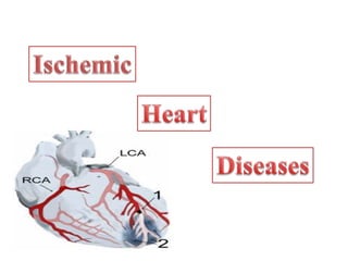
Ischaemic heart disease lecture.
1.
2. ISCHEMIA HEART DISEASE (IHD)
• A group of patho-physiologically related syndromes resulting
from an imbalance between the blood supply and myocardial O2
demand.
• IHD is also called CAD b/c in > 90% of cases, myocardial
ischemia is due to Atherosclerosis in coronary arteries.
Other causes;
Coronary embolism.
Hypotension (shock).
Increased myocardial O2 demand.
(As in cardiac hypertrophy or tachycardia).
3. ETIO-PATHOGENESIS OF IHD
• Atherosclerosis
Causes progressive narrowing (Stenosis) Obstruction myocardial
ischemia Stable angina.
• Acute plaque disruption
Rupture or ulceration in plaque Thrombosis.
thrombus Occlusion acute coronary syndromes( unstable angina,
Acute MI, and sudden death).
• Clinically significant stenosing plaques may be located
anywhere- the LAD, LCX, and RCA.
6. ANGINA PECTORIS
Angina pectoris (chest pain)—characterized by
paroxysmal and usually recurrent attacks of substernal
or precordial chest pain or discomfort caused by
transient (15 seconds to 15 minutes) myocardial
ischemia.
7. TYPES OF ANGINA
STABLE OR TYPICAL ANGINA
• Most common type.
• Chronic stenosing AS.
• Episodic chest pain.
• Physical activity.
• Emotional excitement or other cause of increased
work load.
Relived by:
Rest.
Vasodilators (Nitroglycerin).
8. PRINZMETAL OR VARIANT ANGINA-
Uncommon .
Occurs at rest.
AS with episodes of coronary artery spam.
Not related to any activity.
Responds well to:
Vasodilators (Ng +Ca Channel Blockers)
9. UNSTABLE OR CRESCENDO ANGINA
(PRE INFARCTION ANGINA)
Precursor of MI.
• Increased Frequency of pain.
• Increased intensity.
• Increased duration.
• Precipitated by progressively lower levels of physical
activity or occurs even at rest.
Caused By:
• Disruption of AS plaque with thrombosis.
• Vasospasm.
• Thrombo-embolism.
10. MYOCARDIAL INFARCTION (MI)
“Heart attack,”.
Defined as localized ischemic necrosis of myocardium resulting
from prolonged severe ischemia.
It is by far the most important form of IHD.
Incidence and Risk Factors.
• About 1.5 million people in USA suffer from MI yearly.
• 1/3 die before reaching to the hospital.
• Can occur at any age.
• Frequency rises with increasing age.
11. • Nearly 10% of MI < age 40, and 45% between 40 and
65.
• Blacks & whites equally affected.
• Men at significantly greater risk than women of
reproductive age.
• Rapid development of CAD, and IHD is the most
common cause of death in elderly women.
12. PATHOGENESIS OF MI
Coronary Artery Occlusion
• Acute plaque changes.
(Intraplaque hemorrhage, ulceration & rupture ).
• Platelets aggregation and thrombus formation.
• Mediators released from platelets (Thromboxane A2 ),
stimulate vasospasm.
• Tissue factor (From EC) activates the coagulation
pathway, adding to the bulk of the thrombus.
• Frequently within minutes, the thrombus evolves to
completely occlude the lumen of the vessel with
irreversible injury within 20-40 min.
13. PATHOGENESIS OF MI
• 10% of Transmural MI occurs in the absence of the typical
coronary vascular pathology. Mechanism may be:
• Vasospasm with associated platelet aggregation.
• Cocaine abuse.
• Emboli from the L.Atrium (AF), a left-sided mural thrombus,
infective endocarditis, intracardiac prosthetic material.
• Paradoxical emboli (patent foramen ovale)
• Vasculitis, sickle cell disease, Amyloidosis heart, dissection,
hypotension and inadequate myocardial “protection” during
cardiac surgery.
15. PAIN OF INFARCT
1)Very severe crushing chest pain ± radiation
2) Prolonged pain - at least 30 min.
- often 1 hour
- Some cases 6 hrs
3) Not induced by exertion.
4) Not relieved by rest.
5) Not relieved by nitroglycerin.
16. SUMMARY: ANGINA VS. MI
Myocardial InfarctionAnginaType
Occlusion: complete closure
of the blood vessel
Stenosis: narrowing of the blood
vessel
Type of
Obstruction
Infarct necrosisIschemic fibrosisCardiac lesion
YellowGrayish-white .Post mortem
appearance
1- pain is not induced by
excretion
2-rapid onset
3- last for a long time (30
min-hours)
4- Not Relieved by rest &
Nitroglycerine
1- pain is induced by excretion
2-Paroxysmal (rapid onset and
offset)
Last for about 15 min
3-Relieved by rest & Nitroglycerine
Type of pain
17. Acute myocardial infarction, demonstrating total occlusion of the
distal RCA by an acute thrombus (arrow) and a large zone of
myocardial hypo perfusion involving the posterior left and right
ventricles, as indicated by arrowheads
20. TRANSMURAL VERSUS SUBENDOCARDIAL
INFARCTION.
• TRANSMURAL INFARCTS (“ST ELEVATION INFARCTS” ):
Infarction involves the full or nearly full thickness of the
ventricular wall. Most common type.
Causes:
Chronic coronary atherosclerosis,
Acute plaque change, &
Superimposed thrombosis.
• Subendocardial (nontransmural) infarct
“NON-ST ELEVATION INFARCTS.”
Infarction limited to the inner 1/3 to 1/2one half of the ventricular wall.
As the Subendocardial zone is normally the least perfused
region of myocardium, this area is most vulnerable to any
reduction in coronary flow.
21. MORPHOLOGY
The frequencies of involvement of each of the three main arterial trunks and
the corresponding sites of myocardial lesions resulting in infarction are as
follows.
• LAD (40% to 50%):
Left ventricle, Anterior wall;
ventricular septum (Anterior portion) ;&
Apex circumferentially
• RCA (30% to 40%):
Inferior/posterior wall of left ventricle;
Inferior/posterior part of right ventricle;
Posterior portion of ventricular septum.
• LCA (15% to 20%):
Infarcts involving the lateral wall of left ventricle except the apex.
22. MORPHOLOGY
• Depends on the survival time of the patient after MI.
• Morphological changes
Ischemic coagulative necrosis followed by inflammation
& repair.
• MI < 12 hrs old are usually not apparent on gross
examination.
• If patient died at least 2 to 3 hours after the infarct, it is
possible to highlight the area of necrosis by immersion of
tissue slices in a solution of triphenyl tetrazolium chloride.
• This histochemical stain imparts a brick-red color to
intact, non infarcted myocardium where dehydrogenase
(e.g., lactate dehydrogenase) activity is preserved.
23. • Dehydrogenases leak out through the damaged membranes of
dead cells, an infarct appears as an unstained pale zone.
• By 12 to 24 hours an infarcted area appear as reddish-blue
caused by stagnated, trapped blood.
• There after, the infarct becomes progressively more sharply
defined, yellow-tan, and soft.
• By 10 days to 2 weeks, it is rimmed by a hyperemic zone of
highly vascularized granulation tissue.
• The succeeding weeks, the injured region evolves to a fibrous
scar.
25. • The necrotic muscle elicits acute inflammation
(most prominent between 1 and 3 days).
• Macrophages remove the necrotic myocytes.
(most pronounced at 3 to 7 days).
• Formation of highly vascularized granulation tissue,
(most prominent at 1 to 2 weeks) and its replacement by
fibrous tissue.
• The infarct heals from its margins toward its center due to intact
BV in the margins.
26. GROSS AND MICROSCOPIC FINDINGS
Time Gross L/M Changes
0-½ Hours: None No changes.
½-4 Hours None (Except TZC staining) Waviness of fibers.
4-12 Hours Dark Mottling (Occasional) Coagulation necrosis, edema
and hemorrhages.
12-24 Hours Dark mottling( Marked) Coagulation necrosis, early
neutrophils infiltrate, myocyte
eosinophilia and contraction
bands.
1-3 days: Yellow tan infarct centre. Brisk infiltrate of neutrophils ,loss
of striations and nuclei
3-7 Days Hyperemic border with central yellow tan Myocyte death and
softening early phagocytosis.
10–14 days Red-gray depressed infarct borders Well-established granulation
tissue with new blood vessels
and collagen deposition
> 2 Months Scarring complete
27.
28. Acute myocardial infarct, ( posterolateral left ventricle), demonstrated
histochemically by a lack of staining by triphenyltetrazolium chloride in areas of
necrosis (arrow). The staining defect is due to the enzyme leakage that follows cell
death. Note the myocardial hemorrhage at one edge of the infarct that was
associated with cardiac rupture, and the anterior scar (arrowhead), indicative of
old infarct.
29. Acute myocardial infarct. At 3 days, there is a zone of yellow necrosis
surrounded by darker hyperemic borders. The arrow points to a transmural
infarct in the posterior wall of the left ventricle.
30. Microscopic features of myocardial infarction and its repair.
A, One-day-old infarct showing coagulative necrosis and wavy fibers. (elongated and narrow, as
compared with adjacent normal fibers ). Widened spaces between the dead fibers contain edema fluid
and scattered neutrophils.
B, Dense polymorphonuclear leukocytic infiltrate in area of acute myocardial infarction of 3 to 4
days' duration.
C, Nearly complete removal of necrotic myocytes by phagocytosis (approximately 7 to 10 days).
D, Granulation tissue characterized by loose collagen and abundant capillaries.
E, Well-healed myocardial infarct with replacement of the necrotic fibers by dense collagenous scar.
A few residual cardiac muscle cells are present.
31. weeks –years After, healing is well under way, there is more
extensive collagen deposition.
32. REPERFUSION INJURY.
• Restoration of blood supply can prevent infarction effectively.
• This process is called as REPERFUSION.
• Reperfusion may trigger arrhythmias and myocardial
hemorrhage with contraction bands.
• Loss of myocardial viability in infarction is progressive,
occurring over a period of at least several hours.
• Early reperfusion can salvage myocardium and thereby limit
infarct size, with consequent improvement in both short- and
long-term function and survival.
• Reperfusion should be done with in first 3 to 4 hours.(critical
time).
• Reperfusion of myocardium within 20 minutes of the onset
of ischemia may completely prevent necrosis.
33. CLINICAL MANIFESTATIONS OF MI
• CHEST PAIN (most common)- may radiate to neck, jaw, shoulder, back
or left arm and may be present near the epigastrium; similar to angina but
not relieved by NG.
• Atypical chest, stomach, back or abdominal pain
• Nausea or dizziness
• SOB and difficulty in breathing
• Unexplained anxiety
• Weakness or fatigue
• Palpitations
• Pallor.
• A rapid, weak pulse and profuse sweating.
34. DIAGNOSTIC OF MI
• Electrocardiography (ECG)
• Lab tests
• Troponin-I & T – elevated
• Myoglobin- elevated
• CK-MB- elevated
Unchanged levels of CK-MB and troponin over a period of
2 days excludes the diagnosis of MI.)
• LDH- elevated (Lactate dehydrogenase)
• AST- elevated (Aspartate aminotransferase)
• TLC raised.
• Imaging Studies
• Positron Emission Tomography (PET scan)
• Magnetic Resonance Imaging (MRI)
• Echocardiography (ECHO)
• Transesophageal Echocardiography
35. CONSEQUENCES AND COMPLICATIONS OF MI
• Depend upon the size and location of the infarction & pre-
existing myocardial damage. Include:
• Contractile dysfunction: Myocardial infarcts leads to CHF,
pulmonary edema & Cardiogenic shock in 10-15% of patients.
• Arrhythmias and conduction defects:
MI lead to potentially fatal arrhythmias due to abnormal
current circuit in affected areas.
Sinus bradycardia,
Heart block,
Tachycardia, PVCs, ventricular tachycardia, and ventricular
fibrillation.
36. CONSEQUENCES AND COMPLICATIONS OF MI
Myocardial wall rupture, with possible tamponade
Softening and weakness of the necrotic and subsequently inflamed
myocardium lead to rupture of :
1) Ventricular wall (most common), (cardiac tamponade).
(2) Rupture of the ventricular septum (less common), leading to
an acute VSD and left-to-right shunting.
(3) Papillary muscle rupture (least common), resulting in the acute
onset of severe mitral regurgitation.
Pericarditis: A fibrinous or fibrino hemorrhagic pericarditis
usually develops about the second or third day following a
transmural infarct as a result of underlying myocardial
inflammation.
.
37. • Right ventricular infarction:
Isolated infarction of the RV is unusual, but it may occur after
ischemic injury of the adjacent posterior LV and ventricular
septum.
• Infarct expansion: There may be disproportionate stretching,
thinning, and dilation of the infarct region, which is often
associated with mural thrombus.
• Mural thrombosis, with possible embolization:
Contractile abnormality and endocardial damage leads to stasis &
a thrombogenic surface can initiate mural thrombosis and
thromboembolism.
38. • Ventricular aneurysm formation:
Late complication associated with large transmural infarcts.
The thin scar tissue wall of an aneurysm bulges during systole.
• Complications of ventricular aneurysms include mural
thrombus, arrhythmias, and heart failure.
• Papillary muscle rupture with possible valvular
insufficiency:
• Postinfarct mitral regurgitation results from ischemic
dysfunction of a papillary muscle and underlying myocardium
and later from papillary muscle fibrosis and shortening, or
from ventricular dilation.
• Progressive late heart failure (or chronic IHD)
39. RISK FACTORS FOR COMPLICATIONS &
PROGNOSIS
• Depend primarily on the infarct size, location, and thickness
(subendocardial or transmural).
• Large transmural infarcts: higher probability of cardiogenic shock,
arrhythmias, and late CHF.
• Anterior transmural infarcts: greatest risk for free-wall rupture,
expansion, mural thrombi, and aneurysm.
• Posterior transmural infarcts: conduction blocks, RV involvement, or
both; when acute VSDs occur in this area they are more difficult to
manage.
• Over all, patients with anterior infarcts have a worse
clinical course than those with inferior (posterior) infarcts.
• Pericarditis, rupture, and aneurysms occur rarely with subendocardial
infarcts.
40. • Ventricular remodeling:
• Initially hemodynamically beneficial, but lead to ventricular
dilation aggravating O2 demand, ischemia and depress
cardiac function.
• Changes in ventricular shape & stiffening of the ventricle due
to scar formation and hypertrophy further diminish cardiac out
put.
• Long-term prognosis after MI depends on many factors:
The most important ones are:
• Residual left ventricular function.
• The extent of vascular obstructions that perfuse the viable
myocardium.
41. CHRONIC IHD
• Chronic IHD usually occur after infarction due to the
functional decompensation by hypertrophic non infarcted
myocardium.
• In some cases severe coronary artery obstruction may present
as chronic IHD in the absence of prior infarction.
• Gross:
Heart enlarged and heavy.
LV hypertrophy and dilation.
Evidence of coronary atherosclerosis & mural thrombi..
Discrete scars of healed infarcts.
• Microscopic features:
Findings include myocardial hypertrophy, diffuse
subendocardial vacuolization, and fibrosis.
42. SUDDEN CARDIAC DEATH (SCD)
• SCD is defined as unexpected death from cardiac
causes in individuals without symptomatic heart
disease or early symptom onset (usually within 1 hour).
• It most frequently occurs in the setting of IHD; but it
may be the first clinical manifestation of IHD.
• SCD is usually the consequence of a lethal arrhythmia
(e.g., asystole, ventricular fibrillation) due to acute MI.
• Arrythmogenic foci are often located adjacent to scars
left by old MIs.
43. NONATHEROSCLEROTIC CONDITIONS
ASSOCIATED WITH SCD
• CHD, coronary arterial abnormalities
• Aortic valve stenosis
• Mitral valve prolapse
• Myocarditis
• Dilated or hypertrophic Cardiomyopathy
• Pulmonary hypertension
• Hereditary or acquired cardiac arrhythmias
• Cardiac hypertrophy of any cause (e.g., hypertension)
• Other miscellaneous causes, such as systemic metabolic and hemodynamic
alterations, Catecholamines, and drugs of abuse, particularly cocaine and
methamphetamine.
44. HERITABLE CONDITIONS ASSOCIATED WITH
SCD
• Disorders with recognizable anatomic abnormalities (e.g., congenital anomalies,
hypertrophic cardiomyopathy, MVP).
• Disorder without structural cardiac pathology e.g., Heritable arrhythmias can
precipitate sudden death (primary electrical disorders).
• Long QT syndrome, short QT syndrome, catecholaminergic ventricular
tachycardia, Wolff-Parkinson-White syndrome, congenital sick sinus syndrome,
and isolated cardiac conduction disease.
• The most important are channelopathies, mostly autosomal-dominant, caused by
mutations in genes that are required for normal ion channel function, either
involve genes that encode the ion channels (Na+, K+, and Ca+), or accessory
proteins.
• The prototype is the long QT syndrome, with prolonged QT segment in ECG.
Mutations in 7 different genes account for the majority of cases.
45. TROPONINS
• Specific proteins found in heart muscle.
• Also exist in other muscles.
• Troponins in the heart are called cardiac troponins. There are two main types of cardiac
troponins;
T and I .
• The main difference between troponins I and T is that cardiac troponin I tests measure
only cardiac troponin; tests for cardiac troponin T may cross-react with troponin found
in other muscles and give positive or increased results in the absence of heart damage.
• Normal results
People without heart damage have troponin levels less than 0.5 ng/mL.
• Abnormal results
• Levels greater than 2.0 ng/mL indicate a person has had a significant myocardial injury,
such as an infarction, and is at an increased risk for future serious heart events. Levels
between 0.5 and 2.0 ng/mL indicate a diagnosis of unstable angina, other heart disorders,
or chronic kidney failure.
• Normal troponin levels 12 hours after chest pain has started, mean
NO CARDIAC PROBLEM.
46. • Troponin levels increases in most patients who have
heart attack within 6 hours.
• Reaches its peak level after 12 hours.
• Troponin levels may remain high for 1 to 2 weeks
after a heart attack.
47. CREATININE KINASE (CK, CPK)
• An enzyme found primarily in the heart and skeletal muscles, and to a lesser extent in the
brain.
Elevated in:
• Myocardial infarction
• Crushing muscular trauma
• Brain injury
• Hypothyroidism
• Hypokalemia. Once elevated, CK remains elevated for several days.
CK/CPK Isoenzymes
• There are three Isoenzymes. Measuring them is of value in the presence of elevated levels of
CK or CKP to determine the source of the elevation.
• Isoenzyme MM BB MB
Synonym CK3 CK1 CK2
Foundin: Skeletal M. Brain GI Tract GU Tract Heart M
Heart M.
Normal levels of CK/CPK are almost entirely MM, from skeletal muscle.
• Elevated levels of CK/CPK resulting from acute myocardial infarction are about half MM and
half MB
48. NORMAL VALUES FOR CK, CPK
Total CPK normal values:
• 10 - 120 micrograms per liter (mcg/L)
• Normal Values for CK or CPK Isoenzymes
• MM 97%-100%
• MB 0%-3%
• BB 0%