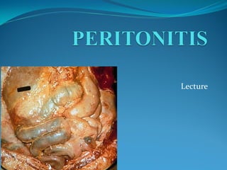
2. Peritonitis
Peritonitis is defined as inflammation of the serosal
membrane that lines the abdominal cavity and the
organs contained there in.
The peritoneum, which is an otherwise sterile
environment, reacts to various pathologic stimuli with
a fairly uniform inflammatory response.
3.
4. Anatomy
The peritoneum is the largest and most complex serous
membrane in the body.
It forms a closed sac by lining the interior surfaces of the
abdominal wall, retroperitoneum, the pelvis and the
diaphragm.
This parietal layer of the peritoneum reflects onto the
abdominal visceral organs to form the visceral peritoneum.
It thereby creates a potential space between the 2 layers (ie,
the peritoneal cavity).
The compartmentalization of the peritoneal cavity, in
conjunction with the greater omentum, influences the
localization and spread of peritoneal inflammation and
infections.
5. Anatomy
Normally, the amount of peritoneal fluid present is
less than 50 mL, and only small volumes are
transferred across the considerable surface area in a
steady state each day.
The peritoneal fluid represents a plasma ultrafiltrate.
In addition, peritoneal fluid contains small numbers
of desquamated mesothelial cells and various numbers
and morphologies of migrating immune cells.
6. Practical significance and clinical actuality
of the pathology
Acute peritonitis reveals in 16% of hospitalized
patients with surgical abdomenal pathology
The cause of death in 50% of moribund after
abdomenal operations is peritonitis.
Severe forms (stages) of this surgical state have
a 25-30% lethality rate.
In cases of multi-organs insufficiency it raises
till 85-90%
Lethality rate was in : 1936 – 1941гг = 23,8%,
1980- 1986г. = 21,9%.
8. Primary (spontaneous) peritonitis
Idiopathic peritonitis is uncommon, constituting
about 1% of all cases of peritonitis and occurs when no
obvious source for the peritoneal infection can be
demonstrated.
It was classically described in young girls where the
port of entry was presumed to be through the fallopian
tubes.
Adult primary peritonitis arises via haematogenous
spread or translocation of bacteria through the bowel
wall, especially in the presence of exogenous or
endogenous immunosuppresssion.
9. Secondary:
Acute suppurative
This is the most common form of peritonitis
encountered by the surgeon and results from
the perforation of a viscus (e.g. appendix, peptic
ulcer, colonic diverticulum, or gallbladder),
ischaemia of an intra-abdominal organ (e.g.
strangulated hernia, volvulus, mesenteric artery
occlusion),
or extension of an existing infection of an
abdominal organ (e.g. appendix abscess, liver
abscess, pyosalpinx).
10. Secondary:
Chemical (aseptic) peritonitis
Aseptic peritonitis refers to the peritoneal inflammation from
substances other than bacteria.
A perforated peptic ulcer provides the chemical peritonitis with
gastric juice and bile contaminating the peritoneal cavity.
Biliary peritonitis alone may follow gangrene and perforation of
the gallbladder, or, after open or laparoscopic cholecystectomy.
Blood in the peritoneum is also a cause of peritoneal irritation
after slow bleeding.
Meconium and urine may also precipitate chemical peritonitis.
Acute pancreatitis causes the release and activation of potent
lipolytic and proteolytic enzymes, which produce a severe
peritonitis and fat necrosis.
11. Secondary:
Interventional peritonitis
Endoscopy of the gastrointestinal tract may precipitate acute
peritonitis through perforation of the hollow organs (stomach,
bowel, oesophageal dilatation, diverticulum, urinary bladder).
The expansion of interventional radiological procedures has
precipitated a multitude of assaults on the abdominal cavity,
such as CT-guided biopsy and drainage of abscesses, mesenteric
angiography and therapeutic embolization, and percutaneous
transhepatic cholangiography and stenting, all providing further
potential for peritonitis.
Peritonitis may follow abdominal surgery where bowel and
gastric contents, blood, and urine escape into the abdominal
cavity following anastomotic dehiscence.
In patients with renal failure treated by continuous ambulatory
peritoneal dialysis, a permanent indwelling catheter in the
abdominal cavity provides a portal of entry for exogenous
bacteria.
12. Secondary:
Traumatic peritonitis
Abdominal trauma may produce acute peritonitis in
several ways. Penetrating wounds of the abdomen
without visceral injury may provide a route for
exogenous bacterial contamination.
Several blunt trauma may disrupt intra-abdominal
organs directly or indirectly through disruption of
their vascular supply.
13. Secondary:
Drug-induced peritonitis
Warfarin anticoagulation can cause peritoneal
irritation and peritonitis through leakage from a
spontaneous retroperitoneal haematoma.
The symptoms of acute peritonitis have also been
described during treatment with the antituberculous
agent, isoniazid.
14. Classification and forms of peritonitis
(continuation)
Simonyan K.S.
reactive phase – 24 hours.,
toxic – 24-72 hours,
terminal – 72 hours.
15. Classification and forms of
peritonitis (continuation)
Кusin M.I.:
I st. – without functional insufficiency of
abdominal organs (compensation)
II st. – with functional deficiency of
abdominal organs (subcompensation)
III st. – with multiple organ failure
(decompensation)
16. Classification and forms of
peritonitis (continuation)
serous peritonitis
fibrinous peritonitis
fibrinopurulent peritonitis
purulent (suppurative) peritonitis
hemorrhagic peritonitis
putrid peritonitis
17. Classification and forms of
peritonitis (continuation)
Fedorov V.D.
localized peritonitis
circumscribed (encloused) peritonitis
(infiltration, abscess)
unlimited (not encloused) peritonitis (no more
than 2 of 9 anatomical region)
generalized peritonitis
diffuse peritonitis (3-5 anatomical region)
general peritonitis
18. Classification and forms of
peritonitis (continuation)
Кusin M.I. (1994)
circumscribed peritonitis (infiltration,
abscess)
diffuse peritonitis
local peritonitis (1 anatomical region)
generalized peritonitis (several anatomical
regions)
general peritonitis (all peritoneum
19. Pathogenesis
Initially, peritoneal inflammation is often localized
and the affected area contained by a wrapping of
greater omentum, adjacent bowel, and fibrinous
adhesions.
If the inflammatory focus is part of an ongoing
process, or if host defences are lowered, localized
peritonitis may progress to life-threatening
generalized peritonitis.
There is massive exudation of inflammatory fluid into
the peritoneal cavity causing hypovolaemia, often
compounded by toxaemia from absorbed products and
septicaemia if infection is present.
Diffuse peritoneal irritation causes peristaltic paralysis
with the cessation of bowel motility.
20. Signs and symptoms
The clinical features of peritonitis are dependent on both
the aetiology and the progression of the inflammation.
The early manifestations of peritonitis following disease of
an abdominal viscus are characterized by the primary
disease process itself.
Irritation of the nearby parietal peritoneum results in
localization of the pain.
Associated symptoms include malaise, nausea and
vomiting, and a low-grade fever.
When the peritonitis is generalized the patient is clearly
unwell, with marked fever, dehydration, and absent bowel
sounds. Pain is diffuse throughout the abdomen and is
exacerbated by even the slightest movement. Shoulder-tip
pain is diagnostic of diaphragmatic inflammation.
21. Signs and symptoms
Physical findings can be divided into abdominal
signs and manifestations of systemic infection.
Local findings include
abdominal pain,
tenderness,
guarding or rigidity,
distention,
free peritoneal air,
and diminished bowel.
22. Signs and symptoms
Systemic findings include
fever,
chills or rigors,
tachycardia,
sweating,
tachypnea,
restlessness,
dehydration,
oliguria,
Disorientation
Shock is due to the combined effects of hypovolemia and
septicemia with multiple organ dysfunction. Recurrent
unexplained shock is highly predictive of serious
intraperitoneal sepsis.
Vital functions depend on the stage of the process: reactive
(or initial), toxic (or late) and terminal.
23. Laboratory findings
blood cell count,
arterial blood gases,
electrolytes,
liver and renal function tests.
A white blood cell count of greater than 200 cells/uL is
indicative of peritonitis, with virtually no false-positive and
minimal false-negative errors.
24. Instrumental investigations
Radiography (X-ray plan of abdomen)
Diagnostic puncture, abdominocentesis
Diagnostic peritoneal lavage
Ultrasonography
Magnetic resonanse tomography
Laparoscopy
25. Treatment
Surgery remains a cornerstone of peritonitis treatment.
Any operation should address the first 2 principles of the
treatment of intraperitoneal infections: early and definitive
source control and elimination of bacteria and toxins from
the abdominal cavity.
The operative approach is directed by the underlying
disease process and the type and severity of the intra-
abdominal infection. The surgeon should always strive to
arrive at a specific diagnosis and delineate the intra-
abdominal anatomy as accurately as possible prior to the
operation.
26. Treatment
In severe abdominal sepsis, however, delays in
operative management may lead to a significantly
higher need for reoperations and to worse outcomes
overall.
To reduce the bacterial load, a lavage of the abdominal
cavity is performed, with particular attention to areas
prone to abscess formation (eg, paracolic gutters,
subphrenic area).
Initial laparoscopic examination of the abdomen can
assist in determination of the etiology of peritonitis.
27. Preoperative preparations
Volume resuscitation and the prevention of secondary organ
system dysfunction are of utmost importance in the treatment of
patients with intra-abdominal infections. Correct existing serum
electrolyte disturbances and coagulation abnormalities as best as
possible before any intervention.
Begin empiric, broad-spectrum, systemic antibiotic therapy as
soon as the diagnosis of intra-abdominal infection is suspected
and tailor therapy according to the underlying disease process
and culture results.
Because patients with peritonitis often have severe abdominal
pain, provide adequate analgesia with parenteral narcotic agents
as soon as possible.
In the setting of significant nausea, vomiting, or abdominal
distension caused by obstruction or ileus, institute nasogastric
decompression as soon as possible.
28. Treatment
The goals of operative treatment of peritonitis are to
eliminate the source of contamination, to reduce the
bacterial inoculum, and to prevent recurrent or
persistent sepsis.
29. Treatment
A vertical midline incision is the incision of choice in
most patients with generalized peritonitis, because it
allows access to the entire peritoneal cavity.
In patients with localized peritonitis (eg, acute
appendicitis, cholecystitis), an incision directly over
the site of pathology (eg, right lower quadrant, right
subcostal) is usually adequate.
In patients with an unclear etiology of the peritonitis,
initial diagnostic laparoscopy may be useful.
Careful dissection and meticulous hemostasis are of
utmost importance.
30. Treatment
When faced with extensive abdominal inflammatory
disease and septic shock, draining the infection
temporarily, controlling the visceral leak quickly, and
deferring any definitive repair until after the patient
has recovered from the initial insult may be better.
31. Intensive care monitoring, often with ventilatory support
Program sanations
Flowing (running) or fractinal peritoneal lavage (Drains should be
removed or advanced once drainage diminishes and becomes more
serous in nature).
Achieving hemodynamic stability to perfuse major organs
(hypogidratation Ht >48%, CVP < 40 mm Hg; hypergidratation Ht <
35% CBP > 100; impaired cardial function Ht = № CVP – enlarged)
and this may entail the use of cardiac inotropic agents besides fluid and
blood product supportive measures.
Antibiotic therapy. Antibiotics are given for 10-14 days, depending on
the severity of peritonitis
Restoration of intestinal motor activity: procaine blocks, enemas,
peridural anesthesia
Enterosorbtion
Postoperative Care
32. Mannheim Peritonitis Index (МРI)
20 points(I degree of peritonitis) lethelity rate-0
20-30 points (II ст) 29%
More than 30 points (III ст) 100%
Risk factor Assessment of
severity (points)
1 Age 50 years 5
2 Sex (female) 5
3 Presence of organ’s failure 7
4 Presence of malignant tumor 4
5 Duration of peritonitis >24ч 4
6 Large bowel is the cause of peritonitis 4
7 Defuse peritonitis 6
8 Exudate (only one answer):
transparent 0
muddy-purulent 6
Fecal and saprogenic 12