More Related Content
Similar to Final UROP poster 2013
Similar to Final UROP poster 2013 (20)
Final UROP poster 2013
- 1. RESEARCH POSTER PRESENTATION DESIGN © 2012
www.PosterPresentations.com
ES (Pluripotent) EZ Sphere Neurosphere Monolayer
Fig 7. Process for the derivation of neural stem cells from hES and hiPS stem cells. These steps allow for direct differentiation and
ensure neural progenitors . The stem cells were first cultured on MEF substrate and then transitioned to either CellStart (1:50) or
StemAdhere (1:25) substrate (XF conditions). The stem cells are neuralized to EZ spheres using an EZ sphere media containing EGF
and bFGF. Neurospheres are generated using a media containing LIF which inhibits the differentiation of stem cells. Neurospheres
were plated on CellStart coated flasks to attach and maintain neural stem cell monolayers using neuroshpere media containing EGF,
bFGF, and LIF.
EZsphere, Neurospheres, and neural stem cell monolayers dissociated into single cells using Trypsan and were plated on 25k/mill on 8
well-chamber slides in DM/B27 for 14 days in-vitro. Cells were fixed with 2% paraformaldehyde.
The cell derivation process for Shef4 is shown below.
iPS69-hNSC (14 div)
IN VITRO (completed)
IN VIVO (in progress)Fig 3. Stem cells are potent meaning they are unspecialized cells
that can give rise to specialized cells. Stem cells also have the
capacity to self-renew; they are able to go through numerous
cycles of cell division while maintaining the undifferentiated
state.
Fig 2. Spinal cord injury (SCI) induces a
series of events that include cell death, the
demyelination of axons, scar formation,
and an inflammatory response.
Fig 1. Various types of accidents account
for the great majority of spinal cord
injuries.
Spinal cord injury (SCI) induces a series of events that include cell death, the demyelination of axons, scar
formation, and an inflammatory response. The ability of human embryonic stem cells (hESCs) and induced
pluripotent stem cells (iPSCs) to self-renew, maintain pluripotency, and differentiate into any cell in the adult body
has made these cells an important resource for regenerative medicine. Our laboratory has previously shown that the
fate and migration of human fetal neural stem cells (hfNSC) are altered by the inflammatory microenvironment,
including the cellular immune response resulting from the infiltration of polymorphonuclear neutrophils (PMN) and
macrophages (MAC) after SCI. In a follow-up study, we have tested the hypothesis that PMN and MAC can affect
the fate of other hNSC populations derived from hES and hiPS cells. As proof of principle, the role of these immune
cells on cell fate was tested in-vitro using PMN and MAC conditioned media (CM). iPS6-9 hNSCs exhibited
decreased expression of GFAP, an astrocyte marker, and increased expression of βeta-tubulin, a neuronal marker,
following 14 days in vitro (div) exposure to PMN-CM; no change in GFAP and βeta-tubulin expression was
observed following 14 div exposure to MAC-CM. In contrast, Shef4 hNSCs exhibited decreased βeta-tubulin
expression following 14 div exposure to either PMN-CM or MAC-CM. Shef4 hNSCs had very low GFAP
expression in PMN-CM and MAC-CM. Further follow-up experiments show a loss of cell number in PMN-CM due
to cell death or detachment after 14 div; however, MAC-CM promoted cell confluency with slight increase or no
change to the number of cells expressing the oligodendrocyte progenitor markers O4 and GalC after 14-div for both
tested cell lines. These results suggest that 14 div may not be optimal to consistently assess the effect of PMN-CM
and MAC-CM in hNSC differentiation. Future experiments will employ earlier time points (4 or 7 div) to minimize
cell death, detachment or excessive confuency in our cell culture. Overall, preliminary data suggest that PMN-CM
and MAC-CM affect the fate of Shef4 and iPS6-9 derived hNSCs. These results also suggest that the differing
intrinsic properties of stem cells may contribute to variable responses to inflammatory conditions. Recent
transplantation of hNSCs resulted in robust cell engraftment and some observable cell migration in the spinal cords
of immunodeficient mice post SCI. Future work will further assess cell fate and migration patterns of these
transplanted hNSC populations, along with assessments of functional recovery via Ladderbeam and CatWalk
analyses.
CONCLUSION
RESULTS
BACKGROUND
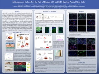
Inflammatory Cells Affect the Fate of Human hES and hiPS Derived Neural Stem Cells
ABSTRACT MATERIALS & METHODS
Shef4- hNSC (14 div)
Fig 4. Microenvironment post-injury analysis. Nuetrophils (PMN) peak in day 1 post
injury, Macrophages (MAC) peak in at days 7 & 60 post injury, and there is a small T-
Cell response.
Fig 5. The results from the in vivo transplantation of fNSC suggest that PMN and
MAC play a role in the fate and migration of fNSC. Immediate transplantation post
injury shows the migration of fNSC towards the injury epicenter. Transplantation of
fNSC 9 & 30 days post injury shows the migration of fNSC away from the injury
epicenter.
Fig 6. To test the role of PMN and MAC in cell fate, an in vitro assay was done using
PMN and MAC condition media. Three markers were used: GFAP (astrocyte marker),
Olig2 (immature oligodendrocyte marker), and βeta-Tubulin III ( neuronal marker).
Greater GFAP expression was observed in PMN-CM and greater β-Tub expression
was observed in MAC-CM in comparison to the control. There was low Olig2
expression in both PMN and MAC CM.
Fig 5 Fig 6
Fig 4
Fig 8
Fig 9
Fig 8. The next step after the derivation of neural stem
cells from hES and hiPS stem cells is to test the effect
of PMN and MAC on cell fate. PMN and MAC-CM
were collected. PMN and MAC are first isolated from
the peritoneal cavity of rats, cultured for 24 hours,
then cultured with the hES and hiPS derived NSCs.
Fig 9. The immunocytochemistry method was used to
asses the fate of the iPS69 and Shef4 derived NSCs.
The fate was assessed in three experimental
conditions: DM, PMN, and MAC. The following
antibodies were used: GFAP (1:500), β-Tub III
(1:500), O4 (1:20), GalC (1: 100).
Fig 10. In vivo experiment steps. Vertebrate at T9 is removed and a moderate injury is induced using the IH impactor (50kDine). In this
experiment 120 agouti rag2 γ hybrid mice were used. They were divided into two groups in order to receive treatment at two different time
points (0 and 30 days post injury). There are 4 different treatment groups (Shef4 hNSC, Shef6 hNSC, iPS 19-9 hNSC, and vehicle negative
control). Using a nano injector, 1 μL of media containing 75,000 hNSCs are transplanted into 4 sites surrounding the injury site. CatWalk, a
program used to record step pattern and coordination, is used at 4 different time points post treatment to assess functional recovery.
Functional recovery is also evaluated using BMS, Ladderbeam, and the Hargreaves test. The injury site is also observed and tested for
engraftment, migration, and cell fate/differentiation.
SSEA β-Tub III β-Tub III β-Tub III
Oct4 GFAP GFAP GFAP
Hoescht Hoescht Hoescht Hoescht
• PMN and MAC-CM affect the fate of ES and iPS derived neural stem cells
•In comparison to the fNSC, there is a difference in the affects that PMN and MAC have on the fate of different hES and
hiPS derived NSCs
•Different responses to microenvironment = different intrinsic properties
•In some experiments cells did not survive or detached in PMN-CM. Moreover, cells in MAC-CM appeared to be overly
confluent which may have affected cell differentiation. In both cases, cell fate is hard to analyze with ICC 14 div. These
observations suggest that 14 div may be too long. In future experiments, earlier time points will be chosen in order to
avoid cell death, detachment, or excess confluency.
•In vitro studies show how the derived NSCs behave upon exposure to factors of the inflammatory microenvironment,
however, what is observed may not translate to what happens in vivo. Because there may be other factors that affect the
fate of iPS6-9 and Shef4 derived NSCs, in vivo studies are necessary. In vivo studies will also allow us to assess functional
recovery and how they may or may not help recovery. Programs like CatWalk and Ladderbeam will be used to observe
functional recovery by observing the stepping pattern of mice.
β-Tub III
GFAP
Hoescht
β-Tub III
GFAP
Hoescht
O4
Hoescht
GalC
Hoescht
O4
Hoescht
GalC
Hoescht
B-Tub III: Increased expression of B-Tub III in PMN-CM but no change of expression in Mac-CM
GFAP: Low expression of GFAP in all conditions
O4: Inconclusive; results indicate that there was cell detachment or cell death in PMN-CM and cells in the Mac-CM appear to
be overly confluent which may affect cell differentiation.
GalC: Inconclusive; results indicate that there was cell detachment or cell death in PMN-CM and cells in the Mac-CM appear to
be overly confluent which may affect cell differentiation.
B-Tub III: Decreased B-Tub III expression in PMN-CM and Mac-CM
GFAP: Very low expression of GFAP in all conditions
O4: Inconclusive; results indicate that there was cell detachment or cell death in PMN-CM and cells in the Mac-CM appear to be
overly confluent which may affect cell differentiation.
GalC: Inconclusive; results indicate that there was cell detachment or cell death in PMN-CM and cells in the Mac-CM appear to
be overly confluent which may affect cell differentiation.
Abdelhalim S4, Nguyen HX1,2,3 & 4, Gohil P4 Funes G4, Nekanti U4 Moreno D4, Kamei N4, Cummings BJ1,2,3 & 4 & Anderson AJ1,2,3 & 4
Physical Medicine & Rehabilitation1, Anatomy and Neurobiology2, Sue and Bill Gross Stem Cell Research Center3, Institute for Memory Impairments
and Neurological Disorders4, University of California, Irvine, CA, USA
References:
Christopher Reeve Spinal Cord Injury and Paralysis Foundation. Christopher & Dana Reeve Foundation. www.christopherreeeve.org
ProQuest. What Are Stem Cells. http://www.csa.com/
Beck, Kevin D., Hal X. Nguyen, Manual D. Galvan, Desiree L. Salazar, Trent M. Woodruff, and Aileen J. Anderson. "Quantitative
Analysis of Cellular Inflammation after Traumatic Spinal Cord Injury: Evidence for a Multiphasic Inflammatory Response in the Acute
to Chronic Environment." Brain 133 (2010): 443-47.
DM/B27
Fig 11. SC121, a human
cytoplasmic marker, was used to test
hNSC engraftment.
DM/B27
PMN-CM
PMN-CM
MAC-CM
MAC-CM
DM/B27
DM/B27
PMN-CM
PMN-CM
MAC-CM
MAC-CM
Mitra Hooshmand, submitted