
Recommended
More Related Content
What's hot
What's hot (20)
Similar to Atlas of Anatomy 4th Edition.Pdf
Similar to Atlas of Anatomy 4th Edition.Pdf (20)
Recently uploaded
Recently uploaded (20)
Atlas of Anatomy 4th Edition.Pdf
- 2. To access the additional media content available with this e-book via Thieme MedOne, please use the code and follow the instructions provided at the back of the e-book.
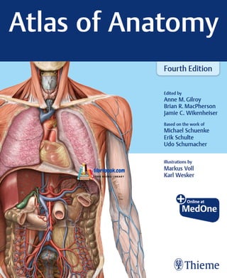
- 5. Based on the work of Michael Schuenke, MD, PhD Institute of Anatomy Christian Albrechts University Kiel Kiel, Germany Erik Schulte, MD Department of Functional and Clinical Anatomy University Medicine Johannes Gutenberg University Mainz, Germany Udo Schumacher, MD, FRCPath, CBiol, FSB, DSc Institute of Anatomy and Experimental Morphology Center for Experimental Medicine University Cancer Center University Medical Center Hamburg-Eppendorf Hamburg, Germany Thieme New York · Stuttgart · Delhi · Rio de Janeiro Atlas of Anatomy Fourth Edition Edited by Anne M.Gilroy, MA Professor Emeritus Department of Radiology University of Massachusetts Medical School Worcester, Massachusetts Brian R.MacPherson, PhD Professor and Vice Chair Department of Neuroscience University of Kentucky College of Medicine Lexington, Kentucky Jamie C. Wikenheiser, PhD Associate Professor Department of Anatomy and Neurobiology UC Irvine School of Medicine Irvine, California Illustrations by Markus Voll Karl Wesker 2113 illustrations
- 6. Illustrators: Markus Voll and Karl Wesker Development Editor: Judith Tomat Production Editor: Barbara Chernow Compositor: Carol Pierson, Chernow Editorial Services, Inc. Library of Congress Cataloging-in-Publication Data Names: Gilroy, Anne M., editor. | MacPherson, Brian R., editor. | Wikenheiser, Jamie C., editor. | Voll, Markus M., illustrator. | Wesker, Karl, illustrator. | Schünke, Michael. Thieme atlas of anatomy. Title: Atlas of anatomy / edited by Anne M. Gilroy, Brian R. MacPherson, Jamie C. Wikenheiser ; based on the work of Michael Schuenke, Erik Schulte, Udo Schumacher ; illustrations by Markus Voll, Karl Wesker. Other titles: Atlas of anatomy (Gilroy) Description: Fourth edition. | New York : Thieme, [2020] | Includes index. | Summary: “An updated atlas that provides a clear, accurate, and fully illustrated guide to human anatomy”— Provided by publisher. Identifiers: LCCN 2019058797 (print) | LCCN 2019058798 (ebook) | ISBN 9781684202034 (paperback) | ISBN 9781684202041 (ebook) Subjects: MESH: Anatomy | Atlas Classification: LCC QM25 (print) | LCC QM25 (ebook) | NLM QS 17 | DDC 611.0022/3—dc23 LC record available at https://lccn.loc.gov/2019058797 LC ebook record available at https://lccn.loc.gov/2019058798 Important note: Medicine is an ever-changing science undergoing continual development. Research and clinical experience are continually expanding our knowledge, in particular our knowledge of proper treat- ment and drug therapy. Insofar as this book mentions any dosage or application, readers may rest assured that the authors, editors, and publishers have made every effort to ensure that such references are in accordance with the state of knowledge at the time of production of the book. Nevertheless, this does not involve, imply, or express any guarantee or responsibility on the part of the publishers in respect to any dosage instructions and forms of applications stated in the book. Every user is requested to examine carefully the manufacturers’ leaflets accompa- nying each drug and to check, if necessary in consultation with a physi- cian or specialist, whether the dosage schedules mentioned therein or the contraindications stated by the manufacturers differ from the state- ments made in the present book. Such examination is particularly im- portant with drugs that are either rarely used or have been newly released on the market. Every dosage schedule or every form of applica- tion used is entirely at the user’s own risk and responsibility. The authors and publishers request every user to report to the publishers any dis- crepancies or inaccuracies noticed. If errors in this work are found after publication, errata will be posted at www.thieme.com on the product description page. Some of the product names, patents, and registered designs referred to in this book are in fact registered trademarks or proprietary names even though specific reference to this fact is not always made in the text. Therefore, the appearance of a name without designation as proprietary is not to be construed as a representation by the publisher that it is in the public domain. This book, including all parts thereof, is legally protected by copyright. Any use, exploitation, or commercialization outside the narrow limits set by copyright legislation without the publisher’s consent is illegal and li- able to prosecution. This applies in particular to photostat reproduction, copying, mimeographing, preparation of microfilms, and electronic data processing and storage. Copyright ©2020 by Thieme Medical Publishers, Inc. Thieme Publishers New York 333 Seventh Avenue, New York, NY 10001 USA +1 800 782 3488, customerservice@thieme.com Thieme Publishers Stuttgart Rüdigerstrasse 14, 70469 Stuttgart, Germany +49 [0]711 8931 421, customerservice@thieme.de Thieme Publishers Delhi A-12, Second Floor, Sector-2, Noida-201301 Uttar Pradesh, India +91 120 45 566 00, customerservice@thieme.in Thieme Revinter Publicações Ltda. Rua do Matoso, 170 – Tijuca Rio de Janeiro RJ 20270-135 – Brasil +55 21 2563-9702 www.thiemerevinter.com.br Cover design: Thieme Publishing Group Printed in India by Replika Press Pvt. Ltd. 5 4 3 2 1 ISBN 978-1-68420-203-4 Also available as an e-book: eISBN 978-1-68420-204-1
- 7. To the thousands of former students who have populated every specialty and migrated to every corner of this country, while dedicating their careers to making life better for thousands more. I am inspired by their empathy and kindness, and grateful to have been a small part of their journey. And as always, to Colin and Bryan. Anne M. Gilroy To my friend and mentor Dr. Ken McFadden, who was responsible for my early training in gross anatomy and was a role model for success in teaching. I deeply appreciate the feedback I have received over the past 40 plus years from the thousands of students I have taught and who have made me an even better teacher. However, none of the success I have enjoyed in my life would have been possible without the constant support, assistance, and encouragement of my late wife, Cynthia Long. Brian R. MacPherson To my wife Jen and my son Quinn. Jamie C. Wikenheiser Dedications
- 9. Table of Contents 1 Surface Anatomy Surface Anatomy. . . . . . . . . . . . . . . . . . . . . . . . . . . . . . . . . . . . . 2 2 Bones, Ligaments & Joints Vertebral Column: Overview . . . . . . . . . . . . . . . . . . . . . . . . . . . 4 Vertebral Column: Elements . . . . . . . . . . . . . . . . . . . . . . . . . . . 6 Cervical Vertebrae . . . . . . . . . . . . . . . . . . . . . . . . . . . . . . . . . . . 8 Thoracic & Lumbar Vertebrae . . . . . . . . . . . . . . . . . . . . . . . . . . 10 Sacrum & Coccyx . . . . . . . . . . . . . . . . . . . . . . . . . . . . . . . . . . . 12 Intervertebral Disks . . . . . . . . . . . . . . . . . . . . . . . . . . . . . . . . . 14 Joints of the Vertebral Column: Overview . . . . . . . . . . . . . . . . 16 Joints of the Vertebral Column: Craniovertebral Region . . . . . 18 Vertebral Ligaments: Overview & Cervical Spine . . . . . . . . . . 20 Vertebral Ligaments: Thoracolumbar Spine . . . . . . . . . . . . . . 22 3 Muscles Muscles of the Back: Overview . . . . . . . . . . . . . . . . . . . . . . . . . 24 Intrinsic Muscles of the Cervical Spine . . . . . . . . . . . . . . . . . . . 26 Intrinsic Muscles of the Back . . . . . . . . . . . . . . . . . . . . . . . . . . 28 Muscle Facts (I) . . . . . . . . . . . . . . . . . . . . . . . . . . . . . . . . . . . . . 30 Muscle Facts (II) . . . . . . . . . . . . . . . . . . . . . . . . . . . . . . . . . . . . 32 Muscle Facts (III) . . . . . . . . . . . . . . . . . . . . . . . . . . . . . . . . . . . . 34 4 Neurovasculature Arteries & Veins of the Back . . . . . . . . . . . . . . . . . . . . . . . . . . . 36 Nerves of the Back . . . . . . . . . . . . . . . . . . . . . . . . . . . . . . . . . . 38 Spinal Cord . . . . . . . . . . . . . . . . . . . . . . . . . . . . . . . . . . . . . . . . 40 Spinal Cord Segments & Spinal Nerves . . . . . . . . . . . . . . . . . . 42 Arteries & Veins of the Spinal Cord . . . . . . . . . . . . . . . . . . . . . 44 Neurovascular Topography of the Back . . . . . . . . . . . . . . . . . . 46 5 Sectional & Radiographic Anatomy Radiographic Anatomy of the Back (I) . . . . . . . . . . . . . . . . . . . 48 Radiographic Anatomy of the Back (II). . . . . . . . . . . . . . . . . . . 50 Back Thorax Acknowledgments. . . . . . . . . . . . . . . . . . . . . . . . . . . . . . . . . . . . . . xiii Foreword. . . . . . . . . . . . . . . . . . . . . . . . . . . . . . . . . . . . . . . . . . . . . . xiv Preface . . . . . . . . . . . . . . . . . . . . . . . . . . . . . . . . . . . . . . . . . . . . . . . xv Preface to the First Edition. . . . . . . . . . . . . . . . . . . . . . . . . . . . . . . xvii 6 Surface Anatomy Surface Anatomy . . . . . . . . . . . . . . . . . . . . . . . . . . . . . . . . . . . 54 7 Thoracic Wall Thoracic Skeleton . . . . . . . . . . . . . . . . . . . . . . . . . . . . . . . . . . . 56 Sternum & Ribs . . . . . . . . . . . . . . . . . . . . . . . . . . . . . . . . . . . . . 58 Joints of the Thoracic Cage . . . . . . . . . . . . . . . . . . . . . . . . . . . 60 Thoracic Wall Muscle Facts . . . . . . . . . . . . . . . . . . . . . . . . . . . . 62 Diaphragm . . . . . . . . . . . . . . . . . . . . . . . . . . . . . . . . . . . . . . . . 64 Neurovasculature of the Diaphragm . . . . . . . . . . . . . . . . . . . . 66 Arteries & Veins of the Thoracic Wall . . . . . . . . . . . . . . . . . . . . 68 Nerves of the Thoracic Wall . . . . . . . . . . . . . . . . . . . . . . . . . . . 70 Neurovascular Topography of the Thoracic Wall . . . . . . . . . . . 72 Female Breast . . . . . . . . . . . . . . . . . . . . . . . . . . . . . . . . . . . . . . 74 Lymphatics of the Female Breast . . . . . . . . . . . . . . . . . . . . . . . 76 8 Thoracic Cavity Divisions of the Thoracic Cavity . . . . . . . . . . . . . . . . . . . . . . . . 78 Arteries of the Thoracic Cavity . . . . . . . . . . . . . . . . . . . . . . . . . 80 Veins of the Thoracic Cavity . . . . . . . . . . . . . . . . . . . . . . . . . . . 82 Lymphatics of the Thoracic Cavity . . . . . . . . . . . . . . . . . . . . . . 84 Nerves of the Thoracic Cavity . . . . . . . . . . . . . . . . . . . . . . . . . 86 vii Table of Contents
- 10. 12 Surface Anatomy Surface Anatomy . . . . . . . . . . . . . . . . . . . . . . . . . . . . . . . . . . 140 13 Abdominal Wall Bony Framework for the Abdominal Wall . . . . . . . . . . . . . . . 142 Muscles of the Anterolateral Abdominal Wall . . . . . . . . . . . . 144 9 Mediastinum Mediastinum: Overview . . . . . . . . . . . . . . . . . . . . . . . . . . . . . . 88 Mediastinum: Structures . . . . . . . . . . . . . . . . . . . . . . . . . . . . . 90 Heart: Functions & Relations . . . . . . . . . . . . . . . . . . . . . . . . . . 92 Pericardium. . . . . . . . . . . . . . . . . . . . . . . . . . . . . . . . . . . . . . . . 94 Heart: Surfaces & Chambers . . . . . . . . . . . . . . . . . . . . . . . . . . 96 Heart: Valves . . . . . . . . . . . . . . . . . . . . . . . . . . . . . . . . . . . . . . . 98 Arteries & Veins of the Heart . . . . . . . . . . . . . . . . . . . . . . . . . 100 Conduction & Innervation of the Heart . . . . . . . . . . . . . . . . . 102 Pre- & Postnatal Circulation . . . . . . . . . . . . . . . . . . . . . . . . . . 104 Esophagus . . . . . . . . . . . . . . . . . . . . . . . . . . . . . . . . . . . . . . . . 106 Neurovasculature of the Esophagus . . . . . . . . . . . . . . . . . . . 108 Lymphatics of the Mediastinum . . . . . . . . . . . . . . . . . . . . . . . 110 10 Pulmonary Cavities Pulmonary Cavities . . . . . . . . . . . . . . . . . . . . . . . . . . . . . . . . . 112 Pleura: Subdivisions, Recesses & Innervation . . . . . . . . . . . . 114 Lungs . . . . . . . . . . . . . . . . . . . . . . . . . . . . . . . . . . . . . . . . . . . . 116 Bronchopulmonary Segments of the Lungs . . . . . . . . . . . . . 118 Trachea & Bronchial Tree . . . . . . . . . . . . . . . . . . . . . . . . . . . . 120 Respiratory Mechanics . . . . . . . . . . . . . . . . . . . . . . . . . . . . . . 122 Pulmonary Arteries & Veins . . . . . . . . . . . . . . . . . . . . . . . . . . 124 Neurovasculature of the Tracheobronchial Tree . . . . . . . . . . 126 Lymphatics of the Pleural Cavity . . . . . . . . . . . . . . . . . . . . . . 128 11 Sectional & Radiographic Anatomy Sectional Anatomy of the Thorax . . . . . . . . . . . . . . . . . . . . . . 130 Radiographic Anatomy of the Thorax (I). . . . . . . . . . . . . . . . . 132 Radiographic Anatomy of the Thorax (II). . . . . . . . . . . . . . . . 134 Radiographic Anatomy of the Thorax (III). . . . . . . . . . . . . . . . 136 Rectus Sheath & Posterior Abdominal Wall . . . . . . . . . . . . . . 146 Abdominal Wall Muscle Facts . . . . . . . . . . . . . . . . . . . . . . . . . 148 Inguinal Region & Canal . . . . . . . . . . . . . . . . . . . . . . . . . . . . . 150 Inguinal Region & Inguinal Hernias. . . . . . . . . . . . . . . . . . . . . 152 Scrotum & Spermatic Cord . . . . . . . . . . . . . . . . . . . . . . . . . . . 154 14 Abdominal Cavity & Spaces Divisions of the Abdominopelvic Cavity . . . . . . . . . . . . . . . . 156 Peritoneum, Mesenteries & Omenta . . . . . . . . . . . . . . . . . . . 158 Mesenteries & Peritoneal Recesses . . . . . . . . . . . . . . . . . . . . 160 Lesser Omentum & Omental Bursa . . . . . . . . . . . . . . . . . . . . 162 Mesenteries & Posterior Abdominal Wall. . . . . . . . . . . . . . . . 164 15 Internal Organs Stomach . . . . . . . . . . . . . . . . . . . . . . . . . . . . . . . . . . . . . . . . . 166 Duodenum . . . . . . . . . . . . . . . . . . . . . . . . . . . . . . . . . . . . . . . 168 Jejunum & Ileum . . . . . . . . . . . . . . . . . . . . . . . . . . . . . . . . . . . 170 Cecum, Appendix & Colon . . . . . . . . . . . . . . . . . . . . . . . . . . . 172 Liver: Overview . . . . . . . . . . . . . . . . . . . . . . . . . . . . . . . . . . . . 174 Liver: Lobes & Segments . . . . . . . . . . . . . . . . . . . . . . . . . . . . . 176 Gallbladder & Bile Ducts . . . . . . . . . . . . . . . . . . . . . . . . . . . . . 178 Pancreas & Spleen . . . . . . . . . . . . . . . . . . . . . . . . . . . . . . . . . . 180 Kidneys & Suprarenal Glands (I) . . . . . . . . . . . . . . . . . . . . . . . 182 Kidneys & Suprarenal Glands (II) . . . . . . . . . . . . . . . . . . . . . . 184 16 Neurovasculature Arteries of the Abdominal Wall & Organs . . . . . . . . . . . . . . . 186 Abdominal Aorta & Renal Arteries . . . . . . . . . . . . . . . . . . . . . 188 Celiac Trunk . . . . . . . . . . . . . . . . . . . . . . . . . . . . . . . . . . . . . . . 190 Superior & Inferior Mesenteric Arteries . . . . . . . . . . . . . . . . . 192 Veins of the Abdominal Wall & Organs . . . . . . . . . . . . . . . . . 194 Inferior Vena Cava & Renal Veins . . . . . . . . . . . . . . . . . . . . . . 196 Portal Vein . . . . . . . . . . . . . . . . . . . . . . . . . . . . . . . . . . . . . . . . 198 Superior & Inferior Mesenteric Veins . . . . . . . . . . . . . . . . . . . 200 Lymphatics of the Abdominal Wall & Organs . . . . . . . . . . . . 202 Lymph Nodes of the Posterior Abdominal Wall . . . . . . . . . . . 204 Lymph Nodes of the Supracolic Organs . . . . . . . . . . . . . . . . . 206 Lymph Nodes of the Infracolic Organs . . . . . . . . . . . . . . . . . . 208 Nerves of the Abdominal Wall . . . . . . . . . . . . . . . . . . . . . . . . 210 Autonomic Innervation: Overview . . . . . . . . . . . . . . . . . . . . . 212 Autonomic Innervation & Referred Pain. . . . . . . . . . . . . . . . . 214 Innervation of the Foregut & Urinary Organs . . . . . . . . . . . . 216 Innervation of the Intestines . . . . . . . . . . . . . . . . . . . . . . . . . 218 17 Sectional & Radiographic Anatomy Sectional Anatomy of the Abdomen . . . . . . . . . . . . . . . . . . . 220 Radiographic Anatomy of the Abdomen (I). . . . . . . . . . . . . . 222 Radiographic Anatomy of the Abdomen (II). . . . . . . . . . . . . . 224 Abdomen viii Table of Contents
- 11. 18 Surface Anatomy Surface Anatomy . . . . . . . . . . . . . . . . . . . . . . . . . . . . . . . . . . 228 19 Bones, Ligaments & Muscles Pelvic Girdle. . . . . . . . . . . . . . . . . . . . . . . . . . . . . . . . . . . . . . . 230 Female & Male Pelvis . . . . . . . . . . . . . . . . . . . . . . . . . . . . . . . 232 Female & Male Pelvic Measurements. . . . . . . . . . . . . . . . . . . 234 Pelvic Ligaments . . . . . . . . . . . . . . . . . . . . . . . . . . . . . . . . . . . 236 Muscles of the Pelvic Floor & Perineum . . . . . . . . . . . . . . . . . 238 Pelvic Floor & Perineal Muscle Facts. . . . . . . . . . . . . . . . . . . . 240 20 Spaces Contents of the Pelvis. . . . . . . . . . . . . . . . . . . . . . . . . . . . . . . 242 Peritoneal Relationships . . . . . . . . . . . . . . . . . . . . . . . . . . . . . 244 Pelvis & Perineum . . . . . . . . . . . . . . . . . . . . . . . . . . . . . . . . . . 246 21 Internal Organs Rectum & Anal Canal. . . . . . . . . . . . . . . . . . . . . . . . . . . . . . . . 248 Ureters . . . . . . . . . . . . . . . . . . . . . . . . . . . . . . . . . . . . . . . . . . . 250 Urinary Bladder & Urethra . . . . . . . . . . . . . . . . . . . . . . . . . . . 252 Overview of the Genital Organs. . . . . . . . . . . . . . . . . . . . . . . 254 Uterus & Ovaries . . . . . . . . . . . . . . . . . . . . . . . . . . . . . . . . . . . 256 Ligaments & Fascia of the Deep Pelvis . . . . . . . . . . . . . . . . . . 258 Vagina . . . . . . . . . . . . . . . . . . . . . . . . . . . . . . . . . . . . . . . . . . . 260 Female External Genitalia . . . . . . . . . . . . . . . . . . . . . . . . . . . . 262 Penis, Testis & Epididymis . . . . . . . . . . . . . . . . . . . . . . . . . . . . 264 Male Accessory Sex Glands. . . . . . . . . . . . . . . . . . . . . . . . . . . 266 22 Neurovasculature Overview of the Blood Supply to Pelvic Organs & Wall. . . . . 268 Arteries & Veins of the Male Pelvis. . . . . . . . . . . . . . . . . . . . . 270 Arteries & Veins of the Female Pelvis. . . . . . . . . . . . . . . . . . . 272 Arteries & Veins of the Rectum & External Genitalia. . . . . . . 274 Lymphatics of the Pelvis . . . . . . . . . . . . . . . . . . . . . . . . . . . . . 276 Lymph Nodes of the Genitalia. . . . . . . . . . . . . . . . . . . . . . . . . 278 Autonomic Innervation of the Genital Organs. . . . . . . . . . . . 280 Autonomic Innervation of the Urinary Organs & Rectum. . . . . . . . . . . . . . . . . . . . . . . . . . . . . . . . . . . . . . . 282 Neurovasculature of the Male & Female Perineum . . . . . . . . 284 23 Sectional & Radiographic Anatomy Sectional Anatomy of the Pelvis & Perineum. . . . . . . . . . . . . 286 Radiographic Anatomy of the Female Pelvis. . . . . . . . . . . . . 288 Radiographic Anatomy of the Male Pelvis . . . . . . . . . . . . . . . 290 Pelvis & Perineum Upper Limb 24 Surface Anatomy Surface Anatomy . . . . . . . . . . . . . . . . . . . . . . . . . . . . . . . . . . 294 25 Shoulder & Arm Bones of the Upper Limb . . . . . . . . . . . . . . . . . . . . . . . . . . . . 296 Clavicle & Scapula . . . . . . . . . . . . . . . . . . . . . . . . . . . . . . . . . . 298 Humerus . . . . . . . . . . . . . . . . . . . . . . . . . . . . . . . . . . . . . . . . . 300 Joints of the Shoulder . . . . . . . . . . . . . . . . . . . . . . . . . . . . . . . 302 Joints of the Shoulder: Glenohumeral Joint . . . . . . . . . . . . . . 304 Subacromial Space & Bursae . . . . . . . . . . . . . . . . . . . . . . . . . 306 Anterior Muscles of the Shoulder & Arm (I) . . . . . . . . . . . . . . 308 Anterior Muscles of the Shoulder & Arm (II) . . . . . . . . . . . . . 310 Posterior Muscles of the Shoulder & Arm (I) . . . . . . . . . . . . . 312 Posterior Muscles of the Shoulder & Arm (II) . . . . . . . . . . . . . 314 Muscle Facts (I) . . . . . . . . . . . . . . . . . . . . . . . . . . . . . . . . . . . . 316 Muscle Facts (II) . . . . . . . . . . . . . . . . . . . . . . . . . . . . . . . . . . . 318 Muscle Facts (III) . . . . . . . . . . . . . . . . . . . . . . . . . . . . . . . . . . . 320 Muscle Facts (IV) . . . . . . . . . . . . . . . . . . . . . . . . . . . . . . . . . . . 322 26 Elbow & Forearm Radius & Ulna . . . . . . . . . . . . . . . . . . . . . . . . . . . . . . . . . . . . . 324 Elbow Joint . . . . . . . . . . . . . . . . . . . . . . . . . . . . . . . . . . . . . . . 326 Ligaments of the Elbow Joint . . . . . . . . . . . . . . . . . . . . . . . . . 328 Radioulnar Joints . . . . . . . . . . . . . . . . . . . . . . . . . . . . . . . . . . . 330 Muscles of the Forearm: Anterior Compartment . . . . . . . . . 332 Muscles of the Forearm: Posterior Compartment . . . . . . . . . 334 Muscle Facts (I) . . . . . . . . . . . . . . . . . . . . . . . . . . . . . . . . . . . . 336 Muscle Facts (II) . . . . . . . . . . . . . . . . . . . . . . . . . . . . . . . . . . . 338 Muscle Facts (III) . . . . . . . . . . . . . . . . . . . . . . . . . . . . . . . . . . . 340 27 Wrist & Hand Bones of the Wrist & Hand . . . . . . . . . . . . . . . . . . . . . . . . . . . 342 Carpal Bones . . . . . . . . . . . . . . . . . . . . . . . . . . . . . . . . . . . . . . 344 Joints of the Wrist & Hand . . . . . . . . . . . . . . . . . . . . . . . . . . . 346 ix Table of Contents
- 12. Ligaments of the Hand . . . . . . . . . . . . . . . . . . . . . . . . . . . . . . 348 Ligaments & Compartments of the Wrist . . . . . . . . . . . . . . . 350 Ligaments of the Fingers . . . . . . . . . . . . . . . . . . . . . . . . . . . . 352 Muscles of the Hand: Superficial & Middle Layers . . . . . . . . . 354 Muscles of the Hand: Middle & Deep Layers . . . . . . . . . . . . . 356 Dorsum of the Hand . . . . . . . . . . . . . . . . . . . . . . . . . . . . . . . . 358 Muscle Facts (I) . . . . . . . . . . . . . . . . . . . . . . . . . . . . . . . . . . . . 360 Muscle Facts (II) . . . . . . . . . . . . . . . . . . . . . . . . . . . . . . . . . . . 362 28 Neurovasculature Arteries of the Upper Limb . . . . . . . . . . . . . . . . . . . . . . . . . . . 364 Veins & Lymphatics of the Upper Limb . . . . . . . . . . . . . . . . . 366 Nerves of the Upper Limb: Brachial Plexus . . . . . . . . . . . . . . 368 Supraclavicular Branches & Posterior Cord . . . . . . . . . . . . . . 370 Posterior Cord: Axillary & Radial Nerves . . . . . . . . . . . . . . . . 372 Medial & Lateral Cords . . . . . . . . . . . . . . . . . . . . . . . . . . . . . . 374 Median & Ulnar Nerves . . . . . . . . . . . . . . . . . . . . . . . . . . . . . . 376 Superficial Veins & Nerves of the Upper Limb . . . . . . . . . . . . 378 Posterior Shoulder & Arm . . . . . . . . . . . . . . . . . . . . . . . . . . . . 380 Anterior Shoulder . . . . . . . . . . . . . . . . . . . . . . . . . . . . . . . . . . 382 Axilla . . . . . . . . . . . . . . . . . . . . . . . . . . . . . . . . . . . . . . . . . . . . 384 Anterior Arm & Cubital Region . . . . . . . . . . . . . . . . . . . . . . . . 386 Anterior & Posterior Forearm . . . . . . . . . . . . . . . . . . . . . . . . . 388 Carpal Region . . . . . . . . . . . . . . . . . . . . . . . . . . . . . . . . . . . . . 390 Palm of the Hand . . . . . . . . . . . . . . . . . . . . . . . . . . . . . . . . . . 392 Dorsum of the Hand . . . . . . . . . . . . . . . . . . . . . . . . . . . . . . . . 394 29 Sectional & Radiographic Anatomy Sectional Anatomy of the Upper Limb. . . . . . . . . . . . . . . . . . 396 Radiographic Anatomy of the Upper Limb (I). . . . . . . . . . . . . 398 Radiographic Anatomy of the Upper Limb (II). . . . . . . . . . . . 400 Radiographic Anatomy of the Upper Limb (III). . . . . . . . . . . . 402 Radiographic Anatomy of the Upper Limb (IV) . . . . . . . . . . . 404 Hip Joint: Ligaments & Capsule . . . . . . . . . . . . . . . . . . . . . . . 416 Anterior Muscles of the Hip, Thigh & Gluteal Region (I) . . . . 418 Anterior Muscles of the Hip, Thigh & Gluteal Region (II) . . . 420 Posterior Muscles of the Hip, Thigh & Gluteal Region (I) . . . 422 Posterior Muscles of the Hip, Thigh & Gluteal Region (II) . . . 424 Muscle Facts (I) . . . . . . . . . . . . . . . . . . . . . . . . . . . . . . . . . . . . 426 Muscle Facts (II) . . . . . . . . . . . . . . . . . . . . . . . . . . . . . . . . . . . 428 Muscle Facts (III) . . . . . . . . . . . . . . . . . . . . . . . . . . . . . . . . . . . 430 32 Knee & Leg Tibia & Fibula . . . . . . . . . . . . . . . . . . . . . . . . . . . . . . . . . . . . . . 432 Knee Joint: Overview . . . . . . . . . . . . . . . . . . . . . . . . . . . . . . . 434 Knee Joint: Capsule, Ligaments & Bursae . . . . . . . . . . . . . . . 436 Knee Joint: Ligaments & Menisci . . . . . . . . . . . . . . . . . . . . . . 438 Cruciate Ligaments . . . . . . . . . . . . . . . . . . . . . . . . . . . . . . . . . 440 Knee Joint Cavity . . . . . . . . . . . . . . . . . . . . . . . . . . . . . . . . . . . 442 Muscles of the Leg: Anterior & Lateral Compartments . . . . . 444 Muscles of the Leg: Posterior Compartment . . . . . . . . . . . . . 446 Muscle Facts (I) . . . . . . . . . . . . . . . . . . . . . . . . . . . . . . . . . . . . 448 Muscle Facts (II) . . . . . . . . . . . . . . . . . . . . . . . . . . . . . . . . . . . 450 33 Ankle & Foot Bones of the Foot . . . . . . . . . . . . . . . . . . . . . . . . . . . . . . . . . . 452 Joints of the Foot (I) . . . . . . . . . . . . . . . . . . . . . . . . . . . . . . . . 454 Joints of the Foot (II) . . . . . . . . . . . . . . . . . . . . . . . . . . . . . . . . 456 Joints of the Foot (III) . . . . . . . . . . . . . . . . . . . . . . . . . . . . . . . 458 Ligaments of the Ankle & Foot . . . . . . . . . . . . . . . . . . . . . . . . 460 Plantar Vault & Arches of the Foot . . . . . . . . . . . . . . . . . . . . . 462 Muscles of the Sole of the Foot . . . . . . . . . . . . . . . . . . . . . . . 464 Muscles & Tendon Sheaths of the Foot . . . . . . . . . . . . . . . . . 466 Muscle Facts (I) . . . . . . . . . . . . . . . . . . . . . . . . . . . . . . . . . . . . 468 Muscle Facts (II) . . . . . . . . . . . . . . . . . . . . . . . . . . . . . . . . . . . 470 34 Neurovasculature Arteries of the Lower Limb . . . . . . . . . . . . . . . . . . . . . . . . . . . 472 Veins & Lymphatics of the Lower Limb . . . . . . . . . . . . . . . . . 474 Lumbosacral Plexus . . . . . . . . . . . . . . . . . . . . . . . . . . . . . . . . 476 Nerves of the Lumbar Plexus . . . . . . . . . . . . . . . . . . . . . . . . . 478 Nerves of the Lumbar Plexus: Obturator & Femoral Nerves . 480 Nerves of the Sacral Plexus . . . . . . . . . . . . . . . . . . . . . . . . . . . 482 Nerves of the Sacral Plexus: Sciatic Nerve . . . . . . . . . . . . . . . 484 Superficial Nerves & Veins of the Lower Limb . . . . . . . . . . . . 486 Topography of the Inguinal Region . . . . . . . . . . . . . . . . . . . . 488 Topography of the Gluteal Region . . . . . . . . . . . . . . . . . . . . . 490 Topography of the Anterior, Medial & Posterior Thigh . . . . . 492 Topography of the Posterior Compartment of the Leg & Foot . . . . . . . . . . . . . . . . . . . . . . . . . . . . . . . . . 494 Topography of the Lateral & Anterior Compartments of the Leg & Dorsum of the Foot . . . . . . . . . . . . . . . . . . . . 496 Topography of the Sole of the Foot . . . . . . . . . . . . . . . . . . . . 498 35 Sectional & Radiographic Anatomy Sectional Anatomy of the Lower Limb . . . . . . . . . . . . . . . . . . 500 Radiographic Anatomy of the Lower Limb (I) . . . . . . . . . . . . 502 Lower Limb 30 Surface Anatomy Surface Anatomy . . . . . . . . . . . . . . . . . . . . . . . . . . . . . . . . . . 408 31 Hip & Thigh Bones of the Lower Limb . . . . . . . . . . . . . . . . . . . . . . . . . . . . 410 Femur . . . . . . . . . . . . . . . . . . . . . . . . . . . . . . . . . . . . . . . . . . . 412 Hip Joint: Overview . . . . . . . . . . . . . . . . . . . . . . . . . . . . . . . . . 414 x Table of Contents
- 13. 36 Surface Anatomy Surface Anatomy . . . . . . . . . . . . . . . . . . . . . . . . . . . . . . . . . . 512 37 Neck Muscle Facts (I) . . . . . . . . . . . . . . . . . . . . . . . . . . . . . . . . . . . . 514 Muscle Facts (II) . . . . . . . . . . . . . . . . . . . . . . . . . . . . . . . . . . . 516 Muscle Facts (III) . . . . . . . . . . . . . . . . . . . . . . . . . . . . . . . . . . . 518 Arteries & Veins of the Neck . . . . . . . . . . . . . . . . . . . . . . . . . . 520 Lymphatics of the Neck . . . . . . . . . . . . . . . . . . . . . . . . . . . . . 522 Innervation of the Neck . . . . . . . . . . . . . . . . . . . . . . . . . . . . . 524 Larynx: Cartilage & Structure . . . . . . . . . . . . . . . . . . . . . . . . . 526 Larynx: Muscles & Levels . . . . . . . . . . . . . . . . . . . . . . . . . . . . . 528 Neurovasculature of the Larynx, Thyroid & Parathyroids . . . 530 Topography of the Neck: Regions & Fascia . . . . . . . . . . . . . . 532 Topography of the Anterior Cervical Region . . . . . . . . . . . . . 534 Topography of the Anterior & Lateral Cervical Regions . . . . 536 Topography of the Lateral Cervical Region . . . . . . . . . . . . . . 538 Topography of the Posterior Cervical Region . . . . . . . . . . . . 540 38 Bones of the Head Anterior & Lateral Skull . . . . . . . . . . . . . . . . . . . . . . . . . . . . . . 542 Posterior Skull & Calvaria . . . . . . . . . . . . . . . . . . . . . . . . . . . . 544 Base of the Skull . . . . . . . . . . . . . . . . . . . . . . . . . . . . . . . . . . . 546 Neurovascular Pathways Exiting or Entering the Cranial Cavity. . . . . . . . . . . . . . . . . . . . . . . . . . . . . . . . . 548 Ethmoid & Sphenoid Bones . . . . . . . . . . . . . . . . . . . . . . . . . . 550 39 Muscles of the Skull & Face Muscles of Facial Expression & of Mastication . . . . . . . . . . . . 552 Muscle Origins & Insertions on the Skull . . . . . . . . . . . . . . . . 554 Muscle Facts (I) . . . . . . . . . . . . . . . . . . . . . . . . . . . . . . . . . . . . 556 Muscle Facts (II) . . . . . . . . . . . . . . . . . . . . . . . . . . . . . . . . . . . 558 40 Cranial Nerves Cranial Nerves: Overview . . . . . . . . . . . . . . . . . . . . . . . . . . . . 560 CN I & II: Olfactory & Optic Nerves . . . . . . . . . . . . . . . . . . . . 562 CN III, IV & VI: Oculomotor, Trochlear & Abducent Nerves . . . 564 CN V: Trigeminal Nerve . . . . . . . . . . . . . . . . . . . . . . . . . . . . . 566 CN VII: Facial Nerve . . . . . . . . . . . . . . . . . . . . . . . . . . . . . . . . . 568 CN VIII: Vestibulocochlear Nerve . . . . . . . . . . . . . . . . . . . . . . 570 CN IX: Glossopharyngeal Nerve . . . . . . . . . . . . . . . . . . . . . . . 572 CN X: Vagus Nerve . . . . . . . . . . . . . . . . . . . . . . . . . . . . . . . . . 574 CN XI & XII: Accessory & Hypoglossal Nerves . . . . . . . . . . . . 576 Autonomic Innervation. . . . . . . . . . . . . . . . . . . . . . . . . . . . . . 578 41 Neurovasculature of the Skull & Face Innervation of the Face . . . . . . . . . . . . . . . . . . . . . . . . . . . . . . 580 Arteries of the Head & Neck . . . . . . . . . . . . . . . . . . . . . . . . . . 582 External Carotid Artery: Anterior, Medial & Posterior Branches . . . . . . . . . . . . . . . . . . . . . . . . . . . . . . . 584 External Carotid Artery: Terminal Branches . . . . . . . . . . . . . . 586 Veins of the Head & Neck . . . . . . . . . . . . . . . . . . . . . . . . . . . . 588 Meninges . . . . . . . . . . . . . . . . . . . . . . . . . . . . . . . . . . . . . . . . . 590 Dural Sinuses . . . . . . . . . . . . . . . . . . . . . . . . . . . . . . . . . . . . . . 592 Topography of the Superficial Face . . . . . . . . . . . . . . . . . . . . 594 Topography of the Parotid Region & Temporal Fossa . . . . . . 596 Topography of the Infratemporal Fossa . . . . . . . . . . . . . . . . . 598 Neurovasculature of the Infratemporal Fossa . . . . . . . . . . . . 600 42 Orbit & Eye Bones of the Orbit . . . . . . . . . . . . . . . . . . . . . . . . . . . . . . . . . . 602 Muscles of the Orbit . . . . . . . . . . . . . . . . . . . . . . . . . . . . . . . . 604 Neurovasculature of the Orbit . . . . . . . . . . . . . . . . . . . . . . . . 606 Topography of the Orbit . . . . . . . . . . . . . . . . . . . . . . . . . . . . . 608 Orbit & Eyelid . . . . . . . . . . . . . . . . . . . . . . . . . . . . . . . . . . . . . 610 Eyeball . . . . . . . . . . . . . . . . . . . . . . . . . . . . . . . . . . . . . . . . . . . 612 Cornea, Iris & Lens . . . . . . . . . . . . . . . . . . . . . . . . . . . . . . . . . 614 43 Nasal Cavity & Nose Bones of the Nasal Cavity . . . . . . . . . . . . . . . . . . . . . . . . . . . . 616 Paranasal Air Sinuses . . . . . . . . . . . . . . . . . . . . . . . . . . . . . . . . 618 Neurovasculature of the Nasal Cavity . . . . . . . . . . . . . . . . . . 620 Pterygopalatine Fossa. . . . . . . . . . . . . . . . . . . . . . . . . . . . . . . 622 44 Temporal Bone & Ear Temporal Bone . . . . . . . . . . . . . . . . . . . . . . . . . . . . . . . . . . . . 624 External Ear & Auditory Canal . . . . . . . . . . . . . . . . . . . . . . . . . 626 Middle Ear: Tympanic Cavity . . . . . . . . . . . . . . . . . . . . . . . . . 628 Middle Ear: Ossicular Chain & Tympanic Membrane . . . . . . . 630 Arteries of the Middle Ear . . . . . . . . . . . . . . . . . . . . . . . . . . . . 632 Inner Ear . . . . . . . . . . . . . . . . . . . . . . . . . . . . . . . . . . . . . . . . . 634 45 Oral Cavity & Pharynx Bones of the Oral Cavity . . . . . . . . . . . . . . . . . . . . . . . . . . . . . 636 Temporomandibular Joint . . . . . . . . . . . . . . . . . . . . . . . . . . . 638 Teeth . . . . . . . . . . . . . . . . . . . . . . . . . . . . . . . . . . . . . . . . . . . . 640 Oral Cavity Muscle Facts . . . . . . . . . . . . . . . . . . . . . . . . . . . . . 642 Innervation of the Oral Cavity . . . . . . . . . . . . . . . . . . . . . . . . 644 Tongue. . . . . . . . . . . . . . . . . . . . . . . . . . . . . . . . . . . . . . . . . . . 646 Topography of the Oral Cavity & Salivary Glands . . . . . . . . . 648 Head & Neck Radiographic Anatomy of the Lower Limb (II) . . . . . . . . . . . . 504 Radiographic Anatomy of the Lower Limb (III). . . . . . . . . . . . 506 Radiographic Anatomy of the Lower Limb (IV). . . . . . . . . . . . 508 xi Table of Contents
- 14. Brain & Nervous System 47 Brain Nervous System: Overview . . . . . . . . . . . . . . . . . . . . . . . . . . . 674 Nervous System: Development . . . . . . . . . . . . . . . . . . . . . . . 676 Brain, Macroscopic Organization . . . . . . . . . . . . . . . . . . . . . . 678 Diencephalon . . . . . . . . . . . . . . . . . . . . . . . . . . . . . . . . . . . . . 680 Brainstem & Cerebellum . . . . . . . . . . . . . . . . . . . . . . . . . . . . . 682 Ventricles & CSF Spaces . . . . . . . . . . . . . . . . . . . . . . . . . . . . . 684 48 Blood Vessels of the Brain Veins & Venous Sinuses of the Brain . . . . . . . . . . . . . . . . . . . 686 Arteries of the Brain . . . . . . . . . . . . . . . . . . . . . . . . . . . . . . . . 688 49 Functional Systems Anatomy & Organization of the Spinal Cord. . . . . . . . . . . . . . 690 Sensory & Motor Pathways . . . . . . . . . . . . . . . . . . . . . . . . . . . 692 50 Autonomic Nervous System Autonomic Nervous System (I): Overview . . . . . . . . . . . . . . . 694 Autonomic Nervous System (II) . . . . . . . . . . . . . . . . . . . . . . . 696 51 Sectional & Radiographic Anatomy Sectional Anatomy of the Nervous System . . . . . . . . . . . . . . 698 Radiographic Anatomy of the Nervous System. . . . . . . . . . . 700 Index . . . . . . . . . . . . . . . . . . . . . . . . . . . . . . . . . . . . . . . . . . . . . . . 703 Tonsils & Pharynx . . . . . . . . . . . . . . . . . . . . . . . . . . . . . . . . . . 650 Pharyngeal Muscles . . . . . . . . . . . . . . . . . . . . . . . . . . . . . . . . 652 Neurovasculature of the Pharynx . . . . . . . . . . . . . . . . . . . . . . 654 46 Sectional & Radiographic Anatomy Sectional Anatomy of the Head & Neck (I). . . . . . . . . . . . . . . 656 Sectional Anatomy of the Head & Neck (II) . . . . . . . . . . . . . . 658 Sectional Anatomy of the Head & Neck (III). . . . . . . . . . . . . . 660 Sectional Anatomy of the Head & Neck (IV). . . . . . . . . . . . . . 662 Sectional Anatomy of the Head & Neck (V). . . . . . . . . . . . . . 664 Radiographic Anatomy of the Head & Neck (I). . . . . . . . . . . . 666 Radiographic Anatomy of the Head & Neck (II). . . . . . . . . . . 668 Radiographic Anatomy of the Head & Neck (III). . . . . . . . . . . 670 xii Table of Contents
- 15. Acknowledgments We would like to thank the authors of the original award-winning Thieme Atlas of Anatomy three-volume series, Michael Schuenke, Erik Schulte, and Udo Schumacher, and the illustrators, Karl Wesker and Marcus Voll, for their work over the course of many years. We thank the many instructors, students, and translators for our non-English versions, who have taken the time to point out to us what we have done well and brought to our attention errors, ambiguities, and new information, or have suggested how we could present a topic more effectively. This input, combined with our experience teaching with the Atlas, have guided our work on this edition. We cordially thank the reviewers of the third edition, especially those who provided in-depth feedback: • Jennifer Brueckner-Collins, PhD University of Louisville School of Medicine Louisville, Kentucky • Jennifer Carr, PhD Salem State University Salem, Massachussetts • C. Cem Denk, MD, PhD Hacettepe University Faculty of Medicine Ankara, Turkey • Gary J. Farkas, PhD University of California, San Francisco School of Medicine San Francisco, California • Derek Harmon, PhD University of California, San Francisco School of Medicine San Francisco, California • Lindsey Kent (Class of 2020) West Virginia School of Osteopathic Medicine Lewisburg, West Virginia • Barbie Klein, PhD University of California, San Francisco School of Medicine San Francisco, California • Nancy Lin (Class of 2021) CUNY School of Medicine New York, New York • Luís Otávio Carvalho de Moraes, PhD Federal University of São Paulo São Paulo, Brazil • F. Baker Mills IV, MS (Class of 2021) University of South Carolina School of Medicine Columbia, South Carolina • Stephen M. Novak, MD, JD Harvard University Cambridge, Massachusetts • Joy R. Patel (Class of 2021) NYIT College of Osteopathic Medicine Old Westbury, New York • Paisley Lynae Pauli, MHA (Class of 2021) University of the Incarnate Word School of Osteopathic Medicine San Antonio, Texas • Guenevere Rae, MS, PhD Tulane University School of Medicine New Orleans, Louisiana • Sherese Richards, MD The College of St. Scholastica Duluth, Minnesota • William J. Swartz, PhD LSU Health Sciences Center New Orleans, Louisiana
- 16. Foreword This Atlas of Anatomy, in my opinion, is the finest single-volume atlas of human anatomy that has ever been created. Two factors make it so: the images and the way they have been organized. The artists, Markus Voll and Karl Wesker, have created a new standard of excellence in anatomical art. Their graceful use of transparency and their sensitive representation of light and shadow give the reader an accurate three-dimensional understanding of every structure. The authors have organized the images so that they give just the flow of information a student needs to build up a clear mental image of the human body. Each two-page spread is a self-contained lesson that un- obtrusively shows the hand of an experienced and thoughtful teacher. I wish I could have held this book in my hands when I was a student; I envy any student who does so now. Robert D.Acland, 1941–2016 Louisville, Kentucky December 2015
- 17. Preface In this new fourth edition of the Atlas of Anatomy, we are proud to offer what we believe is our best effort at presenting a clear and accurate story of human anatomy. A significant part of this effort is the addition of our newest co-author, Dr. Jamie C. Wikenheiser from the University of Cali- fornia, Irvine. Jamie’s love of anatomy, attention to detail, and proud background in teaching excellence in anatomy at all student levels makes him a highly qualified addition to the editorship of the Atlas that will ensure its continued development. As with previous editions, we have made every attempt to respond to the requests, comments, and critiques of our world-wide users. As al- ways, we recognize that anatomy is a changing science. As concepts and terminology evolve, we feel a responsibility to pass this on and keep these aspects of the Atlas updated. Thus, our initial task for this edition was to update and further clarify the material already present in the Atlas. Among these modifications was a major revision of the many au- tonomic innervation wiring schematics. These are now uniformly de- signed to clearly differentiate between sympathetic and parasympathetic components and pre-and post-ganglionic fibers. We improved many tables by reorganizing and rewording the content and enlarging labels. Sectional and radiographic chapters in each unit, established in the third edition, have been expanded with more than forty additional MR and CT images, now accompanied, as are all sectional images throughout the Atlas, by new simplified navigators. Another focus of this edition was to provide more written and schematic- based information that addresses complex anatomic concepts. This in- cludes new schematics that complement other images, expanded legends that accompany images, and most notably, the addition of almost thirty new clinical boxes (most with illustrations) in every unit. These focus on function, pathology, anatomic variations, clinical procedures, diagnostic techniques, embryological development, and aging. We continue to try to make difficult areas of anatomy more easily under- stood through better organization of chapter content and new diagram- matic approaches. The two-page spread that has been so popular in previous editions has been maintained in this edition, but an effort was made to improve their layouts by tabulating some content and adding more than 120 new illustrations and images. In this edition, the reader will notice major changes in two regions. In the abdomen and pelvic units, a greater focus is placed on the peritoneum, mesenteries, and peritoneal spaces. The inguinal region, a difficult area for students, is also expanded with new images and tables, as well as new and revised images of perineal structures. The head and neck unit is the second area of major revisions. In an effort to bring this material into alignment with the way it is usually encountered in the dissection lab, the chapter on the neck now precedes those on the head and includes new artwork that promotes the dissection views. Students will appreciate the reorgani zation and additional clarifying images of areas such as the cavernous sinus, pterygopalatine and infratemporal fossae, and oral and nasal cavi- ties. Finally, a new expanded overview introduces the brain and nervous system chapter. As always, we are extremely grateful for the contributions of the many colleagues and reviewers who provide important feedback on earlier editions, alert us to inaccuracies and ambiguities, and share suggestions for new material. We recognize that our efforts, though important, are just one part of the process that brings this textbook to its final production. The entire Thieme Publishers team has encouraged and supported our efforts throughout this process. Our deep appreciation is extended to the most important contributors: Judith Tomat, Developmental Editor; Delia DeTurris, Acqui- sitions Editor, and Barbara Chernow, PhD, Production Manager, for their dedication and expertise in their respective fields and their confidence in our ability to produce a quality manuscript. Anne M. Gilroy Worcester, Massachusetts Brian R. MacPherson Lexington, Kentucky Jamie C. Wikenheiser Irvine, California December 2019
- 19. Preface to the First Edition Each of the authors was amazed and impressed with the extraordinary detail, accuracy, and beauty of the illustrations that were created for the Thieme Atlas of Anatomy. We feel these images are one of the most significant additions to anatomical education in the past 50 years. It was our intent to use these exceptional illustrations as the cornerstone of our effort in creating a concise single volume Atlas of Anatomy for the curious and eager health science student. Our challenge was first to select from this extensive collection those images that are most instructive and illustrative of current dissec- tion approaches. Along the way, however, we realized that creating a single-volume atlas was much more than choosing images: each im- age has to convey a significant amount of detail while the appeal and labeling need to be clean and soothing to the eye. Therefore, hundreds of illustrations were drawn new or modified to fit the approach of this new atlas. In addition, key schematic diagrams and simplified sum- mary-form tables were added wherever needed. Dozens of applicable radiographic images and important clinical correlates have been added where appropriate. Additionally, surface anatomy illustrations are accompanied by questions designed to direct the student’s atten- tion to anatomic detail that is most relevant in conducting the phys- ical exam. Elements from each of these features are arranged in a regional format to facilitate common dissection approaches. Within each region, the various components are examined systemically, followed by topographical images to tie the systems together within the region. In all of this, a clinical perspective on the anatomical struc- tures is taken. The unique two facing pages “spread” format focuses the user to the area/topic being explored. We hope these efforts — the results of close to 100 combined years experience teaching the discipline of anatomy to bright, enthusiastic students — has resulted in a comprehensive, easy-to-use resource and reference. We would like to thank our colleagues at Thieme Publishers who so professionally facilitated this effort. We cannot thank enough Cathrin E. Schulz, MD, Editorial Director, Educational Products, who so graciously reminded us of deadlines, while always being available to “trouble shoot” problems. More importantly, she encouraged, helped, and complimented our efforts. We also wish to extend very special thanks and appreciation to Bridget Queenan, Developmental Editor, who edited and developed the manuscript with an outstanding talent for visualization and intuitive flow of information. We are very grateful to her for catching many de- tails along the way while always patiently responding to requests for artwork and labeling changes. Cordial thanks to Elsie Starbecker, Senior Production Editor, who with great care and speed produced this atlas with its over 2,200 illustra- tions. Finally, thanks to Rebecca McTavish, Developmental Editor, for joining the team in the correction phase. So very much of their hard work has made the Atlas of Anatomy a reality. Anne M.Gilroy Worcester, Massachusetts Brian R.MacPherson Lexington, Kentucky Lawrence M.Ross Houston, Texas March 2008
- 21. Back 1 Surface Anatomy Surface Anatomy . . . . . . . . . . . . . . . . . . . . . . . . . . . . . . . . . . . . 2 2 Bones, Ligaments & Joints Vertebral Column: Overview . . . . . . . . . . . . . . . . . . . . . . . . . . . 4 Vertebral Column: Elements . . . . . . . . . . . . . . . . . . . . . . . . . . . 6 Cervical Vertebrae . . . . . . . . . . . . . . . . . . . . . . . . . . . . . . . . . . . 8 Thoracic & Lumbar Vertebrae . . . . . . . . . . . . . . . . . . . . . . . . . . 10 Sacrum & Coccyx . . . . . . . . . . . . . . . . . . . . . . . . . . . . . . . . . . . 12 Intervertebral Disks . . . . . . . . . . . . . . . . . . . . . . . . . . . . . . . . . 14 Joints of the Vertebral Column: Overview . . . . . . . . . . . . . . . . 16 Joints of the Vertebral Column: Craniovertebral Region . . . . . 18 Vertebral Ligaments: Overview & Cervical Spine . . . . . . . . . . 20 Vertebral Ligaments: Thoracolumbar Spine . . . . . . . . . . . . . . 22 3 Muscles Muscles of the Back: Overview . . . . . . . . . . . . . . . . . . . . . . . . . 24 Intrinsic Muscles of the Cervical Spine . . . . . . . . . . . . . . . . . . . 26 Intrinsic Muscles of the Back . . . . . . . . . . . . . . . . . . . . . . . . . . 28 Muscle Facts (I) . . . . . . . . . . . . . . . . . . . . . . . . . . . . . . . . . . . . . 30 Muscle Facts (II) . . . . . . . . . . . . . . . . . . . . . . . . . . . . . . . . . . . . 32 Muscle Facts (III) . . . . . . . . . . . . . . . . . . . . . . . . . . . . . . . . . . . . 34 4 Neurovasculature Arteries & Veins of the Back . . . . . . . . . . . . . . . . . . . . . . . . . . . 36 Nerves of the Back . . . . . . . . . . . . . . . . . . . . . . . . . . . . . . . . . . 38 Spinal Cord . . . . . . . . . . . . . . . . . . . . . . . . . . . . . . . . . . . . . . . . 40 Spinal Cord Segments & Spinal Nerves . . . . . . . . . . . . . . . . . . 42 Arteries & Veins of the Spinal Cord . . . . . . . . . . . . . . . . . . . . . 44 Neurovascular Topography of the Back . . . . . . . . . . . . . . . . . . 46 5 Sectional & Radiographic Anatomy Radiographic Anatomy of the Back (I) . . . . . . . . . . . . . . . . . . . 48 Radiographic Anatomy of the Back (II). . . . . . . . . . . . . . . . . . . 50
- 22. Vertebra prominens (C7) Scapular spine Medial border, scapula Inferior angle, scapula Iliac crest Posterior superior iliac spine Ischial tuberosity Greater trochanter, femur Sacrum Anterior superior iliac spine 6th through 12th ribs Greater tubercle, humerus Acromion Teres minor Thoracolumbar fascia Gluteus maximus Gluteus medius External oblique Latissimus dorsi Triceps brachii Teres major Deltoid Trapezius Back 2 1 Surface Anatomy Surface Anatomy Fig. 1.1 Palpable structures of the back Posterior view. B Musculature. A Bony prominences.
- 23. Posterior midline Scapular line Paravertebral line Cervicothoracic junction S2 spinous process Posterior superior iliac spine Iliac crest L4 spinous process T12 spinous process Inferior angle of scapula T7 spinous process Scapular spine T3 spinous process C7 spinous process (vertebra prominens) 12th rib Vertebral region Gluteal region Sacral region Lumbar triangle Infrascapular region Lateral pectoral region Deltoid region Scapular region Interscapular region Suprascapular region Anal region 1 Surface Anatomy 3 Fig. 1.2 Regions of the back and buttocks Posterior view. Fig. 1.3 Spinous processes and landmarks of the back Posterior view. Table 1.1 Reference lines of the back Posterior midline Posterior trunk midline at the level of the spinous processes Paravertebral line Line at the level of the transverse processes Scapular line Line through the inferior angle of the scapula Table 1.2 Spinous processes that provide useful posterior landmarks Vertebral spinous process Posterior landmark C7 Vertebra prominens (the projecting spinous process of C7 is clearly visible and palpable) T3 The scapular spine T7 The inferior angle of the scapula T12 Just below the 12th rib L4 The summit of the iliac crest S2 The posterior superior iliac spine (recognized by small skin depressions directly over the iliac spines)
- 24. C1–C7 vertebrae Coccyx L1–L5 vertebrae Sacrum (S1–S5 vertebrae) T1–T12 vertebrae Spinous process Costal facets Articular processes Interverte- bral foramina Sacral promontory Interverte- bral disk Lumbosacral junction Thoracolumbar junction Cervicothoracic junction Craniocervical junction Sacrum (sacral spine) Lumbar spine Thoracic spine Cervical spine Kyphotic spine of the newborn Transitional phase Adult spinal column Sacral kyphosis Lumbar lordosis Thoracic kyphosis Cervical lordosis 4 2 Bones, Ligaments Joints Vertebral Column: Overview Back Fig. 2.1 Vertebral column Left lateral view. The vertebral column (spine) is divided into four regions: the cervical, thoracic, lumbar, and sacral spines. Both the cervical and lumbar spines demonstrate lordosis (inward curvature); the thoracic and sacral spines demonstrate kyphosis (outward curvature). A Regions of the spine. B Bony vertebral column. Clinical box 2.1 Spinal development The characteristic curvatures of the adult spine appear over the course of postnatal development, being only partially present in a newborn. The newborn has a “kyphotic” spinal curvature (A); lumbar lordosis develops later and becomes stable at puberty (C). A B C
- 25. Whole-body center of gravity Inflection points Line of gravity External auditory canal Dens of axis (C2) Tongue Larynx Trachea Ascending aorta Heart Diaphragm Liver Abdominal aorta Stomach Bladder Coccyx Sacral promontory Cauda equina Conus medullaris Body of L1 Intervertebral disk Spinal cord Spinous process of vertebra prominens (C7) Dens of axis (C2) Rectum Vertebral canal Spinous process Esophagus 5 2 Bones, Ligaments Joints B Midsagittal section through an adult male. Fig. 2.2 Normal anatomical position of the spine Left lateral view. A Line of gravity. The line of gravity passes through certain anatomical landmarks, including the inflection points at the cer- vicothoracic and thoracolumbar junctions. It continues through the center of gravity (anterior to the sacral promontory) before passing through the hip joint, knee, and ankle. Clinical box 2.2 Abnormal Vertebral Column Curvatures A Normal B Excessive kyphosis C Excessive lordosis D Scoliosis Scoliotic curve Asymmetrical waistline E Right convex thoracic scoliosis
- 26. Vertebral body Transverse processes Costal processes Coccyx (Co1—Co4 vertebrae) Sacrum (fused S1—S5 vertebrae) L1—L5 vertebrae T1—T12 vertebrae C1—C7 vertebrae Anterior sacral foramina Intervertebral disk Atlas (C1) Axis (C2) Posterior sacral foramina Coccyx Transverse processes Spinous processes Atlas (C1) Dens of axis (C2) Sacrum Vertebra prominens (C7) L1 6 Vertebral Column: Elements Back Fig. 2.3 Bones of the vertebral column The transverse processes of the lumbar vertebrae are originally rib rudiments and so are named costal processes. A Anterior view. B Posterior view.
- 27. Vertebral arch Pedicle Lamina Inferior articular process Spinous process Superior articular process Transverse process Vertebral body Vertebral foramen Lamina Pedicle Transverse process with groove for spinal n. Body Anterior tubercle Transverse foramen Posterior tubercle Superior articular facet Vertebral arch Spinous process Lamina Pedicle Inferior costal facet Superior costal facet Body Superior articular facet Transverse process Spinous process Costal facet Vertebral foramen Accessory process Vertebral arch Body Superior vertebral notch Transverse process Superior articular process Superior articular facet Spinous process Wing of sacrum Superior articular process Promontory Base of sacrum Lateral part of sacrum Sacral canal Median sacral crest 7 2 Bones, Ligaments Joints Fig. 2.4 Structural elements of a vertebra Left posterosuperior view. With the exception of the atlas (C1) and axis (C2), all vertebrae consist of the same structural elements. Fig. 2.5 Typical vertebrae Superior view. A Cervical vertebra (C4). B Thoracic vertebra (T6). C Lumbar vertebra (L4). D Sacrum. Table 2.1 Structural elements of vertebrae Vertebrae Body Vertebral foramen Transverse processes Articular processes Spinous process Cervical vertebrae C3*–C7 Small (kidney-shaped) Large (triangular) Small (may be absent on C7); anterior and posterior tubercles enclose transverse foramen Superoposteriorly and inferoanteriorly; oblique facets: most nearly horizontal Short (C3–C5); bifid (C3–C6); long (C7) Thoracic vertebrae T1–T12 Medium (heart- shaped); includes costal facets Small (circular) Large and strong; length decreases T1–T12; costal facets (T1–T10) Posteriorly (slightly laterally) and anteriorly (slightly medially); facets in coronal plane Long, sloping postero- inferiorly; tip extends to level of vertebral body below Lumbar vertebrae L1–L5 Large (kidney-shaped) Medium (triangular) Called costal processes, long and slender; accessory process on posterior surface Posteromedially (or medially) and anterolaterally (or laterally); facets nearly in sagittal plane; mammillary process on posterior surface of each superior articular process Short and broad Sacral vertebrae (sacrum) S1–S5 (fused) Decreases from base to apex Sacral canal Fused to rudimentary rib (ribs, see pp. 56–59) Superoposteriorly (SI) superior surface of lateral sacrum- auricular surface Median sacral crest *C1 (atlas) and C2 (axis) are considered atypical (see pp. 8–9).
- 28. Uncinate process Anterior tubercle C1 (atlas) C2 (axis) Groove for spinal n. Vertebral body Anterior tubercle Posterior tubercle Transverse process C7 (vertebra prominens) Transverse foramen Superior articular process Inferior articular process Zygapo- physeal joint Spinous process Posterior arch of atlas Posterior tubercle Spinous process Groove for spinal n. Uncovertebral joint Superior articular facet Anterior tubercle Transverse foramen Inferior articular facet Transverse process Posterior arch of atlas Posterior tubercle Groove for vertebral a. Vertebral arch Anterior articular facet Superior articular facet Transverse foramen Body Transverse process Inferior articular facet Spinous process Posterior articular facet Dens Superior articular process Transverse process Body Groove for spinal n. Inferior articular facet Inferior articular process Spinous process Superior articular facet Transverse foramen C1 (atlas) C2 (axis) C7 spinous process 8 Cervical Vertebrae Back Fig. 2.6 Cervical spine Left lateral view. A Bones of the cervical spine, left lateral view. B Radiograph of the cervical spine, left lateral view. Fig. 2.7 Atlas (C1) Fig. 2.8 Axis (C2) Fig. 2.9 Typical cervical vertebra (C4) A Left lateral view. A Left lateral view. A Left lateral view. The seven vertebrae of the cervical spine differ most conspicuously from the common vertebral morphology. They are specialized to bear the weight of the head and allow the neck to move in all directions. C1 and C2 are known as the atlas and axis, respectively. C7 is called the vertebra prominens for its long, palpable spinous process.
- 29. Anterior arch Superior articular facet Transverse foramen Inferior articular facet Anterior tubercle Transverse process Posterior arch Superior articular facet Anterior arch Anterior tubercle Facet for dens Lateral masses Transverse process Transverse foramen Groove for vertebral a. Posterior tubercle Anterior articular facet Superior articular facet Body Inferior articular facet Transverse process Dens Transverse process Superior articular facet Anterior articular facet Dens Transverse foramen Vertebral foramen Vertebral arch Spinous process Inferior articular process Uncinate process Trans- verse process Spinous process Inferior articular facet Anterior tubercle Groove for spinal n. Posterior tubercle Superior articular process Body Vertebral foramen Lamina Pedicle Transverse process with groove for spinal n. Body Anterior tubercle Transverse foramen Posterior tubercle Superior articular facet Vertebral arch Spinous process Anterior displace- ment of body of C2 vertebra Vertebral body of C3 Spinous process of C1 Spinous process of C2 Fractured vertebral arch of C2 9 2 Bones, Ligaments Joints B Anterior view. C Superior view. B Anterior view. C Superior view. B Anterior view. C Superior view. Clinical box 2.3 Injuries in the cervical spine The cervical spine is prone to hyperextension injuries, such as “whiplash,” which can occur when the head extends back much farther than it normally would. The most common injuries of the cervical spine are fractures of the dens of the axis, traumatic spondylolisthesis (anterior slippage of a vertebral body), and atlas fractures. Patient prognosis is largely dependent on the spinal level of the injuries (see p. 42). This patient hit the dashboard of his car while not wearing a seat belt. The resulting hyperextension caused the traumatic spondylolisthesis of C2 (axis) with fracture of the vertebral arch of C2, as well as tearing of the ligaments between C2 and C3. This injury is often referred to as “hangman’s fracture.”
- 30. Superior costal facet Vertebral body Inter- vertebral foramen Inferior vertebral notch Superior vertebral notch Inferior articular facet Zygapo- physeal joint Costal facet on transverse process Transverse process Superior articular process Inferior articular process Spinous process 1st thoracic vertebra (T1) 12th thoracic vertebra (T12) Inferior costal facet Superior vertebral notch Superior costal facet Body Inferior costal facet Inferior vertebral notch Inferior articular facet Spinous process Costal facet on transverse process Transverse process Superior articular facet Superior articular process Superior costal facet Inferior costal facet Spinous process Body Transverse process Inferior articular facet Costal facet on transverse process Lamina Pedicle Inferior costal facet Superior costal facet Body Superior vertebral notch Superior articular facet Transverse process Spinous process Costal facet on transverse process 10 Fig. 2.10 Thoracic spine Left lateral view. Fig. 2.11 Typical thoracic vertebra (T6) A Left lateral view. B Anterior view. C Superior view. Thoracic Lumbar Vertebrae Back
- 31. Inter- vertebral foramen Inferior vertebral notch Superior vertebral notch Vertebral body 5th lumbar vertebra (L5) Inferior articular process Inferior articular facet Zygapophyseal joint Spinous process Transverse process Superior articular process 1st lumbar vertebra (L1) Body Inferior articular process Inferior articular facet Spinous process Transverse process Superior articular process Mammillary process Inferior vertebral notch Superior articular process Inferior articular process Spinous process Transverse process Body Inferior articular facet Vertebral foramen Accessory process Vertebral arch Body Superior vertebral notch Transverse process Mammillary process Superior articu- lar process Superior articular facet Spinous process 11 Fig. 2.12 Lumbar spine Left lateral view. Fig. 2.13 Typical lumbar vertebra (L4) A Left lateral view. B Anterior view. C Superior view. Clinical box 2.4 A Radiograph of a normal lumbar spine, left lateral view. (Reproduced from Moeller TB, Reif E. Pocket Atlas of Radiographic Anatomy, 3rd ed. New York, NY: Thieme; 2010.) B Radiograph of an osteoporotic lumbar spine with a compression fracture at L1 (arrow). Note that the vertebral bodies are decreased in density, and the internal trabecular structure is coarse. (Reproduced from Jallo J, Vaccaro AR. Neurotrauma and Critical Care of the Spine, 1st ed. New York, NY: Thieme; 2009.) Osteoporosis The spine is the structure most affected by degenerative diseases of the skeleton, such as arthrosis and osteoporosis. In osteoporosis, more bone material gets reabsorbed than built up, resulting in a loss of bone mass. Symptoms include compression fractures and resulting back pain. 2 Bones, Ligaments Joints
- 32. Wing of sacrum Promontory Anterior sacral foramina Coccyx Sacrococcygeal joint Transverse lines Lateral part Superior articular process Apex of sacrum Medial sacral crest Coccygeal cornu Sacrococcygeal joint Sacral cornua Sacral hiatus Median sacral crest Lateral sacral crest Auricular surface Sacral tuberosity Superior articular facet Sacral canal Coccyx Posterior sacral foramina Lateral part 12 Back Sacrum Coccyx Fig. 2.14 Sacrum and coccyx A Anterior view. B Posterior view. The sacrum is formed from five postnatally fused sacral vertebrae. The base of the sacrum articulates with the 5th lumbar vertebra, and the apex articulates with the coccyx, a series of three or four rudimen- tary vertebrae. See Fig. 19.1, p. 230.
- 33. Anterior sacral foramen Coccyx Pelvic surface Lateral part Posterior sacral foramen Median sacral crest Sacral canal Lateral sacral crest Base of sacrum Sacral promon- tory Anterior (pelvic) surface Posterior surface Sacral tuberosity Superior articular process Auricular surface Coccyx Sacro- iliac joint Sacral promon- tory Wing of sacrum Superior articular process Promontory Lateral part of sacrum Sacral canal Median sacral crest 13 2 Bones, Ligaments Joints Fig. 2.15 Sacrum Superior view. B Transverse section through second sacral vertebra demonstrating anterior and posterior sacral foramina, superior view. A Base of sacrum, superior view. C Left lateral view. D Radiograph of sacrum, anteroposterior view. (Reproduced from Moeller TB, Reif E. Pocket Atlas of Radiographic Anatomy, 3rd ed. New York, NY: Thieme; 2010.)
- 34. 14 Intervertebral Disks Back Inter- vertebral disk Anulus fibrosus Nucleus pulposus Spinous process Ligamentum flavum Vertebral arch Superior articular facet Vertebral canal Vertebral body Interspinous lig. Fig. 2.16 Intervertebral disk in the vertebral column Midsagittal section of T11–T12, left lateral view. The intervertebral disks occupy the spaces between vertebrae (intervertebral joints, see p. 16). Intervertebral surface Anulus fibrosus Nucleus pulposus Marginal ridge (epiphyseal ring) Body Hyaline cartilage end plate Transverse process Superior articular process Fig. 2.17 Structure of intervertebral disk Anterosuperior view with the anterior half of the disk and the right half of the end plate removed. The intervertebral disk consists of an external fibrous ring (anulus fibrosus) and a gelatinous core (nucleus pulposus). Superior vertebral notch Nucleus pulposus Anulus fibrosus Transverse process Superior articular process Spinous process Vertebral foramen Intervertebral foramen Inner zone Outer zone Fig. 2.18 Relation of intervertebral disk to vertebral canal Fourth lumbar vertebra, superior view. Superior articular process Marginal ridge (epiphyseal ring) Vertebral bodies Inferior articular process Spinous process Crossing fiber systems of the anulus fibrosus Transverse process Fig. 2.19 Outer zone of the annulus fibrosus Anterior view of L3–L4 with intervertebral disk.
- 35. Fat in the epidural space Herniated disk Cauda equina in CSF*-filled dural sac Sacrum L3 L4 Pedicle (cut surface) Intervertebral disk Dural sac Compressed nerve roots Posterolateral herniation Dural sleeve with spinal n. Central herniation Intervertebral foramen Nucleus pulposus Cauda equina Epidural fat Dural sleeve with spinal n. Spinal dura mater Dural sleeve with spinal n. Posterolateral herniation Spondylophyte Nucleus pulposus 15 2 Bones, Ligaments Joints A Superior view. B Midsagittal T2-weighted MRI (magnetic resonance image). C Superior view. D Posterior view, vertebral arches removed. Clinical box 2.5 As the stress resistance of the anulus fibrosus declines with age, the tissue of the nucleus pulposus may protrude through weak spots under loading. If the fibrous ring of the anulus ruptures completely, the herniated material may compress the contents of the intervertebral foramen (nerve roots and blood vessels—see posterolateral herniation below). These patients often suffer from severe local back pain. Pain is also felt in the associated dermatome (see p. 42). When the motor part of the spinal nerve is affected, the muscles served by that spinal nerve will show weakening. It is an important diagnostic step to test the muscles innervated by a nerve from a certain spinal segment, as well as the sensitivity in the specific dermatome. Example: The first sacral nerve root innervates the gastrocnemius and soleus muscles; thus, standing or walking on toes can be affected (see p. 446). Disk herniation in the lumbar spine Posterior herniation (A, B) In the MRI, a conspicuously herniated disk at the level of L3–L4 protrudes posteriorly (transligamentous herniation). The dural sac is deeply indented at that level. *CSF (cerebrospinal fluid). Posterolateral herniation (C, D) A posterolateral herniation may compress the spinal nerve as it passes through the intervertebral foramen. If more medially positioned, the herniation may spare the nerve at that level but impact nerves at inferior levels. L3 L4 L5 S1 Bone drill Microsurgical instrument Compressed nerve Herniated disc Microdiscectomy surgery (E, F) is performed in order to remove a portion of a herniated disc that is irritating the nerve root. Through a small incision, the erector spinae muscles are reflected laterally to expose the ligamen- tum flavum, which is then removed in order to access the nerve roots in the spinal canal. A small portion of the facet joint may be removed to both facilitate access and relieve pressure on the nerve roots. Only the herniated portion of the disk is removed with the remaining tissue left intact. E F
- 36. Groove for spinal n. Anterior tubercle Posterior tubercle Transverse process Superior articular process Inferior articular process Zygapophyseal joint Spinous process Transverse foramen Transverse process Superior articular facet Zygapophyseal joint Inferior articular facet Costal facet Zygapophyseal joint Transverse process Superior articular process Spinous process Inferior articular process Vertebral foramen ① ② ③ ④ ⑤ 16 Joints of the Vertebral Column: Overview Back Table 2.2 Joints of the vertebral column Craniovertebral joints ① Atlanto-occipital joints Occiput–C1 ② Atlantoaxial joints C1–C2 Joints of the vertebral bodies ③ Uncovertebral joints C3–C7 ④ Intervertebral joints C2–S1 Joints of the vertebral arch ⑤ Zygapophyseal joints C2–S1 Fig. 2.20 Zygapophyseal (intervertebral facet) joints The orientation of the zygapophyseal joints differs between the spinal regions, influencing the degree and direction of movement. A Cervical region, left lateral view. The zygapophyseal joints lie 45 degrees from the horizontal. B Thoracic region, left lateral view. The joints lie in the coronal plane. C Lumbar region, posterior view. The joints lie in the sagittal plane.
- 37. Atlas (C1) Dens Inferior articular facet Groove for spinal n. Intervertebral disk Vertebral body Uncinate processes Axis (C2) Lateral atlantoaxial joint Trans- verse process Posterior tubercle Anterior tubercle C1 spinal n. Vertebral a. in transverse foramen C7 spinal n. Vertebral body (C7) Spinal n. in groove Transverse process Uncinate processes Vertebral a. Axis (C2) Atlas (C1) Spinal n. Vertebral foramen Lamina Spinal cord Superior articular facet Posterior root (spinal) ganglion Vertebral a. Transverse foramen Vertebral body Uncinate process Transverse process Spinous process Anulus fibrosus Nucleus pulposus Uncovertebral joint 17 2 Bones, Ligaments Joints Fig. 2.21 Uncovertebral joints Anterior view. Uncovertebral joints form during childhood between the uncinate processes of C3–C7 and the vertebral bodies immediately superior. The joints may result from fissures in the cartilage of the disks that assume an articular character. If the fissures become complete tears, the risk of nucleus pulposus herniation is increased (see p. 15). A Uncovertebral joints in the cervical spine of an 18-year-old man, anterior view. B Uncovertebral joint (enlarged), anterior view of coronal section. C Uncovertebral joints, split intervertebral disks, anterior view of coronal section. Proximity of the spinal nerve and vertebral artery to the uncinate process The spinal nerve and vertebral artery pass through the intervertebral and transverse foramina, respectively (A and B). Bony outgrowths (osteophytes) on the uncinate process (C) resulting from uncovertebral arthrosis (degeneration) may compress both the nerve and the artery and can lead to chronic pain in the cervical region. Clinical box 2.6 A Cervical spine, anterior view. B Fourth cervical vertebra, superior view. C Advanced uncovertebral arthrosis of the fourth cervical vertebra, superior view. Vertebral body Uncinate process Transverse foramen Superior articular facet Spinous process Spondylo- phytes Inferior articular process
- 38. Transverse process Lateral mass of the atlas Posterior tubercle of the atlas Spinous process of axis Vertebral foramen Superior articular facet Dens Transverse lig. of atlas Apical lig. of the dens Alar ligs. Anterior tubercle Longitudinal fascicles Median atlantoaxial joint Longitudinal fascicles Capsule of lateral atlanto- occipital joint Groove for vertebral a. Spinous process Nuchal lig. Posterior arch of atlas Intertransverse lig. Transverse process Posterior atlanto-occipital membrane Transverse lig. of atlas Tectorial membrane Alar ligs. Apical lig. of the dens Superior articular facet Superior nuchal line Mastoid process (temporal bone) Styloid process (temporal bone) Dens of axis (C2) Atlas (C1) Occipital condyle External occipital protuberance Axis (C2) Groove for vertebral a. Spinous process Transverse process Median atlantoaxial joint Superior articular facet (lateral mass of atlas) Dens of axis (C2) 18 Joints of the Vertebral Column: Craniovertebral Region Back Fig. 2.22 Craniovertebral joints A Posterior view. B Atlas and axis, posterosuperior view. Fig. 2.23 Ligaments of the craniovertebral joints A Ligaments of the median atlantoaxial joint, superior view. The fovea of the atlas is hid- den by the joint capsule. B Ligaments of the craniovertebral joints, posterosuperior view. The dens of the axis is hidden by the tectorial membrane.
- 39. Lateral atlantoaxial joint (capsule) Styloid process Ligamentum flavum Nuchal lig. Atlas (C1) Posterior atlanto- occipital membrane External occipital protuberance Axis (C2) Posterior atlanto- occipital membrane Vertebral arch Tectorial membrane (posterior longitudinal lig.) Posterior arch of atlas Atlanto-occipital joint Nuchal lig. Spinous process Atlanto- occipital capsule Posterior longitudinal lig. Transverse lig. of atlas* Longitudinal fascicles* Alar ligs. Lateral mass of C1 Dens, posterior articular surface Alar lig. Apical lig. of dens 19 2 Bones, Ligaments Joints Fig. 2.24 Dissection of the craniovertebral joint ligaments A Nuchal ligament and posterior atlanto- occipital membrane. B Posterior longitudinal ligament. Removed: Spinal cord; vertebral canal windowed. C Cruciform ligament of atlas (*). Removed: Tectorial membrane, posterior atlanto- occipital membrane, and vertebral arches. D Alar and apical ligaments. Removed: Transverse ligament of atlas. The atlanto-occipital joints are the two articulations between the convex occipital condyles of the occipital bone and the slightly concave superior articular facets of the atlas (C1). The atlantoaxial joints are the two lateral and one medial articulations between the atlas (C1) and axis (C2).
- 40. Anterior longitudinal lig. Posterior longitudinal lig. Vertebral arch Pedicle Lamina Inferior articular process Superior articular process Spinous process Supra- spinous lig. Inter- transverse lig. Transverse process Ligamentum flavum Interspinous lig. P A ① ② ③ ④ Atlanto-occipital capsule Posterior atlanto- occipital membrane Posterior longitudinal lig. Vertebral arch Tectorial membrane Atlanto-occipital joint External occipital protuberance 20 Vertebral Ligaments: Overview Cervical Spine Back The ligaments of the spinal column bind the vertebrae and enable the spine to withstand high mechanical loads and shearing stresses and limit the range of motion. The ligaments are subdivided into vertebral body ligaments and vertebral arch ligaments. Fig. 2.25 Vertebral ligaments Viewed obliquely from the left posterior view. Intervertebral disk Atlanto-occipital joint (atlanto- occipital capsule) Atlas (C1) Transverse foramina Axis (C2) Anterior longitudinal lig. Vertebra prominens (C7) Zygapophyseal joint (capsule) Lateral atlantoaxial joint (capsule) Transverse process Anterior atlanto- occipital membrane Occipital bone, basilar part Internal occipital protuberance Anterior tubercle Posterior tubercle Groove for spinal nerve Fig. 2.26 Anterior longitudinal ligament Anterior view with base of skull removed. Fig. 2.27 Posterior longitudinal ligament Posterior view with vertebral canal opened via laminectomy and spinal cord removed. The tectorial membrane is a broadened expansion of the posterior longitudinal ligament. Table 2.3 Vertebral ligaments Ligament Location Vertebral body ligaments Anterior longitudinal lig. Along anterior surface of vertebral body Posterior longitudinal lig. Along posterior surface of vertebral body Vertebral arch ligaments ① Ligamentum flavum Between laminae ② Interspinous lig. Between spinous process ③ Supraspinous lig. Along posterior ridge of spinous processes ④ Intertransverse lig. Between transverse processes Nuchal lig.* Between external occipital protuberance and spinous process of C7 *Corresponds to a supraspinous ligament that is broadened superiorly. P A
- 41. Posterior atlanto-occipital membrane Sphenoid sinus Maxilla Occipital bone, basilar part Apical lig. of the dens Anterior arch of atlas (C1) Dens of axis (C2) Transverse lig. of atlas Intervertebral disk Anterior longitudinal lig. Posterior longitudinal lig. C7 vertebral body (vertebra prominens) Supraspinous lig. Interspinous lig. Spinous process Ligamenta flava Vertebral arch Zygapophyseal joint capsule Nuchal lig. Posterior arch of atlas, posterior tubercle External occipital protuberance Tectorial membrane Longitu- dinal fascicles Hypoglossal canal Sella turcica Anterior atlanto-occipital membrane Intervertebral foramen Apex of dens Posterior longitu- dinal lig. Vertebral body Intervertebral disk Vertebra prominens (C7) Subarachnoid space Supraspinous lig. Posterior tubercle of atlas Cerebellomedullary cistern Nuchal lig. Spinal cord Body of axis Anterior longitudinal lig. 21 2 Bones, Ligaments Joints Fig. 2.28 Ligaments of the cervical spine Mid-sagittal view. A Midsagittal section, left lateral view. The nuchal ligament is the broadened, sagittally oriented part of the supraspinous ligament that extends from the vertebra prominens (C7) to the external occipital protuberance. B Midsagittal T2-weighted MRI, left lateral view.
- 42. Zygapophyseal joint capsule Posterior longitudinal lig. Intervertebral disk Anulus fibrosus Nucleus pulposus Anterior longitudinal lig. Vertebral body Inferior articular facet Supraspinous lig. Intertransverse ligs. Transverse process Interspinous ligs. Spinous processes Superior articular process Ligamenta flava Vertebral arch Superior articular facet Vertebral canal Clinical box 2.7 Spinal fusion is a surgical procedure used to restore stability to the vertebral column or to eliminate painful motion. The basic idea involves fusing two or more vertebrae so they will heal into a single, solid bone. Fusions can take place at any part of the vertebral column. Spinal fusion procedure 22 Back Vertebral Ligaments: Thoracolumbar Spine Fig. 2.29 Ligaments of the vertebral column: Thoracolumbar junction Left lateral view of T11–L3, with T11–T12 sectioned in the midsagittal plane. A Midline cutaway B Posterior view
- 43. Anterior longitudinal lig. Vertebral body Transverse process Intervertebral disk Transverse process Posterior longitudinal lig. Anterior longitudinal lig. Spinous process Inferior articular facet Superior articular process Ligamenta flava Lamina Inter- transverse ligs. Superior articular process Intervertebral foramen Posterior longitudinal lig. Intervertebral disk Gap in ligamentous reinforcement of the disk Spinous process Inferior articular process Transverse process Superior articular facet Vertebral body Pedicles (cut) of vertebral arches Nutrient foramina Vertebral canal 23 2 Bones, Ligaments Joints Fig. 2.31 Ligamenta flava and intertransverse ligaments Anterior view of opened vertebral canal at level of L2–L5. Removed: L2–L4 vertebral bodies. Fig. 2.32 Posterior longitudinal ligament Posterior view of opened vertebral canal at level of L2–L5. Removed: L2–L4 vertebral arches at pedicular level. Fig. 2.30 Anterior longitudinal ligament Anterior view of L3–L5.
- 44. Lumbar triangle, internal oblique Trapezius (descending part) Trapezius (transverse part) Trapezius (ascending part) Scapular spine Deltoid Teres major Latissimus dorsi Triceps brachii Olecranon Iliac crest Gluteal aponeurosis Gluteus maximus Thoracolumbar fascia, posterior layer Serratus posterior inferior Serratus anterior Teres major Infraspinatus Scapula, medial border Supraspinatus Clavicle Rhomboid major Levator scapulae Rhomboid minor Sternocleido- mastoid Thoracolumbar fascia (= deep layer of nuchal fascia) External oblique Internal oblique Latissimus dorsi (cut) Acromion Aponeurotic origin of latissimus dorsi 24 3 Muscles Muscles of the Back: Overview Back The muscles of the back are divided into two groups, the ex trinsic and the intrinsic muscles, which are separated by the posterior layer of the thoracolumbar fascia. The superficial extrinsic muscles are considered muscles of the upper limb that have migrated to the back; these muscles are discussed in the Upper Limb, pp. 312–317. Fig. 3.1 Superficial extrinsic muscles of the back Posterior view. Removed: Trapezius and latissimus dorsi (right). Revealed: Thoracolumbar fascia. Note: The posterior layer of the thoracolumbar fascia is reinforced by the aponeurotic origin of the latissimus dorsi.
- 45. Carotid sheath Sternocleidomastoid Internal jugular v. Common carotid a. Levator scapulae Superficial layer Deep layer Nuchal fascia Intrinsic back muscles Scalene muscles Prevertebral layer Pretracheal muscular layer Investing (superficial) layer Deep cervical fascia Thyroid gland Pretracheal visceral layer Infrahyoid muscles Trapezius Vagus n. Esophagus Trachea Brachial plexus C6 vertebra Spinal cord Longus colli Intrinsic back muscles Quadratus lumborum Serratus posterior inferior Latissimus dorsi Renal fascia, posterior layer Psoas fascia Fibrous capsule Lateral abdominal wall muscles Renal fascia, anterior layer Parietal peritoneum Kidney Inferior vena cava Abdominal aorta Psoas major L2 vertebra Transverse process of L2 Vertebral arch Spinous process of L1 Posterior layer Middle layer Thoracolumbar fascia Transversalis fascia Anterior layer (quadratus lumborum fascia) A B 25 3 Muscles A Transverse section at level of C6 vertebra, superior view. B Transverse section at level of L2, superior view. Removed: Cauda equina and anterior trunk wall. Fig. 3.2 Thoracolumbar fascia Transverse section, superior view. The intrinsic back muscles are sequestered in an osseofibrous canal, formed by the thoracolumbar fascia, the vertebral arches, and the spinous and transverse processes of associated vertebrae. The thoracolumbar fascia consists of a posterior and middle layer that unite at the lateral margin of the intrinsic back muscles. In the neck, the posterior layer blends with the nuchal fascia (deep layer), becoming continuous with the deep cervical fascia (pre- vertebral layer).
- 46. Splenius capitis Splenius cervicis Semispinalis cervicis Semispinalis capitis Longissimus capitis Obliquus capitis inferior Rectus capitis posterior major Rectus capitis posterior minor Obliquus capitis superior Mastoid process Splenius capitis Semispinalis capitis Atlas (C1), transverse process Axis (C2), spinous process Parietal bone External occipital protuberance Superior nuchal line Semispinalis capitis Sternocleido- mastoid Splenius capitis Trapezius Occipital bone Sternocleido- mastoid 26 Intrinsic Muscles of the Cervical Spine Back Fig. 3.3 Muscles in the nuchal region Posterior view. Removed: Trapezius, sternocleidomastoid, splenius, and semispinalis muscles (right). Revealed: Nuchal muscles (right).
- 47. Transverse process of atlas (C1) Trapezius Sternocleido- mastoid External occipital protuberance Rectus capitis posterior minor Semispinalis capitis Obliquus capitis superior Splenius capitis Longissimus capitis Rectus capitis posterior major Obliquus capitis inferior Spinous process of axis (C2) Intertransversarii cervicis Spinous process of C7 Interspinales cervicis Rectus capitis posterior major Posterior arch of atlas (C1) Posterior atlanto-occipital membrane (pierced by vertebral a.) Mastoid process Obliquus capitis superior Inferior nuchal line Superior nuchal line Transverse process of C7 Longissimus capitis Splenius capitis Semispinalis capitis Interspinales cervicis Obliquus capitis inferior Rectus capitis posterior major Obliquus capitis superior Sternocleido- mastoid Trapezius Rectus capitis posterior minor Intertransversarii cervicis 27 3 Muscles A Course of the short nuchal muscles. B Suboccipital region. Muscle origins are shown in red, insertions in blue. Fig. 3.4 Short nuchal muscles Posterior view. See Fig. 3.6. Three of the short nuchal muscles (obliquus capitis inferior, obliquus capitis superior and the rectus capitis posterior major) form the boundaries of the suboccipital triangle (region).
- 48. Thoracolumbar fascia (= deep layer of nuchal fascia) Internal oblique External oblique (cut) Gluteus maximus Thoraco- lumbar fascia, posterior layer Latissimus dorsi (cut) aponeurosis External oblique Serratus posterior inferior Trapezius (cut) Serratus posterior superior Rhomboids major and minor (cut) Iliac crest External intercostal muscles Semispinalis capitis Splenius capitis Splenius cervicis Spinalis Ilio- costalis Longissi- mus Internal oblique External oblique Thoracolumbar fascia (= deep layer of nuchal fascia) Thoracolumbar fascia, posterior layer Iliac crest External intercostal muscles 28 Intrinsic Muscles of the Back Back The extrinsic muscles of the back (trapezius, latissimus dorsi, leva- tor scapulae, and rhomboids) are discussed in the Upper Limb, pp. 312–313. The serratus posterior, considered an intermediate extrinsic back muscle, has been included with the superficial intrinsic muscles in this unit. Fig. 3.5 Intrinsic muscles of the back Posterior view. Sequential dissection of the thoracolumbar fascia, superficial intrinsic muscles, intermediate intrinsic muscles, and deep intrinsic muscles of the back. A Thoracolumbar fascia. Removed: Shoulder girdles and extrinsic back muscles (except serratus posterior and aponeurotic origin of latis- simus dorsi). Revealed: Posterior layer of thoracolumbar fascia. B Superficial and intermediate intrinsic back muscles. Removed: Tho- racolumbar fascia, posterior layer (left). Revealed: Erector spinae and splenius muscles.
- 49. Splenius capitis (cut) Longissimus capitis Iliocostalis cervicis External intercostal muscles Iliocostalis thoracis Levatores costarum Spinalis Iliocostalis lumborum Transversus abdominis Gluteus maximus Multifidus Longissimus thoracis Splenius cervicis Splenius capitis Semispinalis capitis Iliac crest Thoracolumbar fascia, posterior layer Internal oblique Quadratus lumborum Splenius capitis Obliquus capitis superior Rectus capitis posterior major Longissimus capitis Semispinalis capitis Spinalis cervicis External intercostal muscles Levatores costarum longi Levatores costarum breves Spinalis thoracis Thoracolumbar fascia, posterior layer Iliac crest Multifidus Interspinales lumborum Medial inter- transversarii lumbora Transversus abdominis Lateral inter- transversarii lumbora Rotatores thoracis breves Rotatores thoracis longi Superior nuchal line 12th rib Costal processes Interspinales cervicis Obliquus capitis inferior Rectus capitis posterior minor 29 3 Muscles C Intermediate and deep intrinsic back muscles. Removed: Longissimus thoracis and cervicis, splenius muscles (left); iliocostalis (right). Note: The posterior layer of the thoracolumbar fascia gives origin to the internal oblique and transversus abdominis. Revealed: Deep muscles of the back. D Deep intrinsic back muscles. Removed: Superficial and intermediate intrinsic back muscles (all); deep fascial layer and multifidus (right). Revealed: Intertransversarii and quadratus lumborum (right).
- 50. ① ② ③ ④ Posterior tubercle of atlas (C1) Superior nuchal line Inferior nuchal line Obliquus capitis superior Transverse process of atlas (C1) Obliquus capitis inferior Spinous process of axis (C2) Rectus capitis posterior major Rectus capitis posterior minor Mastoid process Transverse process of atlas (C1) Spinous process of axis (C2) Obliquus capitis inferior Rectus capitis posterior major Rectus capitis posterior minor Obliquus capitis superior External occipital protuberance Mastoid process Mandible Atlas (C1) Axis (C2) 30 Back Muscle Facts (I) Fig. 3.6 Short nuchal and craniovertebral joint muscles A Posterior view, schematic. B Suboccipital muscles, posterior view. C Suboccipital muscles, left lateral view. Table 3.1 Short nuchal and craniovertebral joint muscles Muscle Origin Insertion Innervation Action Rectus capitis posterior ① Rectus capitis posterior major C2 (spinous process) Occipital bone (inferior nuchal line, middle third) C1 (posterior ramus = suboccipital n.) Bilateral: Extends head Unilateral: Rotates head to same side ② Rectus capitis posterior minor C1 (posterior tubercle) Occipital bone (inferior nuchal line, inner third) Obliquus capitis ③ Obliquus capitis superior C1 (transverse process) Occipital bone (inferior nuchal line, middle third; above rectus capitis posterior major) Bilateral: Extends head Unilateral: Flexes head to same side; rotates to opposite side ④ Obliquus capitis inferior C2 (spinous process) C1 (transverse process) Bilateral: Extends head Unilateral: Rotates head to same side
- 51. Atlas (C1) Axis (C2) C7 vertebra T3 vertebra Occipital bone, basilar portion ① ② ③ ④ Anterior tubercle T3 vertebra 1st rib C7 vertebra Transverse process of atlas (C1) Rectus capitis lateralis Rectus capitis anterior Occipital bone, basilar portion Axis (C2) Longus capitis Vertical part Mastoid process Superior oblique part Inferior oblique part Longus colli 31 3 Muscles Fig. 3.7 Prevertebral muscles A Anterior view, schematic. B Prevertebral muscles, anterior view. Removed: Longus capitis (left); cervical viscera. Table 3.2 Prevertebral muscles Muscle Origin Insertion Innervation Action ① Longus capitis C3–C6 (transverse processes, anterior tubercles) Occipital bone (basilar part) Direct branches from cervical plexus (C1–C3) Bilateral: Flexes head Unilateral: Flexes and slightly rotates head to same side ② Longus colli (cervicis) Vertical (medial) part C5–T3 (anterior sides of vertebral bodies) C2–C4 (anterior sides of vertebral bodies) Direct branches from cervical plexus (C2–C6) Bilateral: Flexes cervical spine Unilateral: Flexes and rotates cervical spine to same side Superior oblique part C3–C5 (transverse processes, anterior tubercles) C1 (transverse process, anterior tubercle) Inferior oblique part T1–T3 (anterior sides of vertebral bodies) C5–C6 (transverse processes, anterior tubercles) ③ Rectus capitis anterior C1 (lateral mass) Occipital bone (basilar part) C1 (anterior ramus) Bilateral: Flexion at atlanto-occipital joint Unilateral: Lateral flexion at atlanto- occipital joint ④ Rectus capitis lateralis C1 (transverse process) Occipital bone (basilar part, lateral to occipital condyles)
- 52. ③ ④ ⑤ ⑥ ⑦ ⑧ ⑨ ⑩ ⑪ ⑫ ① ② 32 Back Muscle Facts (II) The intrinsic back muscles are divided into superficial, intermediate, and deep layers. The serratus posterior muscles are extrinsic back muscles, innervated by the anterior rami of intercostal nerves, not the posterior rami, which innervate the intrinsic back muscles. They are included here as they are encountered in dissection of the back musculature. Table 3.3 Superficial intrinsic back muscles Muscle Origin Insertion Innervation Action Serratus posterior ① Serratus posterior superior Nuchal lig.; C7–T3 (spinous processes) 2nd–4th ribs (superior borders) Spinal nn. T2–T5 (anterior rami) Elevates ribs ② Serratus posterior inferior T11–L2 (spinous processes) 8th–12th ribs (inferior borders, near angles) Spinal nn. T9–T12 (anterior rami) Depresses ribs Splenius ③ Splenius capitis Nuchal lig.; C7–T3 or T4 (spinous processes) Lateral 1/3 nuchal line (occipital bone); mastoid process (temporal bone) Spinal nn. C1–C6 (posterior rami, lateral branches) Bilateral: Extends cervical spine and head Unilateral: Laterally flexes and rotates head to the same side ④ Splenius cervicis T3–T6 or T7 (spinous processes) C1–C3/4 (transverse processes) Fig. 3.8 Superficial intrinsic back muscles, schematic Right side, posterior view. Fig. 3.9 Intermediate intrinsic back muscles, schematic Right side, posterior view. These muscles are collectively known as the erector spinae. A Iliocostalis muscles. B Longissimus muscles. C Spinalis muscles. Table 3.4 Intermediate intrinsic back muscles (erector spinae) Muscle Origin Insertion Innervation Action Iliocostalis ⑤ Iliocostalis cervicis 3rd–7th ribs C4–C6 (transverse processes) Spinal nn. C8–L1 (posterior rami, lateral branches) Bilateral: Extends spine Unilateral: Flexes spine laterally to same side ⑥ Iliocostalis thoracis 7th–12th ribs 1st–6th ribs ⑦ Iliocostalis lumborum Sacrum; iliac crest; thoracolumbar fascia (posterior layer) 6th–12th ribs; thoracolumbar fascia (posterior layer); upper lumbar vertebrae (transverse processes) Longissimus ⑧ Longissimus capitis T1–T3 (transverse processes); C4–C7 (transverse and articular processes) Temporal bone (mastoid process) Spinal nn. C1–L5 (posterior rami, lateral branches) Bilateral: Extends head Unilateral: Flexes and rotates head to same side ⑨ Longissimus cervicis T1–T6 (transverse processes) C2–C5 (transverse processes) Bilateral: Extends spine Unilateral: Flexes spine laterally to same side ⑩ Longissimus thoracis Sacrum; iliac crest; lumbar vertebrae (spinous processes); lower thoracic vertebrae (transverse processes) 2nd–12th ribs; thoracic and lumbar vertebrae (transverse processes) Spinalis ⑪ Spinalis cervicis C5–T2 (spinous processes) C2–C5 (spinous processes) Spinal nn. (posterior rami) Bilateral: Extends cervical and thoracic spine Unilateral: Flexes cervical and thoracic spine to same side ⑫ Spinalis thoracis T10–L3 (spinous processes, lateral surfaces) T2–T8 (spinous processes, lateral surfaces) B Splenius muscles. A Serratus posterior.
- 53. Spinalis thoracis Spinalis cervicis Longissimus capitis Iliocostalis cervicis Longissimus cervicis Iliocostalis thoracis Longissimus thoracis Iliocostalis lumborum 33 3 Muscles Fig. 3.10 Superficial and intermediate intrinsic back muscles Posterior view. Superior nuchal line Spinous process of C7 4th rib Splenius cervicis Splenius capitis Mastoid process 12th rib Serratus posterior inferior Serratus posterior superior 8th rib L2 A Superficial back muscles: Splenius and serratus posterior muscles. B Intermediate intrinsic back muscles (erector spinae): Iliocostalis, longissimus, and spinalis muscles.
- 54. ② A ③ ④ ⑤ ⑥ ⑦ ⑧ ⑨ ⑩ ⑪ ⑫ ⑬ 34 Muscle Facts (III) Back The deep intrinsic back muscles are divided into two groups: trans versospinalis and deep segmental muscles. The transversospinalis muscles pass between the transverse and spinous processes of the vertebrae. Table 3.5 Transversospinalis muscles Muscle Origin Insertion Innervation Action Rotatores ① Rotatores breves T1–T12 (between transverse and spinous processes of adjacent vertebrae) Spinal nn. (posterior rami) Bilateral: Extends thoracic spine Unilateral: Rotates thoracic spine to opposite side ② Rotatores longi T1–T12 (between transverse and spinous processes, skipping one vertebra) Multifidus ③ Sacrum, ilium, mamillary processes of L1–L5, transverse and articular processes of T1–T4, C4–C7 Superomedially to spinous processes, skipping two to four vertebrae Bilateral: Extends spine Unilateral: Flexes spine to same side, rotates it to opposite side Semispinalis ④ Semispinalis capitis C4–T7 (transverse and articular processes) Occipital bone (between superior and inferior nuchal lines) Bilateral: Extends thoracic and cervical spines and head (stabilizes craniovertebral joints) Unilateral: Flexes head, cervical and thoracic spines to same side, rotates to opposite side ⑤ Semispinalis cervicis T1–T6 (transverse processes) C2–C5 (spinous processes) ⑥ Semispinalis thoracis T6–T12 (transverse processes) C6–T4 (spinous processes) Fig. 3.11 Transversospinalis muscles Posterior view, schematic. A Rotatores muscles. B Multifidus. C Semispinalis. Fig. 3.12 Deep segmental muscles Posterior view, schematic. Table 3.6 Deep segmental back muscles Muscle Origin Insertion Innervation Action Interspinales* ⑦ Interspinales cervicis C1–C7 (between spinous processes of adjacent vertebrae) Spinal nn. (posterior rami) Extends cervical and lumbar spines ⑧ Interspinales lumbora L1–L5 (between spinous processes of adjacent vertebrae) Bilateral: Stabilizes and extends the cervical and lumbar spines Unilateral: Flexes the cervical and lumbar spines laterally to same side Inter- transversarii* Anterior intertransversarii cervices C2–C7 (between anterior tubercles of adjacent vertebrae) Spinal nn. (anterior rami) ⑨ Posterior intertransversarii cervices C2–C7 (between posterior tubercles of adjacent vertebrae) Spinal nn. (posterior rami) ⑩ Medial intertransversarii lumbora L1–L5 (between mammillary processes of adjacent vertebrae) ⑪ Lateral intertransversarii lumbora L1–L5 (between transverse processes of adjacent vertebrae) Spinal nn. (anterior rami) Levatores costarum ⑫ Levatores costarum breves C7–T11 (transverse processes) Costal angle of next lower rib Spinal nn. (posterior rami) Bilateral: Extends thoracic spine Unilateral: Flexes thoracic spine to same side, rotates to opposite side ⑬ Levatores costarum longi Costal angle of rib two vertebrae below *Both the interspinales and intertransversarii muscles traverse the entire spine; only their clinically relevant components have been included.
- 55. Interspinales lumbora Interspinales cervicis Posterior intertrans- versarii cervices Levatores costarum longi Levatores costarum breves Medial intertransversarii lumbora Lateral intertransversarii lumbora 35 3 Muscles Fig. 3.13 Deep intrinsic back muscles Posterior view. Transverse processes Sacrum Superior nuchal line Inferior nuchal line Spinous process of C7 Rotatores breves Spinous process Rotatores longi Transverse process Semispinalis thoracis Multifidus Semispinalis capitis Semispinalis cervicis A Transversospinalis muscles: Rotatores, multifidus, and semispinalis. B Deep segmental muscles: Interspinales, intertransversarii, and levatores costarum.