SlideShare a Scribd company logo
1 of 122
Download to read offline
0
THE GUIDE TO
LOWER EXTREMITY
ARTHROLOGY
7/31/2015 Table 8
Madeleine Child, Madison Elliott, Jacob Jensen, Deanna Maurer,
Anthony Purviance, Johanna Schanbacher, Amanda Warren and
Chelsea Zemmin Chief Editor
1
Table	of	Contents
The Hip: Regional Overview . . . . . . . . . . . . . . . . . . . . . . . . . . . . . . . . . . . . . . . . . . . . . . . . . . . . . .2
Muscles of the hip . . . . . . . . . . . . . . . . . . . . . . . . . . . . . . . . . . . . . . . . . . . . . . . . . . . . . . . . . .3
Sacroiliac Joint . . . . . . . . . . . . . . . . . . . . . . . . . . . . . . . . . . . . . . . . . . . . . . . . . . . . . . . . . . . . . 9
Pubic Symphysis Joint . . . . . . . . . . . . . . . . . . . . . . . . . . . . . . . . . . . . . . . . . . . . . . . . . . . . . . . 16
Femoroacetabular Joints . . . . . . . . . . . . . . . . . . . . . . . . . . . . . . . . . . . . . . . . . . . . . . . . . . . . .21
The Knee: Regional Overview . . . . . . . . . . . . . . . . . . . . . . . . . . . . . . . . . . . . . . . . . . . . . . . . . . . . 34
Muscles of the knee . . . . . . . . . . . . . . . . . . . . . . . . . . . . . . . . . . . . . . . . . . . . . . . . . . . . . . . . .35
Tibiofemoral Joint . . . . . . . . . . . . . . . . . . . . . . . . . . . . . . . . . . . . . . . . . . . . . . . . . . . . . . . . . . 37
Patellofemoral Joint . . . . . . . . . . . . . . . . . . . . . . . . . . . . . . . . . . . . . . . . . . . . . . . . . . . . . . . . .45
The Foot and Ankle: Regional Overview . . . . . . . . . . . . . . . . . . . . . . . . . . . . . . . . . . . . . . . . . . 54
Muscles of foot and ankle . . . . . . . . . . . . . . . . . . . . . . . . . . . . . . . . . . . . . . . . . . . . . . . . . . . .55
Proximal Tibiofibular Joint . . . . . . . . . . . . . . . . . . . . . . . . . . . . . . . . . . . . . . . . . . . . . . . . . . . .59
Distal Tibiofibular Joint . . . . . . . . . . . . . . . . . . . . . . . . . . . . . . . . . . . . . . . . . . . . . . . . . . . . . . 62
Talocrural Joint . . . . . . . . . . . . . . . . . . . . . . . . . . . . . . . . . . . . . . . . . . . . . . . . . . . . . . . . . . . . 66
Subtalar Joint . . . . . . . . . . . . . . . . . . . . . . . . . . . . . . . . . . . . . . . . . . . . . . . . . . . . . . . . . . . . . . 74
Talonavicular Joint . . . . . . . . . . . . . . . . . . . . . . . . . . . . . . . . . . . . . . . . . . . . . . . . . . . . . . . . . .80
Calcaneocuboid Joint . . . . . . . . . . . . . . . . . . . . . . . . . . . . . . . . . . . . . . . . . . . . . . . . . . . . . . . .86
Cuneonavicular Joint . . . . . . . . . . . . . . . . . . . . . . . . . . . . . . . . . . . . . . . . . . . . . . . . . . . . . . . .90
Cuboideonavicular Joint . . . . . . . . . . . . . . . . . . . . . . . . . . . . . . . . . . . . . . . . . . . . . . . . . . . . . 94
Intercuneiform and Cuneocuboid Complex . . . . . . . . . . . . . . . . . . . . . . . . . . . . . . . . . . . . . .98
Tarsometatarsal Joint . . . . . . . . . . . . . . . . . . . . . . . . . . . . . . . . . . . . . . . . . . . . . . . . . . . . . . . 102
Intermetatarsal Joint . . . . . . . . . . . . . . . . . . . . . . . . . . . . . . . . . . . . . . . . . . . . . . . . . . . . . . . .106
Metatarsophalangeal Joints . . . . . . . . . . . . . . . . . . . . . . . . . . . . . . . . . . . . . . . . . . . . . . . . . . 109
Interphalangeal Joints . . . . . . . . . . . . . . . . . . . . . . . . . . . . . . . . . . . . . . . . . . . . . . . . . . . . . . .114
Appendix: . . . . . . . . . . . . . . . . . . . . . . . . . . . . . . . . . . . . . . . . . . . . . . . . . . . . . . . . . . . . . . . . . . . . . . .
A: Gait Index . . . . . . . . . . . . . . . . . . . . . . . . . . . . . . . . . . . . . . . . . . . . . . . . . . . . . . . . . . . . . . .117
B: Citations . . . . . . . . . . . . . . . . . . . . . . . . . . . . . . . . . . . . . . . . . . . . . . . . . . . . . . . . . . . . . . . .119
2
THE	HIP:	REGIONAL	OVERVIEW
The hip region is composed of three major joints including the pubic symphysis, the paired
sacroiliac joints, and the bilateral femoroacetabular joints. The pubic symphysis and the sacroiliac joints
are both located within the pelvic girdle. The
pelvic girdle is composed of the sacrum and
the two innominate bones which are
comprised of three fused bones: the ilium,
ischium, and pubis. The sacroiliac joint is a
modified synarthrodial joint that is formed by
the articulation between the sacrum and the
ilium and demarcates the transition between
the axial and the appendicular skeleton. The
primary function of the sacroiliac joints is to
provide stability to the pelvic girdle to ensure
effective and efficient transfer of loads
between the spine and the lower extremities.
Due to this relationship, movement occurring
at the lumbar spine has a direct influence on the pelvis moving over the femoral heads resulting in a
synchronization of movement referred to as lumbopelvic rhythm coordinating the upper portion of the
body with the lower extremities.
The pubic symphysis joint is a synarthrodial joint that is comprised of a fibrocartilaginous disc
which joins with the articulations of the medial surfaces of the right and left pubic bones. The primary
function of this joint is to provide stress relief to the anterior portion of the pelvic girdle. Although very
limited motion occurs at both the sacroiliac joints and the pubic symphysis, these joints work together
to allow enough flexibility, stress relief, and stability in the pelvic girdle to allow for sufficient
attenuation of load and preservation of pelvic structure during daily activities such as walking, standing,
and running.
The femoroacetabular joint is closely related to the pelvic girdle as it is formed between the
articulating surfaces of the acetabulum of the innominate and the head of the femur. As a result, this
joint demarcates the link between the pelvic girdle and the lower extremity. This ball and socket
diarthrodial joint allows for a wide range of motion while simultaneously providing a large amount of
stability in order to support the weight of the head, arms, and trunk during a multitude of static and
dynamic weight bearing activities. Stability at this joint is accomplished by specific anatomical
characteristics such as the thick fibrous joint capsule, reinforcing capsular ligaments, and an extensive
amount of musculature about the hip. Further details about each of these joints specific form and
function will be addressed in the following sections.
Figure 1. Joints of the hip region
3
Table 1. Muscles of the hip region
Location Muscle Proximal
Attachment
Distal
Attachment
Action Segmental
Innervation
Peripheral
Innervation
Medial
Thigh
Adductor
Brevis
Body &
inferior pubic
rami
Pectineal line
& proximal
part of linea
aspera of
femur
Adducts
hip, weak
hip flexor
L2-3-4 Obturator
Nerve
Adductor
Longus
Body of
pubis inferior
to pubic
crest
Middle third of
linea aspera of
femur
Adducts
and flexes
hip
L2-3-4 Obturator
Nerve
Adductor
Magnus
Inferior
pubic ramus,
ramus of
ischium
Gluteal
tuberosity,
linea aspera,
medial
supracondylar
line
Hamstring
Part: adductor
tubercle of
femur
Adductor
part:
adducts
and flexes
hip
Hamstring
Part:
extends hip
Adductor
Part: L2-3-4
Hamstring
Part: L4-5,
S1
Adductor
Part:
obturator
nerve
Hamstring
Part: tibial
division of
sciatic
nerve
Gracilis Body and
inferior
ramus of
pubis
Superior part
of medial
surface of tibia
Adducts
hip, flexes
and
medially
rotates
knee
L2-3-4 Obturator
Nerve
Obturator
Externus
Margins of
obturator
foramen,
obturator
membrane
Trochanteric
fossa of femur
Laterally
rotates hip,
stabilizes
head of
femur in
acetabulum
L3-4 Obturator
Nerve
Pectineus Superior
ramus of
pubis
Pectineal line
of femur
Adducts
and flexes
hip
L2-3-4 Femoral
Nerve and
occasionally
Obturator
Nerve
Anterior
Thigh
Iliacus Superior 2/3
of iliac fossa,
iliac crest,
ala of
sacrum,
anterior
sacroiliac
ligaments
Lesser
trochanter of
femur and
shaft inferior,
psoas major
tendon
Flexes hip
and
stabilizes
hip joint
L2-3-4 [L1] Femoral
Nerve
4
Anterior
Thigh
Cont.
Psoas Major Sides of vertebral
bodies of T12-L5
& intervening
intervertebral
discs, transverse
processes of L1-5
Lesser
trochanter
of femur
Flexes
hip and
trunk ,
stabilizes
hip joint
L1-2-
3-4
Femoral Nerve
and Ventral
Rami of L1
Sartorius Anterior superior
iliac spine
Superior
part of
medial
surface of
tibia
Abducts,
laterally
rotates,
and
flexes
hip,
flexes
and
assists
medial
rotation
of knee
L2-3
[4]
Femoral Nerve
Rectus Femoris Anterior inferior
iliac spine and
ilium superior to
acetabulum
Base of
patella and
tibial
tuberosity
via patellar
ligament
Flexes
hip,
extends
knee
L2-3-4 Femoral Nerve
Posterior
Thigh
Biceps Femoris Long head:
Ischial tuberosity,
sacrotuberous
ligament
Short head: Linea
aspera and
lateral
supracondylar
line of femur
Lateral side
of head of
fibula
Long
head:
Extends
hip
Short
and Long
head:
flexes
knee
L5,
S1-2-3
L5,
S1-2
Long head:
Tibial division
of the Sciatic
Nerve
Short head:
Common
fibular division
of the Sciatic
Nerve
Semimembranosus Ischial Tuberosity Posterior
part of
medial
condyle of
tibia
Extends
hip,
flexes &
medially
rotates
knee
L4-5,
S1-2
Tibial division
of Sciatic
Nerve
Semitendinosus Ischial Tuberosity Superior
part of
medial
surface of
tibia
Extends
hip, flex
&
medially
rotate
knee
L4-5,
S1-2
Tibial division
of Sciatic
Nerve
5
Gluteal
Region
Gluteus Minimus Lateral surface
of ilium
between
anterior and
inferior gluteal
lines
Anterior
surface of
greater
trochanter of
femur
Abducts and
medially
rotates hip,
steadies pelvis
on leg when
opposite leg is
raised
L4-5,
S1
Superior
Gluteal
Nerve
Gluteus Medius Lateral surface
of the ilium
between
anterior and
posterior
gluteal lines
Lateral
surface of
greater
trochanter of
femur
Abducts and
medially
rotates hip,
steadies pelvis
on leg when
opposite leg is
raised
L4-5,
S1
Superior
Gluteal
Nerve
Gluteus Maximus Ilium posterior
to posterior
gluteal line,
aponeurosis of
erector spinae,
dorsal surface
of sacrum and
coccyx,
sacrotuberous
ligament
Iliotibial tract
that inserts
into lateral
condyle of
tibia, greater
trochanter
and gluteal
tuberosity of
femur
Extends and
laterally
rotates hip
L5,
S1-2
Inferior
Gluteal
Nerve
Obturator Internus Pelvic surface
of obturator
membrane and
surrounding
bone
Medial
surface of
greater
trochanter of
femur
Extends and
laterally
rotates hip,
abducts flexed
thigh at hip
L5,
S1-2
Nerve to
obturator
internus
Superior Gemellus Outer surface
of ischial spine
Medial
surface of
greater
trochanter of
femur via
obturator
internus
tendon
Laterally
rotate and
extend hip
L5,
S1-2
Nerve to
obturator
internus
Inferior Gemellus Ischial
tuberosity
Medial
surface of
greater
trochanter of
femur via
obturator
internus
tendon
Laterally
rotate and
extend hip
L4-5,
S1
[S2]
Nerve to
quadratus
femoris
6
Gluteal
Region
Cont.
Quadratus Femoris Lateral margin of
ischial tuberosity
Quadrate
tubercle on
inter-
trochanteric
crest of
femur
Laterally
rotates hip
L4-5,
S1
[S2]
Nerve to
quadratus
femoris
Piriformis Anterior surface of
sacral segments 2-
4, posterior
superior iliac spine,
sacrotuberous
ligament
Superior
border of
greater
trochanter of
femur
Laterally
rotates and
abducts hip
Extends hip
Ventral
rami of
L5,
S1-2
Branches
of lumbo-
sacral
plexus
Tensor Fasciae
Latae
Anterior superior
iliac spine and
anterior part of
iliac crest
Iliotibial tract
that attaches
to lateral
condyle of
tibia
Abducts,
medially
rotates, and
flexes hip
and assists
in
maintaining
knee
extension
L4-5,
S1
Superior
Gluteal
Nerve
Pelvic
Floor
Coccygeus Ischial spine,
sacrospinous
ligament
Inferior
sacrum and
coccyx
Supports
pelvic
viscera,
draws
coccyx
forward
S4-5 Ventral
rami S4-5
Levator Ani:
Puborectalis
Pubococcygeus
Iliococcygeus
Body of pubis,
tendinous arch of
obturator fascia,
ischial spine
Perineal
body, coccyx,
ano-
coccygeal
raphe, walls
of prostate
or vagina,
rectum, anal
canal
Supports
pelvic
viscera,
raises pelvic
floor
S2-3-4 Pudendal
nerve and
ventral
rami of S4
Back
Lattisimus Dorsi Spinous processes
of T7-L5,
thoracolumbar
fascia, iliac crest,
and last three ribs
Inter-
tubercular
sulcus of
humerus
Extends,
abducts, and
medially
rotates
humerus
C6-7-8 Thoraco-
dorsal
Nerve
7
Back
Cont.
Erector spinae Posterior sacrum,
iliac crest,
sacrospinous
ligament,
supraspinous
ligament, spinous
processes of lower
lumbar and sacral
vertebrae
Iliocostalis:
angles of
lower ribs,
cervical
transverse
processes
Longissimus:
between
tubercles
and angles of
ribs,
transverse
processes of
thoracic and
cervical
vertebrae,
mastoid
process
Spinalis:
spinous
processes of
upper
thoracic and
midcervical
vertebrae
Extends and
laterally
bends
vertebral
column and
head
Dorsal
rami of
spinal
nerves
Dorsal
rami of
spinal
nerves
Multifidus Sacrum, ilium,
transverse
processes of T1-
12, and articular
processes of C4-7
Spinous
process of
vertebrae
above
spanning 2-4
segments
Stabilizes
spine,
extension
and contra-
lateral
rotation of
spine
Dorsal
rami of
spinal
nerves
Dorsal
rami of
spinal
nerves
Abdominal
Wall
Rectus
abdominus
Pubic Symphysis,
pubic crest
Xiphoid
process,
costal
cartilages 5-7
Flexes
trunk,
compresses
the
abdominal
viscera
T5-T12 Lower
thoracic
ventral
rami
Internal
oblique
Thoracolumbar
fascia, anterior 2/3
of iliac crest,
lateral half of
inguinal ligament
Inferior
borders of
ribs 10-12,
linea alba,
pubis via
Compresses
and
supports
abdominal
viscera,
T7-12,
L1
Lower
thoracic
ventral
rami and
first
8
Abdominal
Wall Cont.
conjoint
tendon
flexes and
rotates
trunk
lumbar
nerves:
iliohypo-
gastric
and ilio-
inguinal
External
oblique
External surface of
ribs 5-12
Linea alba,
pubic
tubercle,
anterior half
of iliac crest
Compresses
and
supports
abdominal
viscera,
flexes and
rotates
trunk
T7-12
(T5-6)
Lower
thoracic
ventral
rami
Transversus
abdominus
Internal surfaces
of costal cartilages
7-12,
thoracolumbar
fascia, iliac crest,
lateral third of the
inguinal ligament
Linea alba
with
aponeurosis
of internal
oblique,
pubic crest,
and pecten
pubis via
conjoint
tendon
Compresses
and
supports
abdominal
viscera
T7-12,
L1
Lower
thoracic
ventral
rami and
first
lumbar
nerves:
iliohypo-
gastric
and ilio-
inguinal
9
Sacroiliac	Joint	(SI	Joint)
Overview
The sacroiliac joints (SI joints) are components of the pelvic girdle that are located anterior to
the PSIS of the ilium. These joints demarcate the site of transition between the axial and inferior
appendicular skeleton. Their primary function is to
provide structural stability to the pelvic girdle in order
to effectively transfer loads of varying magnitudes
between the lumbar spine and the lower extremities.
Formed between the articulating surfaces of the ala
of the sacrum and the ilium of the innominate, the
classification of this joint is unique. Throughout life
the joint changes from a fairly mobile synovial joint in
childhood to a fairly rigid modified synarthrodial joint
by the time of adulthood. Due to these structural
changes, only a small amount of motion occurs at this
joint reportedly measuring at about one to four
degrees of rotation and one to 2mm of translation. In
addition to this structural boney congruity, motion is
restricted at this joint by ligamentous and muscular
contributions.
As reported by Ebraheim et al., the SI joint receives its blood supply from a nutrient artery
branching off of the iliolumbar artery. Due to the location of these arteries anterior and superior to the
SI joint, they are highly susceptible to damage and have the potential to cause large amounts of
bleeding as a result of sacral fractures or surgery that requires an anterior approach to the joint
(Ebraheim, 1997). The sacroiliac joint receives sensory innervation most commonly reported as
contributions from the dorsal rami of L5-S3 spinal nerve roots and less frequently reported as
contributions from the ventral rami of L4-S2 spinal nerve roots however reports in the literature remain
largely variable.
Tissue Layers (Superficial to Deep)
Integumentary
o Epidermis
o Dermis
Fascia
o Superficial Fascia
Subcutaneous adipose
Cutaneous nerves
Superior
cluneal nerves
Medial cluneal
nerves
Posterior
cutaneous
intercostal
nerves
Lateral
cutaneous
Figure 2. The sacroiliac joint
10
intercostal
nerves
Anterior
cutaneous
intercostal
nerves
Superficial blood
vessels
Superficial
epigastric a/v
Superficial
circumflex iliac
a/v
Lymphatic vessels
o Fascia lata of the gluteus
maximus
o Thoracolumbar fascia
Posterior layer
Anterior layer
Muscles
Posterior Approach:
o Gluteus maximus
o Gluteus medius
o Lattisimus dorsi
o External oblique
o Internal oblique
o Erector spinae
o Transversus abdominus
o Multifidus
Anterior Approach:
o External oblique
o Internal oblique
o Rectus abdominus
o Transversus abdominus
o Psoas minor
o Psoas major
o Iliacus
o Quadratus lumborum
o Piriformis
o Coccygeus
o Levator ani (Iliococcygeus)
Neurovasculature
Posterior Approach:
o Inferior gluteal nerve
o Superior gluteal nerve
o Superior gluteal a/v
o Lumbar a/v
o Iliohypogastric nerve
o Ilioinguinal nerve
Anterior Approach:
o Inferior epigastric a/v
o Iliohypogastric nerve
o Ilioinguinal nerve
o Genitofemoral nerve
o Obturator nerve
o Femoral nerve
o Lateral femoral cutaneous
nerve of the thigh
o Common fibular nerve root
o Tibial nerve root
o External Iliac a/v
o Deep circumflex iliac a/v
o Internal Iliac a/v
o Iliolumbar a/v
o Lateral sacral a/v
Ligaments
Posterior Approach:
o Sacrotuberous ligament
o Interosseous ligaments
o Posterior sacroiliac ligaments
Anterior Approach:
o Iliolumbar ligament
o Anterior sacroiliac ligament
o Sacrospinous ligament
Joint Capsule
Bone
o Ala of Sacrum
o Ilium of Innominate
Covered in a hyaline
cartilage
11
Joint Motions and Associated Muscles
Table 2. SI joint motions
Motion Associated Muscles
Nutation Erector Spinae
Multifidi
Recuts Abdominus
Biceps Femoris
Counter nutation Rectus Femoris
Latissimus dorsi thoracolumbar fascia
Joint Configuration and Planes of Motion
The sacroiliac joint is a modified synarthrodial articulation between the C-shaped auricular
surfaces on the lateral aspects of the
sacrum and the matching surfaces of the
right and left ilia. The opening of the “C”
faces the posterior direction. Anteriorly,
the joint is classified as a diarthrodial
articulation, while the posterior aspect is
a fixed synarthrodial connection between
congruent elevations and depressions.
The articular surface of the
sacrum can be found along the lateral
aspect of the sacral foramina of segments S1-S3. This surface is mostly concave, although sexual
dimorphism and variation have been reported throughout the literature. The auricular surface of the ilia
are found to be mostly convex, although again, with variation. Each articulating surface is covered in a
hyaline cartilage layer that thins with aging.
The configuration of the sacroiliac joint changes from birth through adulthood. During
childhood, the SI joint has characteristics of being a synovial joint. The articulating surfaces on both the
sacrum and the ilia are smooth and flat with a pliable capsule surrounding the joint, allowing for slightly
more mobility. With aging, the articulating surfaces
become covered in ridges and grooves that interlock
to create movement resistance between the sacrum
and the ilium. These coordinating connections
create a high amount of friction.
The sacroiliac joint is relatively rigid and
immobile. There is a small amount of translation and
rotation that takes place in the near-sagittal plane
around a near-mediolateral axis. For adults, this can
be anywhere between 1-2mm of translation and up
to 4 degrees of rotation. Because this mechanism of
Figure 3. Articulating surfaces of the sacroiliac joint
Figure 4. Sacroiliac joint cut along transverse plane
12
movement is quite irregular, there are specific terms designated to describe the complex combination of
these movements at the SI joint, which will be discussed in the next section.
Biomechanics and Arthrokinematics
The biomechanics of the sacroiliac joint are difficult to examine due to the complex nature and
location of the joint. The main physiological function of the sacroiliac joint is to provide stability and load
transfer between the axial skeleton and the lower extremities. The SI joints also provide stress relief for
the pelvic ring. If the pelvic ring were a solid structure, it would fracture under the normal stressors of
everyday activity. The SI joints, along with their anterior counterpart the pubic symphysis, provide
enough pliancy and force transmission to prevent breakage from occurring.
During ambulation, the lower extremities move in a reciprocal pattern. At the time of right heel
strike, the left toes remain in contact with the ground. This causes the muscles and ligaments of the hip
to pull on the pelvis and create a torsional force across the right and left innominates. The minimal
flexibility found at the SI joints is enough to be able to attenuate these forces and preserve pelvic
structure.
As mentioned in the previous section, the sacroiliac joint has a unique combination of
translation and rotational movements: nutation and counternutation. Nutation is the relative
anterior/inferior tilt of the sacral promontory while the sacral apex and coccyx move posteriorly. This
motion is similar to sacral flexion of the ilia. Muscular contributions to this movement are from the
erector spinae to rotate the sacrum anteriorly while the rectus abdominis and biceps femoris bring the
ilium posteriorly. In addition to muscle dynamics, the downward force of gravity and the ground
reaction forces through the lower extremities also provide a nutation torque on the joint during double
limb stance. Nutation places the sacrotuberous and interosseous ligaments on tension, creating
compression forces which further increase the stability
of the joint. For these reasons, full nutation is the close-
pack position of the SI joint in which the prominent
compression and shear forces at the joint give the most
articular congruency and most effective load transfer.
Counternutation is the opposite motion, with
the sacral promontory moving posterior/superior while
the apex moves in an anterior direction. To make a
similar comparison, it would be like sacral extension.
Rectus femoris pulls the innominate in the anterior
direction, while the thoracolumbar fascia of the
latissimus dorsi pulls the sacrum posteriorly. The motions of nutation and counternutation can take
place either by the movement of the sacrum on the ilia or of the ilia moving on the sacrum or a
combination of the two. The anteroposterior diameter of the pelvic brim and outlet are impacted based
on whether the sacrum is in nutation or counternutation. In nutation, the pelvic brim diameter is
decreased while the outlet diameter becomes larger. The opposite is true in counternutation. These
changes become especially relevant during pregnancy and childbirth.
Figure 5. Movements of the sacroiliac joint
13
The sacroiliac joint has some level of bony stability due to the interlocking configuration of the
joint itself. Vleeming introduces this concept as form closure in his 1990 article. In perfect form closure,
the articulating surfaces are so
well integrated that they
provide stability without
requiring the assistance of
outside forces to maintain the
load to the joint. However, this
creates a problem of immobility
at the joint. Instead, the type of
stability found at the SI joint is a
combination of form closure
and force closure, which is a
dynamic stability supplied by the combination of friction and the compression forces of the surrounding
ligamentous and muscular structure.
Muscles associated with providing actions that impact the stability found at this joint are the
erector spinae, lumbar multifidi, rectus abdominus, internal oblique, external oblique, transversus
abdominus, biceps femoris, gluteus maximus, lattisimus dorsi, Iliacus, and piriformis. The interosseous
ligaments along with the long and short posterior sacroiliac ligaments also play a large role in stabilizing
the sacroiliac joint region.
Ligaments of the Sacroiliac Joint
Figure 6. Form and force closure of the sacroiliac joint
Figure 7. Posterior view of the sacroiliac joint ligaments
14
Table 3. Ligaments of the sacroiliac joint
Ligament Attachments Function Other associated
joint constraints
Anterior Sacroiliac Thickening of anterior
and inferior portions of
SI joint capsule
Primary stabilizer of SI
joint reinforcing the
anterior portion of the
joint
Reinforces anterior
portion of joint
Interosseous Sacroiliac Posterior sacral
articulating surfaces to
iliac tuberosities
occupying the space
between posterior and
superior margins of the
joint
Primary Stabilizer of SI
joint strongly binding
the sacrum and the
ilium
Provides
multidirectional
structural stability
of the joint,
transfers weight
between axial and
inferior
appendicular
skeleton
Short Posterior Sacroiliac Posterolateral side of the
sacrum to the ilium near
the iliac tuberosity and
posterior superior iliac
spine mixing with the
deeper interosseous
sacroiliac ligament
Primary Stabilizer of SI
joint reinforcing the
posterior portion of the
joint
Assists in force
closure of the joint
Long Posterior Sacroiliac Lateral crest of the third
and fourth sacral
segments to posterior
superior iliac spine of the
ilium mixing with the
sacrotuberous ligament
Primary Stabilizer of SI
joint reinforcing the
posterior portion of the
joint
Restrains
counternutation of
the sacrum, assist in
force closure of the
joint
Iliolumbar Transverse process of L4-
5 to iliac crest of pelvis
Primary Stabilizer of SI
joint reinforcing the
anterior portion of the
joint
Resisting extension,
lateral flexion, and
axial rotation of L5-
S1
Sacrospinous Ischial spine to lateral
borders of sacrum and
coccyx
Secondary Stabilizer of
SI joint
Restrains nutation
of the sacrum
Sacrotuberous Ischial tuberosity to
posterior superior iliac
spine, lateral sacrum,
and coccyx mixing with
the tendon of the biceps
femoris muscle
Secondary Stabilizer of
SI joint
Restrains nutation
of the sacrum
15
Common Pathologies of the Sacroiliac Joint
Sacroiliac Joint Dysfunction (Pain):
Sacroiliac Joint Dysfunction is a general term used to describe impaired load transfer and pain
perceived in the gluteal, lumbar, abdomen, and lower extremity stemming from pathology in the SI
joint. Most pain from the SI region can be attributed to mobility imbalances that arise secondary to
trauma, gradual degeneration, or hormonal changes during pregnancy. Examination includes
provocation tests to elicit a pain response. These provocation tests include distraction, compression,
thigh thrust, Gaenslen’s test, sacral thrust, and motion palpation. Although independently these tests
show little validity, when used together, they give a fairly accurate picture of a symptomatic sacroiliac
joint.
The dysfunction can be in the form of hyper- or hypomobility and treatment follows a course of
focusing on the signs and symptoms present. According to Vanelderen et al., conservative treatment to
reduce pain and improve mobility in the sacroiliac joint is best addressed using a combination of
exercise therapy and manipulation (Vanelderen, 2010). These methods can be used to address the
underlying postural and gait disturbances that are often responsible for SI joint pain. Stabilization
exercises work to strengthen the force closure of the joint, targeting the transversus abdominis,
abdominal oblique muscles, latissimus dorsi, and gluteal muscles to increase myofascial stability. Active
range of motion exercises along with manipulation and mobilizations can be used to improve mobility
on the symptomatic side.
16
Pubic	Symphysis	Joint	
Overview
The pubic symphysis joint is a component of the pelvic girdle acting as the anterior link between
the pubic bones of the paired innominates. This joint is commonly classified as a synarthrosis joint
comprised of a fibrocartilaginous pubic disc that articulates with the medial surfaces of the pubic bones.
The primary function of this joint is to provide
stress relief to the anterior portion of the pelvic
girdle during movement such as walking and
during childbirth. In addition to the pubic disc,
pubic ligaments strongly bind the joint together
allowing only slight motion at the joint
measuring at about 2mm of translation and a
small amount of rotation.
As reported by Becker et al., the pubic
symphysis joint is mainly supplied with blood by
a branch of the obturator artery and a branch of
the inferior epigastric artery. It has also been
suggested that the joint receives additional blood
supply from branches of the external and
internal pudendal arteries and the medial circumflex femoral artery however this supply is more variable
and minimal in amount (Becker, 2010). Also reported by Becker et al., the pubic symphysis is suggested
to be innervated by the pudendal and genitofemoral nerves and branches of the iliohypogastric and
ilioinguinal nerves (Becker, 2010).
Tissue Layers (Superficial to Deep)
Integumentary
o Epidermis
o Dermis
Fascia
o Superficial Fascia
Camper’s Fascia
Scarpa’s Fascia
Cutaneous nerves
Anterior
cutaneous
branch of
subcostal nerve
Anterior
cutaneous
branch of
iliohypogastric
nerve
Anterior branch
of ilioinguinal
nerve
Genital branch
of
genitofemoral
nerve
Superficial blood
vessels
Figure 8. Pubic symphysis joint and associated ligaments
17
Superficial
external
pudendal a/v
Superficial
epigastric a/v
Lymphatic vessels
Anterior rectus sheath
External
oblique
aponeurosis
Internal oblique
aponeurosis
Transversus
abdominus
aponeurosis
Transversalis fascia
Extraperitoneal fascia
Parietal peritoneum
Linea alba
Muscles
o Pyramidalis
o Rectus abdominus
o Ischiocavernosus
o Bulbospongiosus
o Gracilis
o Adductor longus
Neurovasculature
o Ilioinguinal nerve
o Genitofemoral nerve
o Deep external pudendal a/v
o Accessory branches of the
obturator a/v
o Pubic branches of inferior
epigastric a/v
Ligaments
o Anterior pubic ligament
o Inguinal ligament
o Lacunar ligament
o Pectineal ligament
o Superior pubic ligament
o Inferior pubic ligament
o Posterior pubic ligament
Joint
o Fibrocartilaginous disc
Bone
o Paired pubic bones of the
innominate
Covered in a hyaline
cartilage
Joint Motions and Associated Muscles
Table 4. Motions of the pubic symphysis joint
Joint Motions Associated Muscles
Stability Aponeurosis of the Transverse Abdominus,
Rectus Abdominus, Internal Oblique, and
Adductor longus
Translation N/A
Rotation N/A
Joint Configuration and Planes of Motion
The pubic symphysis is typically classified as a synarthrodial articulation and contains a
fibrocartilaginous disc joining the articular surfaces of the right and left pubic bones. There is mixed
literature regarding the width of the symphysis, although most agree that the anterior portion is wider
than the posterior. The interpubic disc has broader superior and inferior edges with a narrow
midsection. Within the superior posterior part of the disc is a narrow slit-like cavity known as the cleft.
18
The articular surfaces of the pubic bones are oriented obliquely in the sagittal plane and of a slightly
convex and oval shape. These ridged articular surfaces are covered in a 1-3mm layer of hyaline cartilage.
This cartilage tends to decrease with aging. The bony surfaces below the cartilage are found to be
irregular in childhood, smoothing and flattening around age 30 and then progressing with degenerative
changes such as joint narrowing and irregularities forming again around the sixth decade of life. The
pubic symphysis is a relatively immobile joint, allowing approximately 1-2mm of translation in the
transverse and sagittal planes and slight rotation in the frontal and sagittal planes.
Biomechanics and Arthrokinematics
Although the pubic symphysis is quite rigid, slight available movements such as translation and
rotation do coordinate with those of the SI joint to attenuate load and provide stability to the pelvic ring
during everyday activities. There are no muscles associated with these movements as they are a product
of the forces acting on the pubic symphysis during various activities. In closed kinetic chain, the
movement at either the pubic symphysis or the sacroiliac joints will create and effect movement at the
other. This movement provides enough flexibility in the ring in order to prevent pelvic fracture during
daily activities.
During double-limb stance, there are tensile forces acting on the inferior part of the pubic
symphysis joint with an equal amount of compression being felt through the superior region. In sitting,
there are compression forces in the pubis that are then transmitted along the pubic rami and dispersed
about the rest of the innominate bones.
Lateral pelvic tilting that occurs during the single limb stance of gait creates a predominantly
shearing force at the pubic symphysis. A typical pubic symphysis joint is able to withstand these forces
with barely discernible amounts of translation and rotation. If dislocation occurs at the joint, the pelvis
becomes unstable during ambulation and additional stress are placed on the sacroiliac and hip joints.
While there are no muscles acting directly
to create movement at the pubic symphysis, there
are a number of tendinous attachments from
surrounding musculature which provide stability for
the anterior innominate. These include the
transversus abdominis, rectus abdominis, internal
oblique and adductor longus. According to Omar et
al, the rectus abdominis and the adductor longus
muscle are the most robust players in contributing
to the stability of the pubic symphysis, as they are
relative antagonists to each other during typical
movement patterns (Omar, 2008).
Figure 9. Muscles acting on the pubic symphysis joint
19
Ligaments of the Pubic Symphysis Joint
Table 5. Ligaments of the pubic symphysis joint
Ligament Attachments Function Other associated
joint constraints
Superior Pubic Pubic tubercle and crest
spanning superiorly to
pubic tubercle and crest
of opposite pubic bone,
connections with the
interpubic disc, pectineal
ligament, linea alba, and
periosteum of superior
pubic rami
Reinforces the superior
aspect of the Pubic
symphysis joint
N/A
Inferior Pubic
(Subpubic or Arcuate
Pubic)
Inferior fibers attach
inferior pubic rami of
one side to inferior pubic
rami of other side
Upper fibers mixing with
interpubic disc and
posterior pubic ligament
Reinforces the inferior
aspect of the Pubic
symphysis Joint
N/A
Anterior Pubic Periosteum of one pubic
bone to periosteum of
other pubic bone
connecting bones
anteriorly
Deep fibers mixing with
interpubic disc,
superficial fibers mixing
with tendinous
insertions of rectus
abdominus and oblique
abdominal muscles
Reinforces the anterior
aspect of the Pubic
symphysis joint
Maintains stability
of the Pubic
symphysis joint
Posterior Pubic Periosteum of one pubic
bone to periosteum of
other pubic bone
connecting bones
posteriorly
Reinforces the posterior
aspect of Pubic
symphysis joint
N/A
Common Pathologies of the Pubis Symphysis Joint
Osteitis Pubis:
Osteitis pubis is an inflammation of the pubic symphysis and surrounding tendons caused by
overuse or shear injury often seen in athletes or pregnant women after trauma or surgery to the pelvic
region. According to Dr. Rob Johnson (2003), in his article about Osteitis pubis, patients with this
disorder often present with a gradual onset of pain in the groin and possibly the lower abdomen, hip,
thigh or perineum. Because there is a lengthy list of differential diagnosis, the testing clinician must be
20
aware of the vague signs and symptoms associated with Osteitis pubis and should keep the pathology
on his or her radar. Specific examination often shows point tenderness to palpation and a positive pubic
spring test, in which the clinician presses the right and left superior pubic rami to elicit a pain response.
Groin pain can also be exacerbated by resisted hip abduction or passive stretch to the hip adductors
(Johnson, 2003). Some loss of hip internal rotation range of motion may occur and there is often an
antalgic gait pattern. Imaging is often used to confirm a diagnosis of Osteitis pubis.
Osteitis pubis is a self-limiting pathology and so treatment often involves modification of activity
and typical exercise therapy. Exercise therapy for this condition should include hip range of motion and
strengthening program for the hip, lumbar and abdominal regions. Prognosis for this disorder is
excellent as 90 - 95% of patients attain full recovery, although that process can take up to 1 year.
Symphysis Pubis Dysfunction (specifically in pregnancy):
Symphysis Pubis Dysfunction, also referred to as symphyseal pain, is a condition that is
commonly characterized by a decrease in pelvic girdle stability resulting in the development of mild to
severe pain (Depledge, 2005). This condition has been reported to affect athletes and patients who
experienced a traumatic pelvic injury. However, due to the high prevalence of this condition reported in
pregnant women, further discussion of this condition will focus specifically on this patient population
(Becker, 2010). One suggested reason for the development of this condition during pregnancy is
attributed to the hormonal changes that occur during this time. These hormonal changes, specifically
the increase of the hormone Relaxin, have been found to be responsible for promoting connective tissue
modifications which result in a more pliable pubic symphysis joint and more relaxed and lengthened
ligaments surrounding the joints of the pelvic girdle (Depledge, 2005; Leadbetter 2004) . While these
hormonal changes do decrease stability in the pelvis, recent literature has reported that Relaxin is not a
significant factor in causing the development of symphyseal pain. As a result, the etiology of this
common condition remains unclear and warrants further investigation into additional factors including
mechanics, metabolism, trauma, and degenerative changes (Aldabe, 2012).
Pain associated with this condition is located in the region of the pubic symphysis with common
referral patterns to the lower abdomen, thigh, back, groin, perineum, and leg and has been reported as
being worst during weight bearing activities (Becker, 2010). Other signs and symptoms include an
audible or palpable clicking or grinding in the joint, atypical waddling gait, tenderness over the pubic
symphysis, and difficulty with daily activities (Depledge, 2005). In an effort to try and diagnose this
condition a couple different sets of diagnostic criteria have been reported. For example, one set of
criteria includes answering “yes” to two of the following inquiries including pain when turning in bed,
walking, lifting a light load, getting up from a chair, or climbing stairs and positive examination findings
suggestive of pain and pelvic dysfunction (Leadbetter, 2004). Conservative management through
physical therapy intervention has been studied to measure its effectiveness in reducing pain and
increasing function for women with this condition. As reported by Depledge et al., exercises targeting
the abdominal stabilizers, pelvic floor, gluteus maximus, lattisimus dorsi muscle, and hip adductor
muscles in addition to patient education, and activity modifications were effective in both (Depledge,
2005).
21
Femoroacetabular	Joints	(Hip	Joints)
Overview
The femoroacetabular joints demarcate the link between the pelvic girdle and the lower
extremities. The commonly classified ball and socket synovial joints are formed by the articulation
between the head of the femur and the acetabulum of the innominate. Due to its anatomical features,
the femoroacetabular joint is a highly mobile joint that allows for three degrees of freedom. Even so, its
primary function is to provide a great amount of stability during a variety of static and dynamic weight-
bearing activities such as ambulation and standing. For this reason, in addition to its thick and fibrous
joint capsule, the joint is also reinforced by ligaments and a large amount of musculature in order to
maintain stability and perform a wide array of movements.
The femoroacetabular joint receives its main blood supply from the retinacular arteries
branching off of the medial and lateral circumflex femoral arteries which originate from the deep artery
of the thigh or less commonly
from the femoral artery. The
joint also receives its blood
supply from the artery to the
head of the femur which
branches off of the obturator
artery and passes through the
ligament of the head of the
femur.
The femoroacetabular
joint is innervated by the same
nerves that are responsible for
innervating adjacent muscles acting on or crossing over the joint. For this reason, the anterior portion of
the capsule is innervated femoral nerve, posterior capsule by the nerve to the quadratus femoris,
inferior capsule by the obturator nerve, and superior capsule by the superior gluteal nerve.
Tissue Layers (Superficial to Deep)
Integumentary
o Epidermis
o Dermis
Fascia
o Superficial fascia
Subcutaneous adipose
Cutaneous nerves
Superior cuneal
nerves
Middle cuneal
nerves
Inferior cuneal
nerves
Lateral femoral
cutaneous
nerve of the
thigh
Anterior
femoral
Figure 10. Blood supply to the femoroacetabular joint
22
cutaneous
nerve of the
thigh
Superficial blood
vessels
Superficial
circumflex iliac
a/v
Superficial
epigastric a/v
Superficial
external
pudendal a/v
Great
saphenous v
Accessory
saphenous v
Lymphatic vessels
Fascia lata of thigh and
gluteus maximus
Muscles (by Compartments)
Anterior Compartment
o Sartorius
o Rectus femoris
o Psoas major
o Iliacus
Medial Compartment
o Gracilis
o Adductor longus
o Adductor magnus
o Adductor brevis
o Pectineus
o Obturator externus
Gluteal Region
o Tensor fasciae latae
o Gluteus maximus
o Gluteus medius
o Gluteus minimus
o Piriformis
o Superior gemellus
o Obturator internus
o Inferior gemellus
o Quadratus femoris
Posterior Compartment
o Biceps Femoris (Long head)
o Semitendinosus
o Semimembranosus
Neurovasculature
Anterior Approach:
o Femoral a/v
o Femoral nerve
o Profunda femoris
o Lateral circumflex femoral a/v
Ascending
Transverse
Descending
o Medial circumflex femoral a/v
o Anterior and posterior branch
of obturator nerve
Posterior Approach:
o Superior gluteal a/v
o Superior gluteal nerve
o Inferior gluteal a/v
o Inferior gluteal nerve
o Sciatic nerve
o Posterior cutaneous nerve of
the thigh
o Pudendal nerve
o Nerve to the obturator internus
o Nerve to the quadratus femoris
Bursa
o Trochanteric
o Iliopectineal
o Ischial
Ligaments
o Iliofemoral
o Pubofemoral
o Ischiofemoral
Joint
o Joint capsule
o Synovial membrane
o Transverse acetabular ligament
23
o Acetabular Labrum
o Ligament of the head of the
femur
Bone
o Head of the femur
o Acetabulum of the innominate
Covered in a layer of
hyaline cartilage
Joint Motions and Associated Muscles
Table 6. Muscles of the femoroacetabular joint
Joint Motion Primary Movers Stabilizing and Helping Movers
Flexion Iliopsoas, Sartorius, Tensor
fasciae latae, Rectus femoris,
Adductor longus, Pectineus
Adductor brevis, Gracilis,
Gluteus minimus (anterior
fibers)
Extension Gluteus maximus, Biceps
femoris (long head),
Semitendinosus,
Semimembranosus, Adductor
magnus (posterior head)
Gluteus medius (posterior
fibers), Adductor magnus
(anterior head)
Abduction Gluteus medius, Gluteus
minimus, Tensor fasciae latae
Piriformis, Sartorius
Adduction Pectineus, Adductor longus,
Gracilis, Adductor brevis,
Adductor magnus
Biceps femoris (long head),
Gluteus maximus (lower fibers),
Quadratus femoris
Internal Rotation N/A Gluteus minimus (anterior
fibers), Gluteus medius
(anterior fibers), Tensor fasciae
latae, Adductor longus,
Adductor brevis, Pectineus
External Rotation Gluteus maximus, Piriformis,
Obturator internus, Superior
Gemellus, Inferior Gemellus,
Quadratus femoris
Gluteus medius (posterior
fibers), Gluteus minimus
(posterior fibers), Obturator
externus, Sartorius, Biceps
Femoris (long head)
24
Joint Configuration and Planes of Motion
The femoroacetabular joint is a classic
ball and socket synovial articulation between the
convex head of the femur and the ipsilateral
concave acetabulum of the pelvis. The head of
the femur is a spherical shape located at the
proximal end of the femur bone. The surface is
almost entirely covered with a layer of hyaline
cartilage, save for the area over the fovea, which
is a small surface cavity located slightly posterior
to the center. The acetabulum is a deep socket
formed from contributions of all three pelvic
bones and is orientated in a lateral, anterior, and
inferior position. The lunate surface of the
acetabulum is a horseshoe shaped area of the rim
covered in hyaline cartilage which contacts directly with the femoral head. The missing inferior segment
of the rim is the acetabular notch which is spanned by the transverse acetabular ligament, connecting
the two ends of the lunate surface. The fibrocartilaginous acetabular labrum surrounds the periphery
and deepens the socket, increasing the concavity of the acetabulum and creating better congruency for
the femoral head. The labrum creates a seal around the joint which maintains a negative intra-articular
pressure and helps encase the synovial fluid.
The entire joint is enclosed within a strong joint capsule, formed by an external fibrous layer and
internal synovial membrane. Thickened segments of the fibrous structure are formed from contributions
of the iliofemoral, pubofemoral, and ischiofemoral ligaments. Most of the capsule fibers spiral from the
hip to the intertrochanteric line of
the femur. The capsule is
reinforced anterosuperiorly where
the joint sustains the most
stresses. Some of the deeper fibers
of the ischiofemoral ligament circle
around the neck of the femur
forming the orbicular zone. Inside
the capsule, synovial fluid is
present, allowing for reduced
friction and fluidity of movement
with hip joint mobility in multiple
directions.
Variations can be noted in the angles created between the head and neck of the femur. The
angle of inclination occurs in the frontal plane between the femoral neck and the medial side of the
femoral shaft. At birth this angle is between 140-150 degrees but changes with weight bearing to about
Figure 11. Femoroacetabular joint configuration
Figure 12. Ligaments of the femoroacetabular joint
25
125 degrees in a normal adult, optimizing the alignment of the joint. This angle coincides with the
greater trochanter being level with the center of the femoral head. Malalignment of this angle creates
altered mechanics down the chain of the lower extremity, influencing the knee, ankle and foot
posturing. Coxa vara is a decreased angle of inclination which leads to increased genus valgus at the
knee and pronation at the foot. This pathological reduction causes an increased moment arm for greater
force production of hip abductor muscles but also simultaneously decreases their functional length.
Thus, this negates the benefits of the longer moment arm. Shear force across the superior portion of the
femoral neck is also amplified. In
children, this condition can lead to
slipped capital femoral epiphysis
(SCFE). Coxa valga is an increased
angle of inclination leading to genu
varus at the knee and supination at
the foot. This enlarged angle has
the opposite effect on the muscles
responsible for hip abduction.
While functional length of the
muscles is increased, the moment
arm for torque production is
diminished. In more extreme cases
of coxa valga, the head of the femur may be positioned in such a way as to favor joint dislocation.
The angle of torsion refers to relative rotation of the femoral head and shaft as viewed from
above. A normal angle is one in which a transverse axis through the femoral head and neck lies about 15
degrees anterior to the mediolateral axis through the femoral condyles. Infants are born with about 30-
40 degrees of anteversion, which normalizes to adult values with continued bone growth and weight
bearing activities. Excessive anteversion is when this angle remains greater than 30 degrees into
adulthood and is associated with an
increased likelihood of anterior hip
dislocation, incongruences of the joint,
and excessive wear on acetabular
cartilage, all of which can predispose an
individual to developing osteoarthritis
of the hip. Range of motion into
external rotation is decreased while
internal rotation ranges are above
average. Pathological levels of femoral
anteversion in children are often seen in
conjunction with a compensatory in-
toeing gait pattern which self-corrects
over time with the structural changes of
Figure 13. Femoral angle of inclination
Figure 14. Femoral anteversion and retroversion
26
the lower extremities. This is not true of children with cerebral palsy who tend to maintain the extreme
60-80 degrees of anteversion and the in-toeing gait pattern.
Retroversion is when the angle of torsion is significantly less than 15 degrees. The availability of
internal rotation at the hip is diminished, while external rotation is excessive of normal values.
Individuals with retroverted femurs may walk with a larger foot progression angle, indicative of a toe out
compensatory posture during gait.
Biomechanics and Arthrokinematics
Arthrokinematics:
As mentioned above, the femoroacetabular joint is formed by the articulation between the
convex head of the femur and the concave surface of the acetabulum. Due to this anatomical structure,
arthrokinematics that occur in the hip joint follow the convex on concave principle when applied to
open chain femur on pelvis motion from a neutral position. This principle states that the intra-articular
motions of roll and glide happen in opposing directions. During abduction, the convex head of the femur
rolls superiorly on the surface of the acetabulum while simultaneously gliding inferiorly, and during
adduction the head of the femur rolls inferiorly and glides superiorly. During external rotation, the head
of the femur rolls posteriorly while the glide occurs anteriorly, and during internal rotation the femur
rolls anteriorly and glides posteriorly. In the case of flexion and extension however, the femur does not
exhibit the same roll and glide motions, but instead the head of the femur spins around a focal point on
the surface of the acetabulum.
Osteokinematics:
Femoroacetabular osteokinematic motion occurs in all three major planes of motion around an
axis of rotation located in the center of the femur. Motion at the hip can be described as either femur
on pelvis motion or pelvis on femur motion. Femur on pelvis motion occurs during open chain activity
where the femur moves freely on a relatively stationary pelvis. Pelvic on femur motion occurs during
closed chain activity where the pelvis moves over relatively stationary femurs. Regardless of which
segment is moving, the osteokinematics that occur at the hip joint are as follows referenced from
anatomical position. Hip flexion and extension occur in the sagittal plane about a medial-lateral axis of
rotation. Average passive range of motion in this plane is 120 degrees of flexion and approximately 20
degrees into extension. Hip abduction and adduction occur in the frontal plane about an anterior-
posterior axis with normal ranges being about 40 degrees of abduction and 25 degrees of adduction. Hip
internal and external rotation occurs in the transverse plane about a longitudinal axis. Normal internal
rotation is about 35 degrees while external rotation reaches approximately 45 degrees.
Open-packed/ Closed-packed position:
The closed pack position of the hip joint is full extension, slight abduction and internal
rotation. In this stable position, the capsular ligaments are taut and pulling the femoral head tightly into
the acetabulum, minimizing the amount of accessory motion that can occur at the joint. This is unique
from other joints in the fact that the closed pack position of the hip is not also the most congruent
position between the articulating surfaces. The position which provides the most congruency is in 90
27
degrees of flexion with abduction and external rotation. It is in this position that a distraction
manipulation would be performed, prior to moving into more limited positions of the joint capsule.
Ligaments/function:
The three principal ligaments associated with femoroacetabular joint capsule are the iliofemoral
and pubofemoral anteriorly, and the ischiofemoral posteriorly. The iliofemoral, also known as the Y-
ligament, is the strongest ligament in the hip. All three ligaments contribute fibers to reinforce the joint
capsule but also provide resistance to prevent the hip from moving into excessive extension. This
ligamentous structure is able to withstand the extension moment created by double limb stance, in
which the body’s natural line of gravity is posterior to the axis of the hip joint. Because of this passive
tension on the ligaments, erect bipedal posture with a slight hyperextension allows for the body weight
to be supported without recruiting muscular activation from the muscles of the hip. This has applicable
value as lower extremity weakness may be detected if this “hanging” stance is observed during standing
posture analysis.
Primary Movers:
The musculature responsible for producing particular movements at the hip is highly dependent
on the hip joint position. It is also important to note here that femoroacetabular joint motion may be
regarded in the context of either the femur moving on the pelvis or vice-versa. Because of the
complexity associated with
discussing specific muscle
contribution at various hip joint
positions, this section will detail
the primary and secondary
movers of each available motion
when starting from anatomical
position and assuming femoral-
on-pelvic motion. Primary movers
are designated based on multiple
factors including cross-sectional
area of the muscle, line of pull,
and moment arm. Stabilizing muscles are helper muscles supporting the primary movers in
accomplishing the movement but those which would be unable to complete the motion independently.
The primary movers of the hip into flexion are the iliopsoas, sartorius, rectus femoris, tensor
fascia latae, adductor longus and pectineus. Helpers of hip flexion include adductor brevis, gracilis and
the anterior fibers of gluteus minimus. The iliopsoas is comprised of two separate muscles. The iliacus
originates from the iliac fossa and the lateral edge of the sacrum. The psoas major originates from the
transverse processes of T12-L5. These two muscles come together to insert via a common tendon which
diverts posteriorly as it crosses the superior pubic ramus to insert at the lesser trochanter of the femur.
The large cross sectional area, combined with the increased leverage created by the tendon diversion,
make the iliopsoas muscle arguably the most significant of all the hip flexors. Sartorius is a long, thin
muscle originating on the ASIS and crosses over the anterior thigh to insert on the medial side of the
Figure 15. Musculature of the femoroacetabular joint
28
proximal tibia. Sartorius has a role in hip flexion, as well as external rotation and abduction. Because it
crosses the knee as well, it is thought that the role of sartorius in hip flexion may be more impactful
when the hip and knee are flexing simultaneously. Rectus femoris attaches to the AIIS and along the
superior portion of the acetabulum. Although the rectus femoris is best known as a primary knee
extensor, its direct line of pull also allows contribution to hip flexion. Tensor fascia latae (TFL) is a small
muscle that originates on the ASIS and anterior part of the iliac crest and extends to join the fibers of the
iliotibial tract, which inserts at the lateral condyle of the tibia. The TFL best flexes the hip in conjunction
with an abduction motion. Finally, adductor longus and pectineus, while involved with hip flexion are
predominantly known for their role in adduction of the hip and so will be discussed further in a later
section.
Extension at the hip is accomplished by the primary contributions of gluteus maximus, biceps
femoris (long head), semitendinosus, semimembranosus and adductor magnus (posterior head). This
motion is helped by the actions of the posterior fibers of gluteus medius along with anterior head of
adductor magnus. The gluteus maximus has a vast origination from the posterior sacrum, coccyx, ilium
and the sacroiliac and sacrotuberous ligaments. This powerful muscle inserts the superior fibers into the
iliotibial tract and the inferior fibers to the gluteal tuberosity of the femur. The gluteus maximus has the
largest cross sectional area of all the lower extremity muscles and a considerable moment arm in
anatomical position, making it the principal hip extensor muscle. The long head of the biceps femoris,
semitendinosus and semimembranosus are lumped together under the name “hamstrings”. This cluster
of muscles collectively originate on the ischial tuberosity and cross the knee joint to insert on either the
lateral side of the fibular head (biceps femoris) or the medial surface of the tibia (semitendinosus and
semimembranosus). The moment arm of the hamstrings group changes as the hip changes position and
it never reaches the extent of the moment arm of the gluteus maximus. The hamstrings serve as primary
knee flexors and their role in hip extension is greatly affected by the position of the knee. As before,
adductor magnus will be discussed in the next paragraph.
The primary movers for hip adduction are adductor longus, adductor brevis, adductor magnus,
pectineus and gracilis. Secondary muscles that assist with this motion are biceps femoris (long head),
gluteus maximus (inferior fibers) and quadratus femoris. The adductor muscles are located on the
medial thigh. The magnus, longus and brevis originate from the body and inferior pubic ramus and
attach at various points along the linea aspera on the posterior femur. Adductor magnus is made up of
the anterior head, which runs with the other adductor muscles, and the posterior head, which attach to
the adductor tubercle on the medial side of the distal femur and acts more as a hip extensor along with
the hamstring group. The pectineus is a small muscle from the superior ramus of the pubis to the
pectineal line of the femur. Gracilis a two-joint muscle that originates with the rest of the adductor
group and courses down to cross the knee and insert medially on the proximal shaft of the tibia.
29
Pectineus and gracilis are in the most superficial
layer of adductors. The bilateral adductors work
together to balance each other out during frontal
plane motion in weight bearing. When adductors
on the right side are working to bring the femur
into adduction, the contralateral adductors are
contracting to bring the pelvis into adduction on
the femur and stabilize the pelvis over the single
limb.
The muscles primarily responsible for
abduction at the hip are gluteus medius, gluteus
minimus and tensor fasciae latae. These are assisted by actions of the piriformis and sartorius muscles.
Gluteus medius is the largest abductor, originating on the lateral surface of the ilium between the
anterior and posterior gluteal lines with its distal attachment on the greater trochanter of the femur,
giving this muscle the longest of the abductor moment arms. The gluteus medius is divided into three
components, each contributing to abduction as well as additional movements of the hip. From a neutral
anatomic position, the anterior and middle fibers are active during internal rotation while posterior
fibers assist with external rotation and extension. Gluteus minimus is a smaller muscle located deep to
the gluteus medius and originates between the anterior and posterior gluteal lines on the ilium to insert
at the greater trochanter and the superior portion of the joint capsule. It’s hypothesized that this
capsular attachment allows gluteus minimus to retract the capsule to avoid impingement and stabilize
the femoral head in the acetabulum. TFL, discussed earlier, also contributes some abductor torque.
The primary movers of external rotation at the hip are gluteus maximus and the group of short
lateral rotators, which consists of piriformis, obturator internus, superior gemellus, inferior gemellus and
quadratus femoris. Associated helper muscles are the posterior fibers of gluteus medius and minimus,
obturator externus, sartorius, and the long head of biceps femoris. The short lateral rotators are
positioned perpendicularly to the vertical axis of the femoral shaft, giving them an effective line of pull
to execute their primary movement. These muscles also provide a compressive force that gives a great
amount of stability to the posterior side of the joint during weight bearing and non-weight bearing
activity at the hip. Obturator externus, although considered one of the six “short external rotators” is
classified as a secondary muscle due to its slightly posterior line of pull from anatomical position.
While there are no primary movers responsible for internal hip rotation, many muscles
previously discussed have anterior segments that work in the horizontal plane to create medial rotation
of the hip. These include gluteus minimus, gluteus medius, TFL, adductor longus, adductor brevis and
pectineus. The torque produced by these muscles into internal rotation is influenced dramatically with
reference to the amount of flexion at the hip. As hip flexion increases, so does the push of these muscles
into their actions as internal rotators of the hip.
Gait:
In analyzing the hip joint motion during ambulation, it is beneficial to reference a single limb as
Figure 16. Adductor coupling
30
it moves through the gait cycle. Magnitude of movements at the hip change quite dramatically based on
walking speed, so for the sake of this brief description, joint angles are approximated in relation to
average gait speed of 1.4m/s. Starting at the moment of initial heel contact, the hip is in approximately
20 degrees of flexion with the extensor muscles engaged in anticipation of accepting the body weight. It
is during this transition from initial contact through loading response that the highest level of activity is
seen from the hip extensors to counter the maximal flexion torque created by the combination of the
large ground reaction force with a long moment arm. From mid-stance through terminal stance, the hip
moves through a neutral position and into roughly 20-30 degrees of extension. The center of mass
moves up and over the base of support during this fairly passive weight transfer, involving very little
muscle activity in the sagittal plane. The abductors are active in the frontal plane to address the peak
adduction force during this phase of single limb stance. At the conclusion of terminal stance is when hip
extension demand is at its height, stabilizing the hip joint in preparation of swing phase. The maximal
contribution of the hip flexors takes place during pre- and initial swing. Extension torque diminishes as
the swinging limb creates a flexion demand, bringing the hip into its most flexed position of 30 degrees
during mid-swing. The hamstrings begin to fire during mid-swing and reach their highest activation
during terminal swing, slowing the leading leg as it approaches the ground. Contraction of the hip
stabilizers occurs as the joint prepares for the succeeding cycle.
Ligaments of the Femoroacetabular Joint
Table 7. Ligaments of the femoroacetabular joint
Ligament Attachments Function Other associated joint
constraints
Iliofemoral
(Y-ligament)
Near the anterior
inferior iliac spine and
adjacent margin of the
acetabulum to the
intertrochanteric line of
the femur
Stabilizing and
strengthening the
anterior aspect of the
joint capsule
Resist excessive
motion into hip
extension and external
rotation
Pubofemoral Anterior and inferior
rim of the acetabulum
and adjacent portions
of superior pubic ramus
and obturator
membrane to mix with
the Iliofemoral
ligament on the
intertrochanteric line of
the femur
Stabilizing and
strengthening the
anterior aspect of the
joint capsule
Resist excessive
motion into hip
abduction, extension,
and lesser amount into
external rotation
Ischiofemoral Posterior, inferior
aspect of the
acetabulum to the
greater trochanter and
femoral neck
Stabilizing and
strengthening the
posterior aspect of the
joint capsule
Resist excessive
motion into internal
rotation, extension,
and adduction
Transverse Acetabular Continuation of the
acetabular labrum
Join the ends of the
acetabular labrum
N/A
31
passing over the
acetabular notch
Ligamentum Teres
(Ligament of the head
of femur)
Both sides of the outer
edge of the acetabular
notch to fovea of the
femur and slight mixing
with transverse
acetabular ligament
Passageway for the
obturator
neurovasculature
Taut in semi-flexion
and adduction
Common Pathologies of the Femoroacetabular Joint
Hip Osteoarthritis (OA):
Hip osteoarthritis is a chronic disease in which the hip joint undergoes progressive degeneration
of the articular cartilage in addition to the manifestation of osteophytes. The etiology of this disease can
be classified as either primary or secondary. Primary OA has no known cause however common risk
factors include increase in age, physical stresses, and genetics. Secondary osteoarthritis occurs when
there has been an identifiable disturbance to the joint such as trauma, overuse, or congenital
abnormalities altering typical joint biomechanics such as slipped capital femoral epiphysis, leg length
differences, avascular necrosis, coxa vara, femoroacetabular impingement, and repetitive dislocation.
Common signs and symptoms of osteoarthritis include anterior groin pain, morning stiffness, weakened
or atrophied hip musculature, atypical gait pattern, and inflammation. In order to diagnosis hip OA, the
American College of Rheumatology has recommended a set of guidelines known as Altman’s Criteria for
Hip OA. These include hip pain, less than 115 degrees of hip flexion, and less than 15 degrees of hip
internal rotation. In the instance that hip internal rotation is greater than 15 degrees an alternative set
of criteria exists which includes painful hip internal rotation, greater than 50 years old, and morning hip
stiffness that lasts less than 60 minutes. In order for a patient to be diagnosed while using these sets of
criteria, all three factors must be present. With the progression of the disease, many activities of daily
living can become challenging including difficulties with squatting, bathing, stair climbing and rising from
sitting to standing. Conservative management through physical therapy intervention can be used to
address these changes and usually includes patient education, activity modification, manual therapy,
therapeutic exercise, and assistive device training. However, if conservative management is not effective
hip OA is an indication for a total hip arthroplasty and post-operative rehabilitation.
Hip Fracture:
A hip fracture is most commonly a break that occurs at the neck, intertrochanteric line, or
subtrochanteric area of the femur. This fracture can occur in younger populations with an aggressive
force of impact through the hip joint while the lower limb is in an extended position. However, this
diagnosis is most common in individuals over the age of 60, due to the weakening bones often
corresponding with osteoporosis and the increased incidence of falls. Hip fracture is associated with a
high rate of mortality in elderly due to secondary complications, such as soft tissue damage and
hemorrhage that arise status post injury. When the fracture is intracapsular, the potential damage to
circumflex arterial blood supply to the proximal end of the femur increases the risk of avascular
necrosis. With the severity of potential associated complications, along with the aging baby boomer
32
population, hip fractures are considered to be a geriatric epidemic, costing over $6 billion a year with
expected rise in the next few decades.
The mechanism of injury is most often a compression trauma with direct impact to the lateral
aspect of the hip. As stated above, this is commonly the result of a fall or high speed impact. Symptoms
include pain with weight bearing and lateral rotation of the affected limb.
Treatment options include open reduction internal fixation (ORIF), external fixation, total hip
arthroplasty, or hemiarthroplasty. Immediately following surgery, it is important for the clinician to
monitor the surgical site and educate patients on the precautions associated with their procedure.
During this time, patients are instructed to utilize the appropriate assistive devices to best follow the
surgeon’s protocols regarding the need for immobilization and/or weight bearing status. Once able, a
treatment program often includes improving range of motion and strength, in conjunction with gait,
balance and functional training. Prognosis is poor for the elderly, with a significant decrease in quality of
life and functional decline following hip fracture. Hip fracture and its associated complications are
indirectly responsible for the greatest number of deaths in the geriatric population.
Femoroacetabular Impingement:
Femoroacetabular impingement (FAI) is a diagnosis common in younger patient presenting with
hip pain stemming from slight variation of bony morphology. Pincer type FAI is an abnormality of the
acetabulum in which there is an over coverage of the femoral head. In this case, extreme hip flexion may
cause the anterior sides of the
femoral and neck to push against
the anterior acetabular rim and
labrum, tearing the cartilage. This
form of FAI tends to be more
prevalent in women. In Cam FAI,
there is an abnormal growth of the
head or neck of the femur, which
then jams into the acetabulum
during extreme flexion leading to
shear forces on labrum and diffuse
articular damage.
Examination reveals sharp groin pain with flexion and internal rotation of the hip. Patients are
usually limited in range for these movements and a FADIR test will be positive for impingement at 90
degrees of flexion with internal rotation. There is posterior and/or lateral hip pain with external rotation
or prolonged sitting and stair climbing. Pain or asymmetry is present with the FADER. Anterior-posterior
and lateral imaging of the pelvis can be used for further diagnosis by showing a femoral head deformity
and the acetabular shape. MRI will highlight labral tears and damage to the cartilage.
Conservative treatment of femoroacetabular impingement may include NSAIDs and limitation of
impingement inducing activities, like those involving extreme hip flexion and excessive compression of
Figure 17. Femoroacetabular impingement classifications
33
the anterior hip joint. Distractions and inferior/lateral glides can decrease pain and are a practical
addition for a self-managed home program. If conservative treatment fails, surgical intervention may be
necessary. Prognosis is good for arthroscopic osteoplasty and most patients are able to return to sports
and activity with good to excellent results.
34
The	Knee:	Regional	Overview
The knee joint is a modified hinge joint and contains two separate joints within the joint capsule.
The first joint is comprised of the articulation between the femoral condyles and the tibial plateau, the
tibiofemoral joint. The second joint, the patellofemoral joint, is made between the articulation between
the posterior articular surface of the patella and the intercondylar groove of the femur. It acts to
increase the moment arm for the quadriceps muscle and increasing the force it is able to exert.
The primary motions of the knee are flexion and extension that are integral to the motion
necessary for gait. These motions are controlled by the hamstring and quadriceps muscle groups
respectively. During the swing phase of gait the knee flexes in order to allow for toe clearance from the
floor. The knee also remains in flexion during the stance phase for shock absorption to minimize wear
on soft tissue structures. Although knee flexion is initially passive, due to rapid ankle plantar flexion and
hip flexion in terminal stance, the hamstrings act eccentrically to slow the rate of passive knee extension
during terminal swing.
The knee is necessary for stability as well as mobility during gait as it resides between the hip
and foot and ankle (discussed in other sections), which are very mobile segments of the lower extremity.
This stability is reliant upon the soft tissue structures of the knee which include ligaments, tendons, joint
capsule, and the meniscus. The knee joint is one of the most commonly injured joints in the body due to
its reliance on soft tissue structures for stability. The anterior cruciate ligament (ACL) and posterior
cruciate ligament (PCL) resist anterior and posterior translation, respectively, of the tibia on the femur in
closed chain. The medial collateral and lateral collateral ligaments resist valgus and varus stress to the
knee joint respectively. All of these ligaments are subject to injury with trauma. Furthermore, unique to
the knee joint is the meniscus that increases the congruent surface area between the femoral condyles
and the tibial plateau allowing for reduced friction
and stability of the joint. This structure is subject to
wear and tear with over use as well as damage due
to trauma.
Blood supply to the knee comes from the
popliteal artery named for the popliteal fossa that
it passes through on the posterior aspect of the
knee. The popliteal artery emerges from the
adductor hiatus on the medial aspect of the thigh
before which is the femoral artery that supplies
blood to the thigh musculature. From the popliteal
artery emerges four branches of genicular arteries;
superior medial, superior lateral, inferior medial
and inferior lateral. The descending genicular
artery branching from the femoral artery
Figure 18. Knee blood supply
35
anastomoses along with the genicular arteries to allow adequate blood supply to the knee despite
occlusion to flow through the popliteal artery.
The knee is innervated by nerves arising from the lumbar and sacral plexi. These nerves include
the femoral, obturator, sciatic and tibial nerves which provide afferent sensory fibers from the joint
capsule and supporting structures. Furthermore, these nerves also provide motor and sensory
innervation to the musculature that control motion of the knee. In general, each nerve supplies a
different compartment of the thigh where the obturator nerve supplies the medial compartment, the
femoral nerve supplies the anterior compartment and the sciatic and tibial nerves innervate the
posterior compartment. Cutaneous innervation of the skin overlying the joint is performed by the
anterior cutaneous branches of the femoral nerve, from the lumbar plexus L2-L3, and the posterior
femoral cutaneous nerve, from the sacral plexus S1-S3.
Table 8. Muscles of the knee joint
Muscle Proximal
Attachment
Distal Attachment Action Innervation
Quadriceps femoris:
Rectus Femoris Anterior inferior
iliac spine and
ilium superior to
acetabulum
Via common
tendinous
(quadriceps
tendon) and
independent
attachments to
base of patella;
indirectly via
patellar ligament
to tibial tuberosity
Extend leg at
knee joint
Femoral
nerve
(L2, L3, L4)
Vastus lateralis Greater
trochanter and
lateral lip of linea
aspera of femur
Vastus medialis Intertrochanteric
line and medial
lip of linea aspera
of femur
Vastus
intermedius
Anterior and
lateral surfaces of
shaft of femur
Hamstrings:
Semitendinosus Ishchial
tuberosity
Medial surface of
superior part of
tibia
Extend thigh;
flex leg and
rotate it
medially
when knee is
flexed; when
thigh and leg
are flexed,
these muscles
Tibial
division of
sciatic nerve
part of tibia
(L5, S1, S2)
Semimembranosus Posterior part of
medial condyle of
tibia; reflected
attachment forms
oblique popliteal
ligament (to
36
lateral femoral
condyle)
can extend
trunk
Biceps femoris Long head: ischial
tuberosity
Short head: linea
aspera and lateral
supracondylar
line of femur
Lateral side of
head of fibula;
tendon is split at
this site by fibular
collateral ligament
of knee
Flexes leg and
rotates it
laterally
when knee is
flexed;
extends thigh
Long head:
tibial
division of
sciatic nerve
(L5, S1, S2)
Short head:
common
fibular
division of
sciatic nerve
(L5, S1, S2)
Sartorius Anterior superior
iliac spine
Medial aspect of
proximal tibia
Flexion and
medial
rotation of
knee
Femoral
nerve
(L2, L3, L4)
Gracilis Body of the pubis
and inferior pubic
ramus
Medial surface of
tibia distal to
condyle, proximal
to insertion of
semitendinosus,
lateral to insertion
of sartorius
Flexion and
medial
rotation of
knee
Obturator
nerve
(L2, L3, L4)
Gastrocnemius Posterior aspect
of femoral
condyles and
joint capsule
Posterior
calcaneal surface
Flexion of
knee
Tibial nerve
(S1, S2)
Plantaris Lateral
supracondylar
line
Posterior
calcaneal surface
Flexion of
knee
Tibial nerve
(L4, L5, S1,
S2)
Popliteus Lateral femoral
condyle and
oblique popliteal
ligament
Soleal line of tibia NWB: medial
rotation of
tibia and
knee flexion
WB: lateral
rotation of
femur and
knee flexion
Tibial nerve
(L4, L5, S1)
Tensor fasciae latae Anterior superior
illiac spine and
external lip iliac
crest
Iliotibial tract Assists in
maintaining
knee
extension
Superior
gluteal
nerve
(L4, L5, S1)
37
Tibiofemoral	Joint	
Overview
The tibiofemoral joint is formed by the articulation of the proximal tibia and distal femur. The
joint is contained within a thick fibrous capsule that provides lubrication and structure. Much of the
stability of the joint comes from the soft tissue structures that will be discussed in later sections.
The tibiofemoral joint is a synovial classification modified hinge joint with 2 degrees of freedom
with motion available in the sagittal and transverse planes. The primary motion of this joint is for flexion
and extension during gait and other functional activities. However, due to the orientation of this joint,
rotation also occurs. Normal range of motion in the healthy adult knee can range between 130-150
degrees of flexion and between 5-10 degrees of extension past 0 degrees. Rotation of this joint varies by
the amount of flexion/extension. At 90 degrees of tibiofemoral flexion, 40-45 degrees of rotation is
available.
Please refer to The Knee: Regional Overview in the previous section for neurovascular supply of
the tibiofemoral joint as it is shared with the patellofemoral joint, within the same joint capsule.
Tissue Layers (Superficial to Deep)
Integumentary
o Epidermis
o Dermis
o Hypodermis
Subcutaneous
o Superficial Fascia
o Adipose tissue
Deep Fascia of the leg/ Fascia lata
o Iliotibial tract
o Intermuscular septa x3
o Patellar Retinaculum (Medial
and Lateral)
Muscles and Tendons
o See table below
Bursa / Fat Pads
o Infrapatellar Fat Pad
o Prepatellar
o Infrapatellar
o Deep Infrapatellar
o Suprapatellar
o Subpopliteal
o Pes Anserine
Neuro Vasculature
o Nerves
Tibial N.
Sural N.
Common Fibular N.
Medial input from
obturator & saphenous
NN.
o Arteries: Genicular
anastomoses
Femoral A.
Popliteal A.
Anterior/ posterior
recurrent A.
Extracapsular and capsular ligaments
o MCL (superficial and deep)
o LCL
o Joint capsule
o ACL
o PCL
o Menisci
Synovial Joint
38
o Joint Capsule
o Synovial membrane
o Joint space
o Articular Cartilage
o Osseous joint surface
Joint Motions and Associated Muscles
Table 9. Motions and muscles of the knee joint
Motion Primary Mover Stabilizing and Helping
Synergists
Extension Quadriceps Femoris Popliteus
Flexion Hamstring (Semitendinosus, Semimembranosus,
Biceps Femoris)
Gastrocnemius, Popliteus
Internal
Rotation
Flexed knee: Semimembranosis and
Semitendinosis.
Knee extended: Popliteus
Sartortius, Gracilis
External
Rotation
Biceps femoris N/A
Joint Configuration and Planes of Motion
The tibiofemoral joint is a bi-condyloid joint that allows for flexion and extension in the sagittal
plane and internal and external rotation in the transverse plane. The joint itself has medial and lateral
compartments each of which contain a femoral condyle, meniscus and tibial plateau. As the name
implies, the tibiofemoral joint is made from the articulation of the femur and tibia.
The distal end of the femur is comprised
of medial and lateral femoral condyles
containing the articular cartilage that contacts
the articular cartilage of the proximal tibia,
known as the tibial plateau. The femur itself is
not anatomically vertical, rather it lies on an
oblique angle from the hip joint to the knee.
This angle necessitates the medial femoral
condyle to protrude more inferiorly than the
lateral condyle for maximal congruency of the
joint. Conversely, the lateral condyle protrudes
more anterior. Between the two condyles is the
intercondylar notch where the cruciate
ligaments cross. The anterior-superior aspect of
the condyles is the patellar groove containing
the articular cartilage that contacts the patella (to be discussed later). Superior to the femoral condyles
are medial and lateral epicondyles that serve as attachment points for the collateral ligaments.
Figure 19. Tibial plateau and femoral condyles
39
The articular surfaces of the proximal tibia lie on a structure known as the tibial plateau. These
surfaces are quite shallow when compared to their femoral counterparts, necessitating the meniscus to
increase congruence between the articular surfaces. The medial and lateral tibial plateaus are separated
by intercondylar tubercles. Just inferior to the tibial plateau is the tibial tuberosity that serves as the
attachment point for the patellar tendon and the quadriceps femoris muscle group.
As previously mentioned, the tibiofemoral joint relies heavily on soft tissue structures for
stability. There are four primary ligaments that act to support the structure of the knee joint. Two paired
collateral ligaments lie on the medial and lateral aspects of the knee coursing from distal femur to
proximal tibia. Within the joint capsule are two cruciate ligaments that course from the tibial plateau to
the medial aspects of the femoral condyles.
The paired collateral ligaments, medial collateral (MCL) and lateral collateral (LCL) serve to
protect the knee from excessive coronal plane motion. The MCL is a relatively wide and thin structure on
the medial aspect of the knee spanning from the medial femoral epicondyle to the medial surface of the
proximal tibia and resists valgus forces. Due to the thin, paper-like structure of this ligament, it is
commonly injured and will be discussed further in subsequent sections. The LCL is a narrower, cord-like
structure on the lateral aspect of the knee coursing from the lateral epicondyle of the femur to the head
of the fibula resisting varus forces.
More internal to the joint are
two cruciate ligaments, anterior
cruciate (ACL) and posterior cruciate
(PCL), which cross each other and are
named based on their attachment to
the tibia. These ligaments primarily
resist sagittal plane motion, however,
due to the obliquity of their course
through the knee, they resist almost
all motions of the knee. The ACL
courses from the medial part of the
anterior intercondylar area of the
tibia to the posterior part of the
medial surface of the lateral condyle
of the femur. This ligament has two
bundles, anterior-medial and
posterior-lateral, which are named for their relative attachment points on the tibia. The anterior-medial
bundle The ACL acts to resist tibiofemoral extension as well as anterior displacement of the tibia on the
femur or posterior glide of the femur on the tibia. The PCL runs from the posterior intercondylar area of
the tibia to the lateral surface of the medial condyle of the femur. This ligament acts to resist posterior
translation of the tibia on the femur or anterior translation of the femur on the tibia. All of the ligaments
of the knee are tight in extension.
Figure 20. Cruciate ligaments
40
The meniscus is a supporting structure that is unique to the tibiofemoral joint. It is a
fibrocartilaginous structure on the tibial plateau that allows for shock absorption, lubrication, and
increased congruency of the joint. The meniscus itself is composed of medial and lateral portions that
are shaped differently in order to accommodate the dissimilar shapes of the medial and lateral femoral
condyles. Comparatively, the medial meniscus is more oval shaped while the lateral meniscus is more
circular shaped. Each meniscus is anchored to the tibial intercondylar region by way of their anterior and
posterior horns. They are further anchored to the joint capsule via the coronary ligaments. Finally, the
two menisci are connected together with a transverse ligament on the anterior side.
The meniscus has three
vascular zones, from superficial to
deep they are red-red, red-white
and white-white. These zones
correlate with the amount of
blood supply that is received by
that region. The most superficial
zone, red-red, has the most blood
supply, where the deep zone,
white-white, has no vascular
supply. This has implications on
the healing of meniscal injuries, to
be discussed later.
The tibiofemoral joint
would not be complete without the joint capsule. The capsule is fairly lax to allow for the necessary
amount of range of motion. The capsule has two layers that differ functionally. The outer layer is fibrous
and gives support to the joint and the inner layer is a synovial membrane, as this joint is a synovial joint,
this membrane acts to produce the lubricating synovial encased within the joint itself.
Biomechanics and Arthrokinematics
The tibiofemoral joint allows for two degrees of freedom. In the sagittal plane, flexion and
extension occur about the medio-lateral axis that runs through the medial and lateral femoral condyles.
The greatest range of motion for this joint occurs on this plane and is therefore the most important for
function, namely gait. In the transverse plane, internal and external rotation occur about the
longitudinal axis. There is limited range of motion available on this plane. The motion of this joint can be
discussed in either closed chain, where the femur moves on a stationary tibia, or in open chain, where
the tibial moves on a stationary femur. Closed chain motion is considered more functional when
discussing the lower extremity as most functional activities are performed in weight bearing.
Tibiofemoral flexion occurs when the angle between the tibia and femur is decreased when
measured from the posterior direction. In order for this motion to occur, in closed chain, the convex
articular surface of the distal femur rolls posterior upon the concave articular surface of the proximal
tibia. Therefore, in order to maintain congruency of the joint and allow for the greatest range of motion,
Figure 21. Tibiofemoral soft tissue structures
41
the femur must also glide anterior on the tibia. Conversely, in open chain, the concave surface of the
tibia rolls posterior on the convex condyles of the femur and also must glide posterior to maintain
congruency.
The hamstring muscle group, containing semitendinosus, semimembranosus and biceps femoris,
are the primary movers for tibiofemoral flexion. This muscle group resides on the posterior thigh and is
innervated by the sciatic nerve. The semitendinosus and semimembranosus occupy the medial
hamstring and the biceps femoris occupies the lateral hamstring. As these muscles share their proximal
attachment on the ischial tuberosity, therefore
crossing the hip joint as well, this indicates that they
also function in hip extension (see femoroacetabular
joint). The distal attachment of the semitendinosus
and semimembranosus is on the medial aspect of
the proximal tibia where the distal attachment for
the biceps femoris is on the fibular head oriented
laterally to the proximal tibia (details in table 1).
Extension of the tibiofemoral joint occurs
when the angle between the femur and tibia is
increased. In closed chain, the convex femur rolls
anteriorly and glides posteriorly upon the concave tibia (seen in the right picture in Figure 22). The
opposite is true in open chain where the concave tibia rolls anterior and glides anteriorly on the convex
femur. In both of these cases, the meniscus is pulled anteriorly by the quadriceps muscle.
The primary mover for extension of the tibiofemoral joint is the quadriceps femoris group
containing the vastus muscles (vastus lateralis, vastus intermedius and vastus medialis) and the rectus
femoris muscle. These muscles have variable proximal
attachments, however, they share their distal attachment at
the quadriceps tendon at the base of the patella and ultimately
the tibial tuberosity via the patellar tendon. The rectus femoris
muscle originates at the anterior inferior iliac spine (AIIS) and
courses on the middle anterior superficial thigh. The vastus
lateralis originates from the greater trochanter and linea
aspera of the femur and course along the lateral thigh. It is the
vastus lateralis that has the largest cross-sectional area of the
knee extensors, making it the primary mover in this direction.
The vastus intermedius lies deep to the rectus femoris and
originates from the anterior and lateral shaft of the femur.
Finally, the vastus medialis originates from the
intertrochanteric line and linea aspera and courses along the
medial thigh. Due to their positions on the thigh, these muscles
have variable lines of pull acting at the tibiofemoral joint,
however, when acting together, the summed force creates the
Figure 22. Tibiofemoral arthrokinematics
Figure 23. Quadriceps pull on patella
42
optimal line of pull for knee extension. The vastus lateralis and medialis also counteract each other to
stabilize the patella (discussed in detail later).
Axial rotation of the tibiofemoral joint involves primarily spin at the joint surface. As the joint is
most congruent in extension, due to the stability provided by the soft tissue structures mentioned
earlier, very little, if any rotation occurs when the knee is fully extended. More rotation is available as
the tibiofemoral joint is flexed towards 90 degrees. It is at 90 degrees that greatest amount of rotation is
available.
Primary movers for axial rotation are the hamstrings. Although their proximal attachments are
on the ischial tuberosity, giving them function at the hip as well, internal rotation is primarily provided
by the semitendinosus and semimembranosus when the knee is flexed. When the knee is extended,
however, the popliteus acts to internally rotate the knee to unlock the knee joint and allow flexion to
occur. As its distal attachment is on the lateral side of the tibia, the biceps femoris muscle acts to
externally rotate the tibiofemoral joint. The short head of the biceps femoris is in the most optimal
position for this due to its proximal attachment on the posterior femur itself. The proximal attachment
of the long head arises from the ischial tuberosity allowing it to function as a hip extensor as well.
Much of the orientation and function of the knee
joint is due to the shape of the femoral condyles. Although
flexion and extension occur about a medial-lateral axis, the
axis is not fixed because the condyles of the femur have an
eccentric curvature that causes the axis of rotation to
migrate, known as the evolute. Functionally the evolute
causes the moment arm of the quadriceps and hamstrings
to change with varying degrees of flexion/extension.
Another consequence of the shape of the condyles is the
screw-home mechanism. This is the conjunct motion of
external rotation of the tibia on the femur during
tibiofemoral extension (roughly the last 30 degrees). This
motion allows for maximal bony congruency of the
tibiofemoral joint when the knee is extended.
Medial and lateral rotation, also known as axial
rotation, are also available occurring on the transverse plane
about the longitudinal axis. The amount of rotation at this
joint is variable depending on the degree of knee flexion. With the knee flexed to 90 degrees, roughly
40-45 degrees of rotation is available. There is typically twice as much external rotation as internal
rotation.
Mobilization of the tibiofemoral joint is an effective intervention to increase the range of motion
when range is limited. Considering the arthrokinematics is an effective way to remember which glides to
perform and improve each motion. To improve tibiofemoral flexion, perform an anterior to posterior
Figure 24. Evolute
43
glide mobilization of the tibia on the femur. Conversely, to improve tibiofemoral extension, performing a
posterior to anterior glide of the tibia on the femur is effective.
Ligaments of the Tibiofemoral Joint
Table 10. Ligaments of the tibiofemoral joint
Ligament Proximal
Attachment
Distal Attachment Function Other
associated
joint
constraints
Anterior
Cruciate
Ligament
medial part of
the anterior
intercodylar area
posterior part of
medial surface of
lateral condyle of
femur
prevent posterior
displacement of femur on
tibia and hyperextension
of knee
Also resists
rotation RA
Posterior
Cruciate
Ligament
posterior
intercondylar
area of tibia
lateral surface of
medial condyle of
femur
prevents anterior
displacement of the
femur on the tibia and
hyperflexion of the knee
Also resistes
rotation
Medial
Collateral
Ligament
medial femoral
epicondyle
Medial condyle
and shaft of tibia
Stabilizes medial aspect
of joint
Prevents genu
valgum,
abduction of
knee
Lateral
Collateral
Ligament
Lateral femoral
epicondyle
Head of fibula Stabilizes lateral aspect of
joint
Resists genu
varum,
adduction of
knee
Common Pathologies of Tibiofemoral Joint
ACL Tear/Rupture:
The ACL is the most frequently ruptured ligament of the knee. Injury to this ligament typically
occurs with trauma. Due to the oblique angle by which the ACL courses through the knee, it is at high
risk for injury because it resists many different directions of movement. Injury to the ACL leads to
impaired joint mechanics and stability. A common mechanism of injury is rupture or tear during rapid
deceleration, cutting, or landing from a jump. Others can include severe rotation of the knee with the
foot planted and severe tibiofemoral hyperextension. The anterior drawer test is specific for testing the
amount of laxity in the ACL. Although conservative treatment can be effective, common intervention for
ACL injury is reconstructive surgery in which the surgeon grafts a new ligament from either the
hamstring or patellar ligament of the patient.
44
MCL Tear:
Due to its broad and thin structure, the MCL is another ligament that is commonly injured in the
knee. Injury to this ligament commonly occurs with ACL injuries. Like the ACL, trauma is typically
involved with MCL tears. A common mechanism of injury is contact to the lateral side and placing a
valgus force on the knee with the foot planted. Also, damage can occur with severe hyperextension of
the knee. Injury to this ligament can be detected
with valgus force to the knee joint when compared
bilaterally.
Meniscal Tear:
The meniscus is another commonly injured
structure of the tibiofemoral joint. Approximately
49% of sports related ACL tears also involve a tear
to the meniscus. There are four different types of
meniscal tears including bucket handle, flap,
transverse, and horn tear. Of these, the bucket
handle tear has the worst prognosis as it is
completely within the avascular zone. Treatment
of a meniscal tear is difficult as much of the
structure is avascular. With a peripheral injury,
surgical repair can be successful. However, if concurrent with an ACL injury, surgery to the meniscus can
have implications on the healing of the ACL because with a post meniscal repair is necessary to remain
non weight bearing for a period of time. Apley’s and McMurray tests are specific for a meniscal injury.
Figure 25. Meniscal instability
45
Patellofemoral	Joint	
Overview
The patellofemoral joint is characterized by the articulation of the patella and the intercondylar
(trochlear) groove of the femur. The function of the patella is to increase the torque output created by
the quadriceps femoris muscles at the knee joint and to decrease friction forces in the anterior knee
that are associated with knee flexion and extension. The patella is an inverted-triangle shaped sesamoid
bone that is imbedded within the quadriceps femoris tendon. Superiorly, at its base, is the insertion of
the quadriceps tendon, and inferiorly, at its apex, is the proximal attachment of the patellar ligament
which attaches distally to the tibial tuberosity. The patella has 2 main facets on its posterior surface
which is covered in smooth articular cartilage (thickest in the body). These facets interact with their
corresponding medial and lateral femoral condyle to promote proper patellar tracking as it moves
superiorly and inferiorly in relation the femur,
demonstrated in Figure 26. It is important to note
that the patella remains relatively motionless in
relation to the tibia and they act as a unit during
flexion and extension due to their solid
attachment via the patellar ligament.
The patellofemoral joint is part of the
greater knee joint complex sharing its synovial
structure and neurovascular supply with the
tibiofibular joint (see in The Knee: Regional
Overview). It is one of the most incongruent joints
in the body due to the nature of its function and
mobility. Due to this incongruence, the patella is dependent on local structures and forces to provide
stability. The stability is provided by both active and passive stabilization, and joint surface interaction.
The patellofemoral joint is an arthrodial/plane joint which functions in a multiplanar space and is non-
axial. Movement at this joint is guided by the intercondylar groove of the femur. This joint is often
compared to a train on the track, with the patella moving within the intercondylar groove. The patella is
more mobile when restricted only by passive restraints such as the surrounding retinacular fibers and
the joint capsule. However, during active range of motion the patella receives dynamic stability from the
quadriceps femoris approximating it into the trochlear groove. This increases stability while the patella
glides superiorly and inferiorly and limits its mobility especially, in side to side motion. Major deviations
from its normal tracking motion can and often do lead to patellofemoral joint pathology.
Tissue Layers (Superficial to Deep)
Integumentary
o Epidermis
o Dermis
o Hypodermis
Subcutaneous
o Superficial Fascia
o Adipose tissue
Deep Fascia of the leg/ Fascia lata
Figure 26. Patellar motion
46
o Iliotibial tract
o Intermuscular septa x3
o Patellar Retinaculum (Medial
and Lateral)
Muscles and Tendons
o See table below
Bursa / Fat Pads
o Infrapatellar Fat Pad
o Prepatellar
o Infrapatellar
o Deep Infrapatellar
o Suprapatellar
o Subpopliteal
o Pes Anserine
Neuro Vasculature
o Nerves
Tibial N.
Sural N.
Common Fibular N.
Medial input from
obturator & saphenous
NN.
o Arteries: Genicular
anastomoses
Femoral A.
Popliteal A.
Anterior/ posterior
recurrent A.
Extracapsular and capsular ligaments
o MCL (superficial and deep)
o LCL
o Joint capsule
o ACL
o PCL
o Menisci
Synovial Joint
o Joint Capsule
o Synovial membrane
o Joint space
o Articular Cartilage
o Osseous joint surface
Patellofemoral Joint Motions and Associated Muscles
Table 11. Patellofemoral muscles and motions
Motion Primary Mover Stabilizing and Helping
Synergists
Superior glide of the patella on
a fixed femur (associated with
knee extension) OKC
Quadriceps femoris: Rectus
femoris, Vastus lateralis, Vastus
medialis, Vastus intermedius
Tensor fasciae latae via the
Iliotibial band
Inferior glide of the patella on
a fixed femur (associated with
knee flexion) OKC
Quadriceps femoris Hamstrings: Biceps femoris,
Semitendinosus,
Semimembranosus
Superior glide of the femur on
a fixed patella CKC
Quadriceps femoris Gluteus maximus, Gluteus
medius, Gluteus minimus
Inferior glide of the femur on a
fixed patella CKC
Quadriceps femoris Gluteus maximus, Gluteus
medius, Gluteus minimus
Joint Configuration and Planes of Motion
The medial and lateral femoral condyles are separated by the anterior intercondylar groove that
articulates with the posterior aspect of the patella. The intercondylar groove is concave from side to side
and slightly convex in the sagittal plane. The intercondylar groove of the femur includes a medial and
LE arthrology guide_final_pdf

More Related Content

What's hot (20)

Six Common Joints in the Human Body
Six Common Joints in the Human Body Six Common Joints in the Human Body
Six Common Joints in the Human Body
 
Pelvic, hip knee, Bones, Joints and Muscles
Pelvic, hip knee, Bones, Joints and MusclesPelvic, hip knee, Bones, Joints and Muscles
Pelvic, hip knee, Bones, Joints and Muscles
 
Stay apparatus HORSE
Stay apparatus HORSEStay apparatus HORSE
Stay apparatus HORSE
 
Appendicular skeleton
Appendicular skeletonAppendicular skeleton
Appendicular skeleton
 
Muscular system skeletal
Muscular system  skeletal Muscular system  skeletal
Muscular system skeletal
 
Muscles 3
Muscles 3Muscles 3
Muscles 3
 
Muscles of abdomen, pelvis and lower limbs
Muscles of abdomen, pelvis and lower limbsMuscles of abdomen, pelvis and lower limbs
Muscles of abdomen, pelvis and lower limbs
 
Musculoskeletal system – movements of the lower limb technologies
Musculoskeletal system – movements of the lower limb technologiesMusculoskeletal system – movements of the lower limb technologies
Musculoskeletal system – movements of the lower limb technologies
 
Thigh muscles
Thigh musclesThigh muscles
Thigh muscles
 
The knee and related structures f09
The knee and related structures f09The knee and related structures f09
The knee and related structures f09
 
Slideshow: Ankle Joint
Slideshow: Ankle JointSlideshow: Ankle Joint
Slideshow: Ankle Joint
 
The lower leg and ankle f09
The lower leg and ankle f09The lower leg and ankle f09
The lower leg and ankle f09
 
Appendicular skeleton
Appendicular skeletonAppendicular skeleton
Appendicular skeleton
 
Muscular intro
Muscular introMuscular intro
Muscular intro
 
Slideshow: Shoulder Joint
Slideshow: Shoulder JointSlideshow: Shoulder Joint
Slideshow: Shoulder Joint
 
Muscle anatomy
Muscle anatomy Muscle anatomy
Muscle anatomy
 
Appendicula skeleton
Appendicula skeletonAppendicula skeleton
Appendicula skeleton
 
Muscular anatomy
Muscular anatomyMuscular anatomy
Muscular anatomy
 
The appendicular skeleton
The appendicular skeletonThe appendicular skeleton
The appendicular skeleton
 
MusCYQ
MusCYQMusCYQ
MusCYQ
 

Similar to LE arthrology guide_final_pdf

Normal ultrasound anatomy of the musculoskeletal system 2012
Normal ultrasound anatomy of the musculoskeletal system 2012Normal ultrasound anatomy of the musculoskeletal system 2012
Normal ultrasound anatomy of the musculoskeletal system 2012MikelSilva1
 
Iron shirt chi_kung_iii
Iron shirt chi_kung_iiiIron shirt chi_kung_iii
Iron shirt chi_kung_iiimcGraveface
 
Stretching and flexibility
Stretching and flexibilityStretching and flexibility
Stretching and flexibilityEliseu Correa
 
Stretching and flexibility
Stretching and flexibilityStretching and flexibility
Stretching and flexibilityEliseu Correa
 
Anatomy of the horse
Anatomy of the horseAnatomy of the horse
Anatomy of the horseRaquel Rabelo
 
Atlas of Anatomy 4th Edition.Pdf
Atlas of Anatomy 4th Edition.PdfAtlas of Anatomy 4th Edition.Pdf
Atlas of Anatomy 4th Edition.PdfShounakChatterjee4
 
Diagnosis healthy anglais_3-07-2013_bd
Diagnosis healthy anglais_3-07-2013_bdDiagnosis healthy anglais_3-07-2013_bd
Diagnosis healthy anglais_3-07-2013_bdnatura medicatrix
 
marathi 100 greatest science
marathi 100 greatest science marathi 100 greatest science
marathi 100 greatest science dattatray godase
 
current orthopedics.pdf
current orthopedics.pdfcurrent orthopedics.pdf
current orthopedics.pdfDrGill3
 
Antomy and physiology (1355p)
Antomy and physiology (1355p)Antomy and physiology (1355p)
Antomy and physiology (1355p)Kiệm Phan
 
Advanced cardiovascular exercites
Advanced cardiovascular exercitesAdvanced cardiovascular exercites
Advanced cardiovascular exercitesMaria Lopez
 
Anatomic_Basis_of_Neurologic_Diagnosis.pdf
Anatomic_Basis_of_Neurologic_Diagnosis.pdfAnatomic_Basis_of_Neurologic_Diagnosis.pdf
Anatomic_Basis_of_Neurologic_Diagnosis.pdfDayanaBecerra13
 
Diagnosis healthy anglais_flipbook_9-04-2014_bd
Diagnosis healthy anglais_flipbook_9-04-2014_bdDiagnosis healthy anglais_flipbook_9-04-2014_bd
Diagnosis healthy anglais_flipbook_9-04-2014_bdnatura medicatrix
 
Pathophysiology a practical approach (Lachel Story) (z-lib.org).pdf
Pathophysiology a practical approach (Lachel Story) (z-lib.org).pdfPathophysiology a practical approach (Lachel Story) (z-lib.org).pdf
Pathophysiology a practical approach (Lachel Story) (z-lib.org).pdfHectorSantiagoLopezA2
 
_functional-neuroanatomy-text-and-atlas-2nd-ed-9780071408127-0071408126.pdf
_functional-neuroanatomy-text-and-atlas-2nd-ed-9780071408127-0071408126.pdf_functional-neuroanatomy-text-and-atlas-2nd-ed-9780071408127-0071408126.pdf
_functional-neuroanatomy-text-and-atlas-2nd-ed-9780071408127-0071408126.pdfwalelgn1
 
Cfj seminars training_guide_012013-s_dy-2
Cfj seminars training_guide_012013-s_dy-2Cfj seminars training_guide_012013-s_dy-2
Cfj seminars training_guide_012013-s_dy-2Zach Holms
 
College Physics for AP courses
College Physics for AP coursesCollege Physics for AP courses
College Physics for AP coursesSarah Sue Calbio
 

Similar to LE arthrology guide_final_pdf (20)

Normal ultrasound anatomy of the musculoskeletal system 2012
Normal ultrasound anatomy of the musculoskeletal system 2012Normal ultrasound anatomy of the musculoskeletal system 2012
Normal ultrasound anatomy of the musculoskeletal system 2012
 
Iron shirt chi_kung_iii
Iron shirt chi_kung_iiiIron shirt chi_kung_iii
Iron shirt chi_kung_iii
 
Stretching and flexibility
Stretching and flexibilityStretching and flexibility
Stretching and flexibility
 
Stretching and flexibility
Stretching and flexibilityStretching and flexibility
Stretching and flexibility
 
Anatomy of the horse
Anatomy of the horseAnatomy of the horse
Anatomy of the horse
 
Atlas of Anatomy 4th Edition.Pdf
Atlas of Anatomy 4th Edition.PdfAtlas of Anatomy 4th Edition.Pdf
Atlas of Anatomy 4th Edition.Pdf
 
Diagnosis healthy anglais_3-07-2013_bd
Diagnosis healthy anglais_3-07-2013_bdDiagnosis healthy anglais_3-07-2013_bd
Diagnosis healthy anglais_3-07-2013_bd
 
marathi 100 greatest science
marathi 100 greatest science marathi 100 greatest science
marathi 100 greatest science
 
Biology book1
Biology book1Biology book1
Biology book1
 
EPILEPSY A public health imperative
EPILEPSY A public health imperativeEPILEPSY A public health imperative
EPILEPSY A public health imperative
 
current orthopedics.pdf
current orthopedics.pdfcurrent orthopedics.pdf
current orthopedics.pdf
 
Antomy and physiology (1355p)
Antomy and physiology (1355p)Antomy and physiology (1355p)
Antomy and physiology (1355p)
 
Advanced cardiovascular exercites
Advanced cardiovascular exercitesAdvanced cardiovascular exercites
Advanced cardiovascular exercites
 
Anatomic_Basis_of_Neurologic_Diagnosis.pdf
Anatomic_Basis_of_Neurologic_Diagnosis.pdfAnatomic_Basis_of_Neurologic_Diagnosis.pdf
Anatomic_Basis_of_Neurologic_Diagnosis.pdf
 
Diagnosis healthy anglais_flipbook_9-04-2014_bd
Diagnosis healthy anglais_flipbook_9-04-2014_bdDiagnosis healthy anglais_flipbook_9-04-2014_bd
Diagnosis healthy anglais_flipbook_9-04-2014_bd
 
Pathophysiology a practical approach (Lachel Story) (z-lib.org).pdf
Pathophysiology a practical approach (Lachel Story) (z-lib.org).pdfPathophysiology a practical approach (Lachel Story) (z-lib.org).pdf
Pathophysiology a practical approach (Lachel Story) (z-lib.org).pdf
 
_functional-neuroanatomy-text-and-atlas-2nd-ed-9780071408127-0071408126.pdf
_functional-neuroanatomy-text-and-atlas-2nd-ed-9780071408127-0071408126.pdf_functional-neuroanatomy-text-and-atlas-2nd-ed-9780071408127-0071408126.pdf
_functional-neuroanatomy-text-and-atlas-2nd-ed-9780071408127-0071408126.pdf
 
Cfj seminars training_guide_012013-s_dy-2
Cfj seminars training_guide_012013-s_dy-2Cfj seminars training_guide_012013-s_dy-2
Cfj seminars training_guide_012013-s_dy-2
 
College Physics for AP courses
College Physics for AP coursesCollege Physics for AP courses
College Physics for AP courses
 
Free high-school-science-texts-physics
Free high-school-science-texts-physicsFree high-school-science-texts-physics
Free high-school-science-texts-physics
 

Recently uploaded

Russian Call Girls in Pune Tanvi 9907093804 Short 1500 Night 6000 Best call g...
Russian Call Girls in Pune Tanvi 9907093804 Short 1500 Night 6000 Best call g...Russian Call Girls in Pune Tanvi 9907093804 Short 1500 Night 6000 Best call g...
Russian Call Girls in Pune Tanvi 9907093804 Short 1500 Night 6000 Best call g...Miss joya
 
Call Girls Service Jaipur Grishma WhatsApp ❤8445551418 VIP Call Girls Jaipur
Call Girls Service Jaipur Grishma WhatsApp ❤8445551418 VIP Call Girls JaipurCall Girls Service Jaipur Grishma WhatsApp ❤8445551418 VIP Call Girls Jaipur
Call Girls Service Jaipur Grishma WhatsApp ❤8445551418 VIP Call Girls Jaipurparulsinha
 
VIP Call Girls Pune Vani 9907093804 Short 1500 Night 6000 Best call girls Ser...
VIP Call Girls Pune Vani 9907093804 Short 1500 Night 6000 Best call girls Ser...VIP Call Girls Pune Vani 9907093804 Short 1500 Night 6000 Best call girls Ser...
VIP Call Girls Pune Vani 9907093804 Short 1500 Night 6000 Best call girls Ser...Miss joya
 
Call Girls Service Surat Samaira ❤️🍑 8250192130 👄 Independent Escort Service ...
Call Girls Service Surat Samaira ❤️🍑 8250192130 👄 Independent Escort Service ...Call Girls Service Surat Samaira ❤️🍑 8250192130 👄 Independent Escort Service ...
Call Girls Service Surat Samaira ❤️🍑 8250192130 👄 Independent Escort Service ...CALL GIRLS
 
Call Girls Yelahanka Just Call 7001305949 Top Class Call Girl Service Available
Call Girls Yelahanka Just Call 7001305949 Top Class Call Girl Service AvailableCall Girls Yelahanka Just Call 7001305949 Top Class Call Girl Service Available
Call Girls Yelahanka Just Call 7001305949 Top Class Call Girl Service Availablenarwatsonia7
 
Call Girl Coimbatore Prisha☎️ 8250192130 Independent Escort Service Coimbatore
Call Girl Coimbatore Prisha☎️  8250192130 Independent Escort Service CoimbatoreCall Girl Coimbatore Prisha☎️  8250192130 Independent Escort Service Coimbatore
Call Girl Coimbatore Prisha☎️ 8250192130 Independent Escort Service Coimbatorenarwatsonia7
 
Russian Call Girls Chickpet - 7001305949 Booking and charges genuine rate for...
Russian Call Girls Chickpet - 7001305949 Booking and charges genuine rate for...Russian Call Girls Chickpet - 7001305949 Booking and charges genuine rate for...
Russian Call Girls Chickpet - 7001305949 Booking and charges genuine rate for...narwatsonia7
 
Low Rate Call Girls Ambattur Anika 8250192130 Independent Escort Service Amba...
Low Rate Call Girls Ambattur Anika 8250192130 Independent Escort Service Amba...Low Rate Call Girls Ambattur Anika 8250192130 Independent Escort Service Amba...
Low Rate Call Girls Ambattur Anika 8250192130 Independent Escort Service Amba...narwatsonia7
 
Call Girls Colaba Mumbai ❤️ 9920874524 👈 Cash on Delivery
Call Girls Colaba Mumbai ❤️ 9920874524 👈 Cash on DeliveryCall Girls Colaba Mumbai ❤️ 9920874524 👈 Cash on Delivery
Call Girls Colaba Mumbai ❤️ 9920874524 👈 Cash on Deliverynehamumbai
 
CALL ON ➥9907093804 🔝 Call Girls Baramati ( Pune) Girls Service
CALL ON ➥9907093804 🔝 Call Girls Baramati ( Pune)  Girls ServiceCALL ON ➥9907093804 🔝 Call Girls Baramati ( Pune)  Girls Service
CALL ON ➥9907093804 🔝 Call Girls Baramati ( Pune) Girls ServiceMiss joya
 
Aspirin presentation slides by Dr. Rewas Ali
Aspirin presentation slides by Dr. Rewas AliAspirin presentation slides by Dr. Rewas Ali
Aspirin presentation slides by Dr. Rewas AliRewAs ALI
 
Sonagachi Call Girls Services 9907093804 @24x7 High Class Babes Here Call Now
Sonagachi Call Girls Services 9907093804 @24x7 High Class Babes Here Call NowSonagachi Call Girls Services 9907093804 @24x7 High Class Babes Here Call Now
Sonagachi Call Girls Services 9907093804 @24x7 High Class Babes Here Call NowRiya Pathan
 
Call Girls In Andheri East Call 9920874524 Book Hot And Sexy Girls
Call Girls In Andheri East Call 9920874524 Book Hot And Sexy GirlsCall Girls In Andheri East Call 9920874524 Book Hot And Sexy Girls
Call Girls In Andheri East Call 9920874524 Book Hot And Sexy Girlsnehamumbai
 
Call Girls Service Chennai Jiya 7001305949 Independent Escort Service Chennai
Call Girls Service Chennai Jiya 7001305949 Independent Escort Service ChennaiCall Girls Service Chennai Jiya 7001305949 Independent Escort Service Chennai
Call Girls Service Chennai Jiya 7001305949 Independent Escort Service ChennaiNehru place Escorts
 
College Call Girls Pune Mira 9907093804 Short 1500 Night 6000 Best call girls...
College Call Girls Pune Mira 9907093804 Short 1500 Night 6000 Best call girls...College Call Girls Pune Mira 9907093804 Short 1500 Night 6000 Best call girls...
College Call Girls Pune Mira 9907093804 Short 1500 Night 6000 Best call girls...Miss joya
 
Housewife Call Girls Hoskote | 7001305949 At Low Cost Cash Payment Booking
Housewife Call Girls Hoskote | 7001305949 At Low Cost Cash Payment BookingHousewife Call Girls Hoskote | 7001305949 At Low Cost Cash Payment Booking
Housewife Call Girls Hoskote | 7001305949 At Low Cost Cash Payment Bookingnarwatsonia7
 
VIP Mumbai Call Girls Hiranandani Gardens Just Call 9920874524 with A/C Room ...
VIP Mumbai Call Girls Hiranandani Gardens Just Call 9920874524 with A/C Room ...VIP Mumbai Call Girls Hiranandani Gardens Just Call 9920874524 with A/C Room ...
VIP Mumbai Call Girls Hiranandani Gardens Just Call 9920874524 with A/C Room ...Garima Khatri
 
Call Girls Chennai Megha 9907093804 Independent Call Girls Service Chennai
Call Girls Chennai Megha 9907093804 Independent Call Girls Service ChennaiCall Girls Chennai Megha 9907093804 Independent Call Girls Service Chennai
Call Girls Chennai Megha 9907093804 Independent Call Girls Service ChennaiNehru place Escorts
 

Recently uploaded (20)

Russian Call Girls in Pune Tanvi 9907093804 Short 1500 Night 6000 Best call g...
Russian Call Girls in Pune Tanvi 9907093804 Short 1500 Night 6000 Best call g...Russian Call Girls in Pune Tanvi 9907093804 Short 1500 Night 6000 Best call g...
Russian Call Girls in Pune Tanvi 9907093804 Short 1500 Night 6000 Best call g...
 
Call Girls Service Jaipur Grishma WhatsApp ❤8445551418 VIP Call Girls Jaipur
Call Girls Service Jaipur Grishma WhatsApp ❤8445551418 VIP Call Girls JaipurCall Girls Service Jaipur Grishma WhatsApp ❤8445551418 VIP Call Girls Jaipur
Call Girls Service Jaipur Grishma WhatsApp ❤8445551418 VIP Call Girls Jaipur
 
VIP Call Girls Pune Vani 9907093804 Short 1500 Night 6000 Best call girls Ser...
VIP Call Girls Pune Vani 9907093804 Short 1500 Night 6000 Best call girls Ser...VIP Call Girls Pune Vani 9907093804 Short 1500 Night 6000 Best call girls Ser...
VIP Call Girls Pune Vani 9907093804 Short 1500 Night 6000 Best call girls Ser...
 
Call Girls Service Surat Samaira ❤️🍑 8250192130 👄 Independent Escort Service ...
Call Girls Service Surat Samaira ❤️🍑 8250192130 👄 Independent Escort Service ...Call Girls Service Surat Samaira ❤️🍑 8250192130 👄 Independent Escort Service ...
Call Girls Service Surat Samaira ❤️🍑 8250192130 👄 Independent Escort Service ...
 
Call Girls Yelahanka Just Call 7001305949 Top Class Call Girl Service Available
Call Girls Yelahanka Just Call 7001305949 Top Class Call Girl Service AvailableCall Girls Yelahanka Just Call 7001305949 Top Class Call Girl Service Available
Call Girls Yelahanka Just Call 7001305949 Top Class Call Girl Service Available
 
sauth delhi call girls in Bhajanpura 🔝 9953056974 🔝 escort Service
sauth delhi call girls in Bhajanpura 🔝 9953056974 🔝 escort Servicesauth delhi call girls in Bhajanpura 🔝 9953056974 🔝 escort Service
sauth delhi call girls in Bhajanpura 🔝 9953056974 🔝 escort Service
 
Call Girl Coimbatore Prisha☎️ 8250192130 Independent Escort Service Coimbatore
Call Girl Coimbatore Prisha☎️  8250192130 Independent Escort Service CoimbatoreCall Girl Coimbatore Prisha☎️  8250192130 Independent Escort Service Coimbatore
Call Girl Coimbatore Prisha☎️ 8250192130 Independent Escort Service Coimbatore
 
Russian Call Girls Chickpet - 7001305949 Booking and charges genuine rate for...
Russian Call Girls Chickpet - 7001305949 Booking and charges genuine rate for...Russian Call Girls Chickpet - 7001305949 Booking and charges genuine rate for...
Russian Call Girls Chickpet - 7001305949 Booking and charges genuine rate for...
 
Low Rate Call Girls Ambattur Anika 8250192130 Independent Escort Service Amba...
Low Rate Call Girls Ambattur Anika 8250192130 Independent Escort Service Amba...Low Rate Call Girls Ambattur Anika 8250192130 Independent Escort Service Amba...
Low Rate Call Girls Ambattur Anika 8250192130 Independent Escort Service Amba...
 
Call Girls Colaba Mumbai ❤️ 9920874524 👈 Cash on Delivery
Call Girls Colaba Mumbai ❤️ 9920874524 👈 Cash on DeliveryCall Girls Colaba Mumbai ❤️ 9920874524 👈 Cash on Delivery
Call Girls Colaba Mumbai ❤️ 9920874524 👈 Cash on Delivery
 
CALL ON ➥9907093804 🔝 Call Girls Baramati ( Pune) Girls Service
CALL ON ➥9907093804 🔝 Call Girls Baramati ( Pune)  Girls ServiceCALL ON ➥9907093804 🔝 Call Girls Baramati ( Pune)  Girls Service
CALL ON ➥9907093804 🔝 Call Girls Baramati ( Pune) Girls Service
 
Aspirin presentation slides by Dr. Rewas Ali
Aspirin presentation slides by Dr. Rewas AliAspirin presentation slides by Dr. Rewas Ali
Aspirin presentation slides by Dr. Rewas Ali
 
Sonagachi Call Girls Services 9907093804 @24x7 High Class Babes Here Call Now
Sonagachi Call Girls Services 9907093804 @24x7 High Class Babes Here Call NowSonagachi Call Girls Services 9907093804 @24x7 High Class Babes Here Call Now
Sonagachi Call Girls Services 9907093804 @24x7 High Class Babes Here Call Now
 
Call Girls In Andheri East Call 9920874524 Book Hot And Sexy Girls
Call Girls In Andheri East Call 9920874524 Book Hot And Sexy GirlsCall Girls In Andheri East Call 9920874524 Book Hot And Sexy Girls
Call Girls In Andheri East Call 9920874524 Book Hot And Sexy Girls
 
Russian Call Girls in Delhi Tanvi ➡️ 9711199012 💋📞 Independent Escort Service...
Russian Call Girls in Delhi Tanvi ➡️ 9711199012 💋📞 Independent Escort Service...Russian Call Girls in Delhi Tanvi ➡️ 9711199012 💋📞 Independent Escort Service...
Russian Call Girls in Delhi Tanvi ➡️ 9711199012 💋📞 Independent Escort Service...
 
Call Girls Service Chennai Jiya 7001305949 Independent Escort Service Chennai
Call Girls Service Chennai Jiya 7001305949 Independent Escort Service ChennaiCall Girls Service Chennai Jiya 7001305949 Independent Escort Service Chennai
Call Girls Service Chennai Jiya 7001305949 Independent Escort Service Chennai
 
College Call Girls Pune Mira 9907093804 Short 1500 Night 6000 Best call girls...
College Call Girls Pune Mira 9907093804 Short 1500 Night 6000 Best call girls...College Call Girls Pune Mira 9907093804 Short 1500 Night 6000 Best call girls...
College Call Girls Pune Mira 9907093804 Short 1500 Night 6000 Best call girls...
 
Housewife Call Girls Hoskote | 7001305949 At Low Cost Cash Payment Booking
Housewife Call Girls Hoskote | 7001305949 At Low Cost Cash Payment BookingHousewife Call Girls Hoskote | 7001305949 At Low Cost Cash Payment Booking
Housewife Call Girls Hoskote | 7001305949 At Low Cost Cash Payment Booking
 
VIP Mumbai Call Girls Hiranandani Gardens Just Call 9920874524 with A/C Room ...
VIP Mumbai Call Girls Hiranandani Gardens Just Call 9920874524 with A/C Room ...VIP Mumbai Call Girls Hiranandani Gardens Just Call 9920874524 with A/C Room ...
VIP Mumbai Call Girls Hiranandani Gardens Just Call 9920874524 with A/C Room ...
 
Call Girls Chennai Megha 9907093804 Independent Call Girls Service Chennai
Call Girls Chennai Megha 9907093804 Independent Call Girls Service ChennaiCall Girls Chennai Megha 9907093804 Independent Call Girls Service Chennai
Call Girls Chennai Megha 9907093804 Independent Call Girls Service Chennai
 

LE arthrology guide_final_pdf

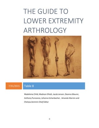
  • 1. 0 THE GUIDE TO LOWER EXTREMITY ARTHROLOGY 7/31/2015 Table 8 Madeleine Child, Madison Elliott, Jacob Jensen, Deanna Maurer, Anthony Purviance, Johanna Schanbacher, Amanda Warren and Chelsea Zemmin Chief Editor
  • 2. 1 Table of Contents The Hip: Regional Overview . . . . . . . . . . . . . . . . . . . . . . . . . . . . . . . . . . . . . . . . . . . . . . . . . . . . . .2 Muscles of the hip . . . . . . . . . . . . . . . . . . . . . . . . . . . . . . . . . . . . . . . . . . . . . . . . . . . . . . . . . .3 Sacroiliac Joint . . . . . . . . . . . . . . . . . . . . . . . . . . . . . . . . . . . . . . . . . . . . . . . . . . . . . . . . . . . . . 9 Pubic Symphysis Joint . . . . . . . . . . . . . . . . . . . . . . . . . . . . . . . . . . . . . . . . . . . . . . . . . . . . . . . 16 Femoroacetabular Joints . . . . . . . . . . . . . . . . . . . . . . . . . . . . . . . . . . . . . . . . . . . . . . . . . . . . .21 The Knee: Regional Overview . . . . . . . . . . . . . . . . . . . . . . . . . . . . . . . . . . . . . . . . . . . . . . . . . . . . 34 Muscles of the knee . . . . . . . . . . . . . . . . . . . . . . . . . . . . . . . . . . . . . . . . . . . . . . . . . . . . . . . . .35 Tibiofemoral Joint . . . . . . . . . . . . . . . . . . . . . . . . . . . . . . . . . . . . . . . . . . . . . . . . . . . . . . . . . . 37 Patellofemoral Joint . . . . . . . . . . . . . . . . . . . . . . . . . . . . . . . . . . . . . . . . . . . . . . . . . . . . . . . . .45 The Foot and Ankle: Regional Overview . . . . . . . . . . . . . . . . . . . . . . . . . . . . . . . . . . . . . . . . . . 54 Muscles of foot and ankle . . . . . . . . . . . . . . . . . . . . . . . . . . . . . . . . . . . . . . . . . . . . . . . . . . . .55 Proximal Tibiofibular Joint . . . . . . . . . . . . . . . . . . . . . . . . . . . . . . . . . . . . . . . . . . . . . . . . . . . .59 Distal Tibiofibular Joint . . . . . . . . . . . . . . . . . . . . . . . . . . . . . . . . . . . . . . . . . . . . . . . . . . . . . . 62 Talocrural Joint . . . . . . . . . . . . . . . . . . . . . . . . . . . . . . . . . . . . . . . . . . . . . . . . . . . . . . . . . . . . 66 Subtalar Joint . . . . . . . . . . . . . . . . . . . . . . . . . . . . . . . . . . . . . . . . . . . . . . . . . . . . . . . . . . . . . . 74 Talonavicular Joint . . . . . . . . . . . . . . . . . . . . . . . . . . . . . . . . . . . . . . . . . . . . . . . . . . . . . . . . . .80 Calcaneocuboid Joint . . . . . . . . . . . . . . . . . . . . . . . . . . . . . . . . . . . . . . . . . . . . . . . . . . . . . . . .86 Cuneonavicular Joint . . . . . . . . . . . . . . . . . . . . . . . . . . . . . . . . . . . . . . . . . . . . . . . . . . . . . . . .90 Cuboideonavicular Joint . . . . . . . . . . . . . . . . . . . . . . . . . . . . . . . . . . . . . . . . . . . . . . . . . . . . . 94 Intercuneiform and Cuneocuboid Complex . . . . . . . . . . . . . . . . . . . . . . . . . . . . . . . . . . . . . .98 Tarsometatarsal Joint . . . . . . . . . . . . . . . . . . . . . . . . . . . . . . . . . . . . . . . . . . . . . . . . . . . . . . . 102 Intermetatarsal Joint . . . . . . . . . . . . . . . . . . . . . . . . . . . . . . . . . . . . . . . . . . . . . . . . . . . . . . . .106 Metatarsophalangeal Joints . . . . . . . . . . . . . . . . . . . . . . . . . . . . . . . . . . . . . . . . . . . . . . . . . . 109 Interphalangeal Joints . . . . . . . . . . . . . . . . . . . . . . . . . . . . . . . . . . . . . . . . . . . . . . . . . . . . . . .114 Appendix: . . . . . . . . . . . . . . . . . . . . . . . . . . . . . . . . . . . . . . . . . . . . . . . . . . . . . . . . . . . . . . . . . . . . . . . A: Gait Index . . . . . . . . . . . . . . . . . . . . . . . . . . . . . . . . . . . . . . . . . . . . . . . . . . . . . . . . . . . . . . .117 B: Citations . . . . . . . . . . . . . . . . . . . . . . . . . . . . . . . . . . . . . . . . . . . . . . . . . . . . . . . . . . . . . . . .119
  • 3. 2 THE HIP: REGIONAL OVERVIEW The hip region is composed of three major joints including the pubic symphysis, the paired sacroiliac joints, and the bilateral femoroacetabular joints. The pubic symphysis and the sacroiliac joints are both located within the pelvic girdle. The pelvic girdle is composed of the sacrum and the two innominate bones which are comprised of three fused bones: the ilium, ischium, and pubis. The sacroiliac joint is a modified synarthrodial joint that is formed by the articulation between the sacrum and the ilium and demarcates the transition between the axial and the appendicular skeleton. The primary function of the sacroiliac joints is to provide stability to the pelvic girdle to ensure effective and efficient transfer of loads between the spine and the lower extremities. Due to this relationship, movement occurring at the lumbar spine has a direct influence on the pelvis moving over the femoral heads resulting in a synchronization of movement referred to as lumbopelvic rhythm coordinating the upper portion of the body with the lower extremities. The pubic symphysis joint is a synarthrodial joint that is comprised of a fibrocartilaginous disc which joins with the articulations of the medial surfaces of the right and left pubic bones. The primary function of this joint is to provide stress relief to the anterior portion of the pelvic girdle. Although very limited motion occurs at both the sacroiliac joints and the pubic symphysis, these joints work together to allow enough flexibility, stress relief, and stability in the pelvic girdle to allow for sufficient attenuation of load and preservation of pelvic structure during daily activities such as walking, standing, and running. The femoroacetabular joint is closely related to the pelvic girdle as it is formed between the articulating surfaces of the acetabulum of the innominate and the head of the femur. As a result, this joint demarcates the link between the pelvic girdle and the lower extremity. This ball and socket diarthrodial joint allows for a wide range of motion while simultaneously providing a large amount of stability in order to support the weight of the head, arms, and trunk during a multitude of static and dynamic weight bearing activities. Stability at this joint is accomplished by specific anatomical characteristics such as the thick fibrous joint capsule, reinforcing capsular ligaments, and an extensive amount of musculature about the hip. Further details about each of these joints specific form and function will be addressed in the following sections. Figure 1. Joints of the hip region
  • 4. 3 Table 1. Muscles of the hip region Location Muscle Proximal Attachment Distal Attachment Action Segmental Innervation Peripheral Innervation Medial Thigh Adductor Brevis Body & inferior pubic rami Pectineal line & proximal part of linea aspera of femur Adducts hip, weak hip flexor L2-3-4 Obturator Nerve Adductor Longus Body of pubis inferior to pubic crest Middle third of linea aspera of femur Adducts and flexes hip L2-3-4 Obturator Nerve Adductor Magnus Inferior pubic ramus, ramus of ischium Gluteal tuberosity, linea aspera, medial supracondylar line Hamstring Part: adductor tubercle of femur Adductor part: adducts and flexes hip Hamstring Part: extends hip Adductor Part: L2-3-4 Hamstring Part: L4-5, S1 Adductor Part: obturator nerve Hamstring Part: tibial division of sciatic nerve Gracilis Body and inferior ramus of pubis Superior part of medial surface of tibia Adducts hip, flexes and medially rotates knee L2-3-4 Obturator Nerve Obturator Externus Margins of obturator foramen, obturator membrane Trochanteric fossa of femur Laterally rotates hip, stabilizes head of femur in acetabulum L3-4 Obturator Nerve Pectineus Superior ramus of pubis Pectineal line of femur Adducts and flexes hip L2-3-4 Femoral Nerve and occasionally Obturator Nerve Anterior Thigh Iliacus Superior 2/3 of iliac fossa, iliac crest, ala of sacrum, anterior sacroiliac ligaments Lesser trochanter of femur and shaft inferior, psoas major tendon Flexes hip and stabilizes hip joint L2-3-4 [L1] Femoral Nerve
  • 5. 4 Anterior Thigh Cont. Psoas Major Sides of vertebral bodies of T12-L5 & intervening intervertebral discs, transverse processes of L1-5 Lesser trochanter of femur Flexes hip and trunk , stabilizes hip joint L1-2- 3-4 Femoral Nerve and Ventral Rami of L1 Sartorius Anterior superior iliac spine Superior part of medial surface of tibia Abducts, laterally rotates, and flexes hip, flexes and assists medial rotation of knee L2-3 [4] Femoral Nerve Rectus Femoris Anterior inferior iliac spine and ilium superior to acetabulum Base of patella and tibial tuberosity via patellar ligament Flexes hip, extends knee L2-3-4 Femoral Nerve Posterior Thigh Biceps Femoris Long head: Ischial tuberosity, sacrotuberous ligament Short head: Linea aspera and lateral supracondylar line of femur Lateral side of head of fibula Long head: Extends hip Short and Long head: flexes knee L5, S1-2-3 L5, S1-2 Long head: Tibial division of the Sciatic Nerve Short head: Common fibular division of the Sciatic Nerve Semimembranosus Ischial Tuberosity Posterior part of medial condyle of tibia Extends hip, flexes & medially rotates knee L4-5, S1-2 Tibial division of Sciatic Nerve Semitendinosus Ischial Tuberosity Superior part of medial surface of tibia Extends hip, flex & medially rotate knee L4-5, S1-2 Tibial division of Sciatic Nerve
  • 6. 5 Gluteal Region Gluteus Minimus Lateral surface of ilium between anterior and inferior gluteal lines Anterior surface of greater trochanter of femur Abducts and medially rotates hip, steadies pelvis on leg when opposite leg is raised L4-5, S1 Superior Gluteal Nerve Gluteus Medius Lateral surface of the ilium between anterior and posterior gluteal lines Lateral surface of greater trochanter of femur Abducts and medially rotates hip, steadies pelvis on leg when opposite leg is raised L4-5, S1 Superior Gluteal Nerve Gluteus Maximus Ilium posterior to posterior gluteal line, aponeurosis of erector spinae, dorsal surface of sacrum and coccyx, sacrotuberous ligament Iliotibial tract that inserts into lateral condyle of tibia, greater trochanter and gluteal tuberosity of femur Extends and laterally rotates hip L5, S1-2 Inferior Gluteal Nerve Obturator Internus Pelvic surface of obturator membrane and surrounding bone Medial surface of greater trochanter of femur Extends and laterally rotates hip, abducts flexed thigh at hip L5, S1-2 Nerve to obturator internus Superior Gemellus Outer surface of ischial spine Medial surface of greater trochanter of femur via obturator internus tendon Laterally rotate and extend hip L5, S1-2 Nerve to obturator internus Inferior Gemellus Ischial tuberosity Medial surface of greater trochanter of femur via obturator internus tendon Laterally rotate and extend hip L4-5, S1 [S2] Nerve to quadratus femoris
  • 7. 6 Gluteal Region Cont. Quadratus Femoris Lateral margin of ischial tuberosity Quadrate tubercle on inter- trochanteric crest of femur Laterally rotates hip L4-5, S1 [S2] Nerve to quadratus femoris Piriformis Anterior surface of sacral segments 2- 4, posterior superior iliac spine, sacrotuberous ligament Superior border of greater trochanter of femur Laterally rotates and abducts hip Extends hip Ventral rami of L5, S1-2 Branches of lumbo- sacral plexus Tensor Fasciae Latae Anterior superior iliac spine and anterior part of iliac crest Iliotibial tract that attaches to lateral condyle of tibia Abducts, medially rotates, and flexes hip and assists in maintaining knee extension L4-5, S1 Superior Gluteal Nerve Pelvic Floor Coccygeus Ischial spine, sacrospinous ligament Inferior sacrum and coccyx Supports pelvic viscera, draws coccyx forward S4-5 Ventral rami S4-5 Levator Ani: Puborectalis Pubococcygeus Iliococcygeus Body of pubis, tendinous arch of obturator fascia, ischial spine Perineal body, coccyx, ano- coccygeal raphe, walls of prostate or vagina, rectum, anal canal Supports pelvic viscera, raises pelvic floor S2-3-4 Pudendal nerve and ventral rami of S4 Back Lattisimus Dorsi Spinous processes of T7-L5, thoracolumbar fascia, iliac crest, and last three ribs Inter- tubercular sulcus of humerus Extends, abducts, and medially rotates humerus C6-7-8 Thoraco- dorsal Nerve
  • 8. 7 Back Cont. Erector spinae Posterior sacrum, iliac crest, sacrospinous ligament, supraspinous ligament, spinous processes of lower lumbar and sacral vertebrae Iliocostalis: angles of lower ribs, cervical transverse processes Longissimus: between tubercles and angles of ribs, transverse processes of thoracic and cervical vertebrae, mastoid process Spinalis: spinous processes of upper thoracic and midcervical vertebrae Extends and laterally bends vertebral column and head Dorsal rami of spinal nerves Dorsal rami of spinal nerves Multifidus Sacrum, ilium, transverse processes of T1- 12, and articular processes of C4-7 Spinous process of vertebrae above spanning 2-4 segments Stabilizes spine, extension and contra- lateral rotation of spine Dorsal rami of spinal nerves Dorsal rami of spinal nerves Abdominal Wall Rectus abdominus Pubic Symphysis, pubic crest Xiphoid process, costal cartilages 5-7 Flexes trunk, compresses the abdominal viscera T5-T12 Lower thoracic ventral rami Internal oblique Thoracolumbar fascia, anterior 2/3 of iliac crest, lateral half of inguinal ligament Inferior borders of ribs 10-12, linea alba, pubis via Compresses and supports abdominal viscera, T7-12, L1 Lower thoracic ventral rami and first
  • 9. 8 Abdominal Wall Cont. conjoint tendon flexes and rotates trunk lumbar nerves: iliohypo- gastric and ilio- inguinal External oblique External surface of ribs 5-12 Linea alba, pubic tubercle, anterior half of iliac crest Compresses and supports abdominal viscera, flexes and rotates trunk T7-12 (T5-6) Lower thoracic ventral rami Transversus abdominus Internal surfaces of costal cartilages 7-12, thoracolumbar fascia, iliac crest, lateral third of the inguinal ligament Linea alba with aponeurosis of internal oblique, pubic crest, and pecten pubis via conjoint tendon Compresses and supports abdominal viscera T7-12, L1 Lower thoracic ventral rami and first lumbar nerves: iliohypo- gastric and ilio- inguinal
  • 10. 9 Sacroiliac Joint (SI Joint) Overview The sacroiliac joints (SI joints) are components of the pelvic girdle that are located anterior to the PSIS of the ilium. These joints demarcate the site of transition between the axial and inferior appendicular skeleton. Their primary function is to provide structural stability to the pelvic girdle in order to effectively transfer loads of varying magnitudes between the lumbar spine and the lower extremities. Formed between the articulating surfaces of the ala of the sacrum and the ilium of the innominate, the classification of this joint is unique. Throughout life the joint changes from a fairly mobile synovial joint in childhood to a fairly rigid modified synarthrodial joint by the time of adulthood. Due to these structural changes, only a small amount of motion occurs at this joint reportedly measuring at about one to four degrees of rotation and one to 2mm of translation. In addition to this structural boney congruity, motion is restricted at this joint by ligamentous and muscular contributions. As reported by Ebraheim et al., the SI joint receives its blood supply from a nutrient artery branching off of the iliolumbar artery. Due to the location of these arteries anterior and superior to the SI joint, they are highly susceptible to damage and have the potential to cause large amounts of bleeding as a result of sacral fractures or surgery that requires an anterior approach to the joint (Ebraheim, 1997). The sacroiliac joint receives sensory innervation most commonly reported as contributions from the dorsal rami of L5-S3 spinal nerve roots and less frequently reported as contributions from the ventral rami of L4-S2 spinal nerve roots however reports in the literature remain largely variable. Tissue Layers (Superficial to Deep) Integumentary o Epidermis o Dermis Fascia o Superficial Fascia Subcutaneous adipose Cutaneous nerves Superior cluneal nerves Medial cluneal nerves Posterior cutaneous intercostal nerves Lateral cutaneous Figure 2. The sacroiliac joint
  • 11. 10 intercostal nerves Anterior cutaneous intercostal nerves Superficial blood vessels Superficial epigastric a/v Superficial circumflex iliac a/v Lymphatic vessels o Fascia lata of the gluteus maximus o Thoracolumbar fascia Posterior layer Anterior layer Muscles Posterior Approach: o Gluteus maximus o Gluteus medius o Lattisimus dorsi o External oblique o Internal oblique o Erector spinae o Transversus abdominus o Multifidus Anterior Approach: o External oblique o Internal oblique o Rectus abdominus o Transversus abdominus o Psoas minor o Psoas major o Iliacus o Quadratus lumborum o Piriformis o Coccygeus o Levator ani (Iliococcygeus) Neurovasculature Posterior Approach: o Inferior gluteal nerve o Superior gluteal nerve o Superior gluteal a/v o Lumbar a/v o Iliohypogastric nerve o Ilioinguinal nerve Anterior Approach: o Inferior epigastric a/v o Iliohypogastric nerve o Ilioinguinal nerve o Genitofemoral nerve o Obturator nerve o Femoral nerve o Lateral femoral cutaneous nerve of the thigh o Common fibular nerve root o Tibial nerve root o External Iliac a/v o Deep circumflex iliac a/v o Internal Iliac a/v o Iliolumbar a/v o Lateral sacral a/v Ligaments Posterior Approach: o Sacrotuberous ligament o Interosseous ligaments o Posterior sacroiliac ligaments Anterior Approach: o Iliolumbar ligament o Anterior sacroiliac ligament o Sacrospinous ligament Joint Capsule Bone o Ala of Sacrum o Ilium of Innominate Covered in a hyaline cartilage
  • 12. 11 Joint Motions and Associated Muscles Table 2. SI joint motions Motion Associated Muscles Nutation Erector Spinae Multifidi Recuts Abdominus Biceps Femoris Counter nutation Rectus Femoris Latissimus dorsi thoracolumbar fascia Joint Configuration and Planes of Motion The sacroiliac joint is a modified synarthrodial articulation between the C-shaped auricular surfaces on the lateral aspects of the sacrum and the matching surfaces of the right and left ilia. The opening of the “C” faces the posterior direction. Anteriorly, the joint is classified as a diarthrodial articulation, while the posterior aspect is a fixed synarthrodial connection between congruent elevations and depressions. The articular surface of the sacrum can be found along the lateral aspect of the sacral foramina of segments S1-S3. This surface is mostly concave, although sexual dimorphism and variation have been reported throughout the literature. The auricular surface of the ilia are found to be mostly convex, although again, with variation. Each articulating surface is covered in a hyaline cartilage layer that thins with aging. The configuration of the sacroiliac joint changes from birth through adulthood. During childhood, the SI joint has characteristics of being a synovial joint. The articulating surfaces on both the sacrum and the ilia are smooth and flat with a pliable capsule surrounding the joint, allowing for slightly more mobility. With aging, the articulating surfaces become covered in ridges and grooves that interlock to create movement resistance between the sacrum and the ilium. These coordinating connections create a high amount of friction. The sacroiliac joint is relatively rigid and immobile. There is a small amount of translation and rotation that takes place in the near-sagittal plane around a near-mediolateral axis. For adults, this can be anywhere between 1-2mm of translation and up to 4 degrees of rotation. Because this mechanism of Figure 3. Articulating surfaces of the sacroiliac joint Figure 4. Sacroiliac joint cut along transverse plane
  • 13. 12 movement is quite irregular, there are specific terms designated to describe the complex combination of these movements at the SI joint, which will be discussed in the next section. Biomechanics and Arthrokinematics The biomechanics of the sacroiliac joint are difficult to examine due to the complex nature and location of the joint. The main physiological function of the sacroiliac joint is to provide stability and load transfer between the axial skeleton and the lower extremities. The SI joints also provide stress relief for the pelvic ring. If the pelvic ring were a solid structure, it would fracture under the normal stressors of everyday activity. The SI joints, along with their anterior counterpart the pubic symphysis, provide enough pliancy and force transmission to prevent breakage from occurring. During ambulation, the lower extremities move in a reciprocal pattern. At the time of right heel strike, the left toes remain in contact with the ground. This causes the muscles and ligaments of the hip to pull on the pelvis and create a torsional force across the right and left innominates. The minimal flexibility found at the SI joints is enough to be able to attenuate these forces and preserve pelvic structure. As mentioned in the previous section, the sacroiliac joint has a unique combination of translation and rotational movements: nutation and counternutation. Nutation is the relative anterior/inferior tilt of the sacral promontory while the sacral apex and coccyx move posteriorly. This motion is similar to sacral flexion of the ilia. Muscular contributions to this movement are from the erector spinae to rotate the sacrum anteriorly while the rectus abdominis and biceps femoris bring the ilium posteriorly. In addition to muscle dynamics, the downward force of gravity and the ground reaction forces through the lower extremities also provide a nutation torque on the joint during double limb stance. Nutation places the sacrotuberous and interosseous ligaments on tension, creating compression forces which further increase the stability of the joint. For these reasons, full nutation is the close- pack position of the SI joint in which the prominent compression and shear forces at the joint give the most articular congruency and most effective load transfer. Counternutation is the opposite motion, with the sacral promontory moving posterior/superior while the apex moves in an anterior direction. To make a similar comparison, it would be like sacral extension. Rectus femoris pulls the innominate in the anterior direction, while the thoracolumbar fascia of the latissimus dorsi pulls the sacrum posteriorly. The motions of nutation and counternutation can take place either by the movement of the sacrum on the ilia or of the ilia moving on the sacrum or a combination of the two. The anteroposterior diameter of the pelvic brim and outlet are impacted based on whether the sacrum is in nutation or counternutation. In nutation, the pelvic brim diameter is decreased while the outlet diameter becomes larger. The opposite is true in counternutation. These changes become especially relevant during pregnancy and childbirth. Figure 5. Movements of the sacroiliac joint
  • 14. 13 The sacroiliac joint has some level of bony stability due to the interlocking configuration of the joint itself. Vleeming introduces this concept as form closure in his 1990 article. In perfect form closure, the articulating surfaces are so well integrated that they provide stability without requiring the assistance of outside forces to maintain the load to the joint. However, this creates a problem of immobility at the joint. Instead, the type of stability found at the SI joint is a combination of form closure and force closure, which is a dynamic stability supplied by the combination of friction and the compression forces of the surrounding ligamentous and muscular structure. Muscles associated with providing actions that impact the stability found at this joint are the erector spinae, lumbar multifidi, rectus abdominus, internal oblique, external oblique, transversus abdominus, biceps femoris, gluteus maximus, lattisimus dorsi, Iliacus, and piriformis. The interosseous ligaments along with the long and short posterior sacroiliac ligaments also play a large role in stabilizing the sacroiliac joint region. Ligaments of the Sacroiliac Joint Figure 6. Form and force closure of the sacroiliac joint Figure 7. Posterior view of the sacroiliac joint ligaments
  • 15. 14 Table 3. Ligaments of the sacroiliac joint Ligament Attachments Function Other associated joint constraints Anterior Sacroiliac Thickening of anterior and inferior portions of SI joint capsule Primary stabilizer of SI joint reinforcing the anterior portion of the joint Reinforces anterior portion of joint Interosseous Sacroiliac Posterior sacral articulating surfaces to iliac tuberosities occupying the space between posterior and superior margins of the joint Primary Stabilizer of SI joint strongly binding the sacrum and the ilium Provides multidirectional structural stability of the joint, transfers weight between axial and inferior appendicular skeleton Short Posterior Sacroiliac Posterolateral side of the sacrum to the ilium near the iliac tuberosity and posterior superior iliac spine mixing with the deeper interosseous sacroiliac ligament Primary Stabilizer of SI joint reinforcing the posterior portion of the joint Assists in force closure of the joint Long Posterior Sacroiliac Lateral crest of the third and fourth sacral segments to posterior superior iliac spine of the ilium mixing with the sacrotuberous ligament Primary Stabilizer of SI joint reinforcing the posterior portion of the joint Restrains counternutation of the sacrum, assist in force closure of the joint Iliolumbar Transverse process of L4- 5 to iliac crest of pelvis Primary Stabilizer of SI joint reinforcing the anterior portion of the joint Resisting extension, lateral flexion, and axial rotation of L5- S1 Sacrospinous Ischial spine to lateral borders of sacrum and coccyx Secondary Stabilizer of SI joint Restrains nutation of the sacrum Sacrotuberous Ischial tuberosity to posterior superior iliac spine, lateral sacrum, and coccyx mixing with the tendon of the biceps femoris muscle Secondary Stabilizer of SI joint Restrains nutation of the sacrum
  • 16. 15 Common Pathologies of the Sacroiliac Joint Sacroiliac Joint Dysfunction (Pain): Sacroiliac Joint Dysfunction is a general term used to describe impaired load transfer and pain perceived in the gluteal, lumbar, abdomen, and lower extremity stemming from pathology in the SI joint. Most pain from the SI region can be attributed to mobility imbalances that arise secondary to trauma, gradual degeneration, or hormonal changes during pregnancy. Examination includes provocation tests to elicit a pain response. These provocation tests include distraction, compression, thigh thrust, Gaenslen’s test, sacral thrust, and motion palpation. Although independently these tests show little validity, when used together, they give a fairly accurate picture of a symptomatic sacroiliac joint. The dysfunction can be in the form of hyper- or hypomobility and treatment follows a course of focusing on the signs and symptoms present. According to Vanelderen et al., conservative treatment to reduce pain and improve mobility in the sacroiliac joint is best addressed using a combination of exercise therapy and manipulation (Vanelderen, 2010). These methods can be used to address the underlying postural and gait disturbances that are often responsible for SI joint pain. Stabilization exercises work to strengthen the force closure of the joint, targeting the transversus abdominis, abdominal oblique muscles, latissimus dorsi, and gluteal muscles to increase myofascial stability. Active range of motion exercises along with manipulation and mobilizations can be used to improve mobility on the symptomatic side.
  • 17. 16 Pubic Symphysis Joint Overview The pubic symphysis joint is a component of the pelvic girdle acting as the anterior link between the pubic bones of the paired innominates. This joint is commonly classified as a synarthrosis joint comprised of a fibrocartilaginous pubic disc that articulates with the medial surfaces of the pubic bones. The primary function of this joint is to provide stress relief to the anterior portion of the pelvic girdle during movement such as walking and during childbirth. In addition to the pubic disc, pubic ligaments strongly bind the joint together allowing only slight motion at the joint measuring at about 2mm of translation and a small amount of rotation. As reported by Becker et al., the pubic symphysis joint is mainly supplied with blood by a branch of the obturator artery and a branch of the inferior epigastric artery. It has also been suggested that the joint receives additional blood supply from branches of the external and internal pudendal arteries and the medial circumflex femoral artery however this supply is more variable and minimal in amount (Becker, 2010). Also reported by Becker et al., the pubic symphysis is suggested to be innervated by the pudendal and genitofemoral nerves and branches of the iliohypogastric and ilioinguinal nerves (Becker, 2010). Tissue Layers (Superficial to Deep) Integumentary o Epidermis o Dermis Fascia o Superficial Fascia Camper’s Fascia Scarpa’s Fascia Cutaneous nerves Anterior cutaneous branch of subcostal nerve Anterior cutaneous branch of iliohypogastric nerve Anterior branch of ilioinguinal nerve Genital branch of genitofemoral nerve Superficial blood vessels Figure 8. Pubic symphysis joint and associated ligaments
  • 18. 17 Superficial external pudendal a/v Superficial epigastric a/v Lymphatic vessels Anterior rectus sheath External oblique aponeurosis Internal oblique aponeurosis Transversus abdominus aponeurosis Transversalis fascia Extraperitoneal fascia Parietal peritoneum Linea alba Muscles o Pyramidalis o Rectus abdominus o Ischiocavernosus o Bulbospongiosus o Gracilis o Adductor longus Neurovasculature o Ilioinguinal nerve o Genitofemoral nerve o Deep external pudendal a/v o Accessory branches of the obturator a/v o Pubic branches of inferior epigastric a/v Ligaments o Anterior pubic ligament o Inguinal ligament o Lacunar ligament o Pectineal ligament o Superior pubic ligament o Inferior pubic ligament o Posterior pubic ligament Joint o Fibrocartilaginous disc Bone o Paired pubic bones of the innominate Covered in a hyaline cartilage Joint Motions and Associated Muscles Table 4. Motions of the pubic symphysis joint Joint Motions Associated Muscles Stability Aponeurosis of the Transverse Abdominus, Rectus Abdominus, Internal Oblique, and Adductor longus Translation N/A Rotation N/A Joint Configuration and Planes of Motion The pubic symphysis is typically classified as a synarthrodial articulation and contains a fibrocartilaginous disc joining the articular surfaces of the right and left pubic bones. There is mixed literature regarding the width of the symphysis, although most agree that the anterior portion is wider than the posterior. The interpubic disc has broader superior and inferior edges with a narrow midsection. Within the superior posterior part of the disc is a narrow slit-like cavity known as the cleft.
  • 19. 18 The articular surfaces of the pubic bones are oriented obliquely in the sagittal plane and of a slightly convex and oval shape. These ridged articular surfaces are covered in a 1-3mm layer of hyaline cartilage. This cartilage tends to decrease with aging. The bony surfaces below the cartilage are found to be irregular in childhood, smoothing and flattening around age 30 and then progressing with degenerative changes such as joint narrowing and irregularities forming again around the sixth decade of life. The pubic symphysis is a relatively immobile joint, allowing approximately 1-2mm of translation in the transverse and sagittal planes and slight rotation in the frontal and sagittal planes. Biomechanics and Arthrokinematics Although the pubic symphysis is quite rigid, slight available movements such as translation and rotation do coordinate with those of the SI joint to attenuate load and provide stability to the pelvic ring during everyday activities. There are no muscles associated with these movements as they are a product of the forces acting on the pubic symphysis during various activities. In closed kinetic chain, the movement at either the pubic symphysis or the sacroiliac joints will create and effect movement at the other. This movement provides enough flexibility in the ring in order to prevent pelvic fracture during daily activities. During double-limb stance, there are tensile forces acting on the inferior part of the pubic symphysis joint with an equal amount of compression being felt through the superior region. In sitting, there are compression forces in the pubis that are then transmitted along the pubic rami and dispersed about the rest of the innominate bones. Lateral pelvic tilting that occurs during the single limb stance of gait creates a predominantly shearing force at the pubic symphysis. A typical pubic symphysis joint is able to withstand these forces with barely discernible amounts of translation and rotation. If dislocation occurs at the joint, the pelvis becomes unstable during ambulation and additional stress are placed on the sacroiliac and hip joints. While there are no muscles acting directly to create movement at the pubic symphysis, there are a number of tendinous attachments from surrounding musculature which provide stability for the anterior innominate. These include the transversus abdominis, rectus abdominis, internal oblique and adductor longus. According to Omar et al, the rectus abdominis and the adductor longus muscle are the most robust players in contributing to the stability of the pubic symphysis, as they are relative antagonists to each other during typical movement patterns (Omar, 2008). Figure 9. Muscles acting on the pubic symphysis joint
  • 20. 19 Ligaments of the Pubic Symphysis Joint Table 5. Ligaments of the pubic symphysis joint Ligament Attachments Function Other associated joint constraints Superior Pubic Pubic tubercle and crest spanning superiorly to pubic tubercle and crest of opposite pubic bone, connections with the interpubic disc, pectineal ligament, linea alba, and periosteum of superior pubic rami Reinforces the superior aspect of the Pubic symphysis joint N/A Inferior Pubic (Subpubic or Arcuate Pubic) Inferior fibers attach inferior pubic rami of one side to inferior pubic rami of other side Upper fibers mixing with interpubic disc and posterior pubic ligament Reinforces the inferior aspect of the Pubic symphysis Joint N/A Anterior Pubic Periosteum of one pubic bone to periosteum of other pubic bone connecting bones anteriorly Deep fibers mixing with interpubic disc, superficial fibers mixing with tendinous insertions of rectus abdominus and oblique abdominal muscles Reinforces the anterior aspect of the Pubic symphysis joint Maintains stability of the Pubic symphysis joint Posterior Pubic Periosteum of one pubic bone to periosteum of other pubic bone connecting bones posteriorly Reinforces the posterior aspect of Pubic symphysis joint N/A Common Pathologies of the Pubis Symphysis Joint Osteitis Pubis: Osteitis pubis is an inflammation of the pubic symphysis and surrounding tendons caused by overuse or shear injury often seen in athletes or pregnant women after trauma or surgery to the pelvic region. According to Dr. Rob Johnson (2003), in his article about Osteitis pubis, patients with this disorder often present with a gradual onset of pain in the groin and possibly the lower abdomen, hip, thigh or perineum. Because there is a lengthy list of differential diagnosis, the testing clinician must be
  • 21. 20 aware of the vague signs and symptoms associated with Osteitis pubis and should keep the pathology on his or her radar. Specific examination often shows point tenderness to palpation and a positive pubic spring test, in which the clinician presses the right and left superior pubic rami to elicit a pain response. Groin pain can also be exacerbated by resisted hip abduction or passive stretch to the hip adductors (Johnson, 2003). Some loss of hip internal rotation range of motion may occur and there is often an antalgic gait pattern. Imaging is often used to confirm a diagnosis of Osteitis pubis. Osteitis pubis is a self-limiting pathology and so treatment often involves modification of activity and typical exercise therapy. Exercise therapy for this condition should include hip range of motion and strengthening program for the hip, lumbar and abdominal regions. Prognosis for this disorder is excellent as 90 - 95% of patients attain full recovery, although that process can take up to 1 year. Symphysis Pubis Dysfunction (specifically in pregnancy): Symphysis Pubis Dysfunction, also referred to as symphyseal pain, is a condition that is commonly characterized by a decrease in pelvic girdle stability resulting in the development of mild to severe pain (Depledge, 2005). This condition has been reported to affect athletes and patients who experienced a traumatic pelvic injury. However, due to the high prevalence of this condition reported in pregnant women, further discussion of this condition will focus specifically on this patient population (Becker, 2010). One suggested reason for the development of this condition during pregnancy is attributed to the hormonal changes that occur during this time. These hormonal changes, specifically the increase of the hormone Relaxin, have been found to be responsible for promoting connective tissue modifications which result in a more pliable pubic symphysis joint and more relaxed and lengthened ligaments surrounding the joints of the pelvic girdle (Depledge, 2005; Leadbetter 2004) . While these hormonal changes do decrease stability in the pelvis, recent literature has reported that Relaxin is not a significant factor in causing the development of symphyseal pain. As a result, the etiology of this common condition remains unclear and warrants further investigation into additional factors including mechanics, metabolism, trauma, and degenerative changes (Aldabe, 2012). Pain associated with this condition is located in the region of the pubic symphysis with common referral patterns to the lower abdomen, thigh, back, groin, perineum, and leg and has been reported as being worst during weight bearing activities (Becker, 2010). Other signs and symptoms include an audible or palpable clicking or grinding in the joint, atypical waddling gait, tenderness over the pubic symphysis, and difficulty with daily activities (Depledge, 2005). In an effort to try and diagnose this condition a couple different sets of diagnostic criteria have been reported. For example, one set of criteria includes answering “yes” to two of the following inquiries including pain when turning in bed, walking, lifting a light load, getting up from a chair, or climbing stairs and positive examination findings suggestive of pain and pelvic dysfunction (Leadbetter, 2004). Conservative management through physical therapy intervention has been studied to measure its effectiveness in reducing pain and increasing function for women with this condition. As reported by Depledge et al., exercises targeting the abdominal stabilizers, pelvic floor, gluteus maximus, lattisimus dorsi muscle, and hip adductor muscles in addition to patient education, and activity modifications were effective in both (Depledge, 2005).
  • 22. 21 Femoroacetabular Joints (Hip Joints) Overview The femoroacetabular joints demarcate the link between the pelvic girdle and the lower extremities. The commonly classified ball and socket synovial joints are formed by the articulation between the head of the femur and the acetabulum of the innominate. Due to its anatomical features, the femoroacetabular joint is a highly mobile joint that allows for three degrees of freedom. Even so, its primary function is to provide a great amount of stability during a variety of static and dynamic weight- bearing activities such as ambulation and standing. For this reason, in addition to its thick and fibrous joint capsule, the joint is also reinforced by ligaments and a large amount of musculature in order to maintain stability and perform a wide array of movements. The femoroacetabular joint receives its main blood supply from the retinacular arteries branching off of the medial and lateral circumflex femoral arteries which originate from the deep artery of the thigh or less commonly from the femoral artery. The joint also receives its blood supply from the artery to the head of the femur which branches off of the obturator artery and passes through the ligament of the head of the femur. The femoroacetabular joint is innervated by the same nerves that are responsible for innervating adjacent muscles acting on or crossing over the joint. For this reason, the anterior portion of the capsule is innervated femoral nerve, posterior capsule by the nerve to the quadratus femoris, inferior capsule by the obturator nerve, and superior capsule by the superior gluteal nerve. Tissue Layers (Superficial to Deep) Integumentary o Epidermis o Dermis Fascia o Superficial fascia Subcutaneous adipose Cutaneous nerves Superior cuneal nerves Middle cuneal nerves Inferior cuneal nerves Lateral femoral cutaneous nerve of the thigh Anterior femoral Figure 10. Blood supply to the femoroacetabular joint
  • 23. 22 cutaneous nerve of the thigh Superficial blood vessels Superficial circumflex iliac a/v Superficial epigastric a/v Superficial external pudendal a/v Great saphenous v Accessory saphenous v Lymphatic vessels Fascia lata of thigh and gluteus maximus Muscles (by Compartments) Anterior Compartment o Sartorius o Rectus femoris o Psoas major o Iliacus Medial Compartment o Gracilis o Adductor longus o Adductor magnus o Adductor brevis o Pectineus o Obturator externus Gluteal Region o Tensor fasciae latae o Gluteus maximus o Gluteus medius o Gluteus minimus o Piriformis o Superior gemellus o Obturator internus o Inferior gemellus o Quadratus femoris Posterior Compartment o Biceps Femoris (Long head) o Semitendinosus o Semimembranosus Neurovasculature Anterior Approach: o Femoral a/v o Femoral nerve o Profunda femoris o Lateral circumflex femoral a/v Ascending Transverse Descending o Medial circumflex femoral a/v o Anterior and posterior branch of obturator nerve Posterior Approach: o Superior gluteal a/v o Superior gluteal nerve o Inferior gluteal a/v o Inferior gluteal nerve o Sciatic nerve o Posterior cutaneous nerve of the thigh o Pudendal nerve o Nerve to the obturator internus o Nerve to the quadratus femoris Bursa o Trochanteric o Iliopectineal o Ischial Ligaments o Iliofemoral o Pubofemoral o Ischiofemoral Joint o Joint capsule o Synovial membrane o Transverse acetabular ligament
  • 24. 23 o Acetabular Labrum o Ligament of the head of the femur Bone o Head of the femur o Acetabulum of the innominate Covered in a layer of hyaline cartilage Joint Motions and Associated Muscles Table 6. Muscles of the femoroacetabular joint Joint Motion Primary Movers Stabilizing and Helping Movers Flexion Iliopsoas, Sartorius, Tensor fasciae latae, Rectus femoris, Adductor longus, Pectineus Adductor brevis, Gracilis, Gluteus minimus (anterior fibers) Extension Gluteus maximus, Biceps femoris (long head), Semitendinosus, Semimembranosus, Adductor magnus (posterior head) Gluteus medius (posterior fibers), Adductor magnus (anterior head) Abduction Gluteus medius, Gluteus minimus, Tensor fasciae latae Piriformis, Sartorius Adduction Pectineus, Adductor longus, Gracilis, Adductor brevis, Adductor magnus Biceps femoris (long head), Gluteus maximus (lower fibers), Quadratus femoris Internal Rotation N/A Gluteus minimus (anterior fibers), Gluteus medius (anterior fibers), Tensor fasciae latae, Adductor longus, Adductor brevis, Pectineus External Rotation Gluteus maximus, Piriformis, Obturator internus, Superior Gemellus, Inferior Gemellus, Quadratus femoris Gluteus medius (posterior fibers), Gluteus minimus (posterior fibers), Obturator externus, Sartorius, Biceps Femoris (long head)
  • 25. 24 Joint Configuration and Planes of Motion The femoroacetabular joint is a classic ball and socket synovial articulation between the convex head of the femur and the ipsilateral concave acetabulum of the pelvis. The head of the femur is a spherical shape located at the proximal end of the femur bone. The surface is almost entirely covered with a layer of hyaline cartilage, save for the area over the fovea, which is a small surface cavity located slightly posterior to the center. The acetabulum is a deep socket formed from contributions of all three pelvic bones and is orientated in a lateral, anterior, and inferior position. The lunate surface of the acetabulum is a horseshoe shaped area of the rim covered in hyaline cartilage which contacts directly with the femoral head. The missing inferior segment of the rim is the acetabular notch which is spanned by the transverse acetabular ligament, connecting the two ends of the lunate surface. The fibrocartilaginous acetabular labrum surrounds the periphery and deepens the socket, increasing the concavity of the acetabulum and creating better congruency for the femoral head. The labrum creates a seal around the joint which maintains a negative intra-articular pressure and helps encase the synovial fluid. The entire joint is enclosed within a strong joint capsule, formed by an external fibrous layer and internal synovial membrane. Thickened segments of the fibrous structure are formed from contributions of the iliofemoral, pubofemoral, and ischiofemoral ligaments. Most of the capsule fibers spiral from the hip to the intertrochanteric line of the femur. The capsule is reinforced anterosuperiorly where the joint sustains the most stresses. Some of the deeper fibers of the ischiofemoral ligament circle around the neck of the femur forming the orbicular zone. Inside the capsule, synovial fluid is present, allowing for reduced friction and fluidity of movement with hip joint mobility in multiple directions. Variations can be noted in the angles created between the head and neck of the femur. The angle of inclination occurs in the frontal plane between the femoral neck and the medial side of the femoral shaft. At birth this angle is between 140-150 degrees but changes with weight bearing to about Figure 11. Femoroacetabular joint configuration Figure 12. Ligaments of the femoroacetabular joint
  • 26. 25 125 degrees in a normal adult, optimizing the alignment of the joint. This angle coincides with the greater trochanter being level with the center of the femoral head. Malalignment of this angle creates altered mechanics down the chain of the lower extremity, influencing the knee, ankle and foot posturing. Coxa vara is a decreased angle of inclination which leads to increased genus valgus at the knee and pronation at the foot. This pathological reduction causes an increased moment arm for greater force production of hip abductor muscles but also simultaneously decreases their functional length. Thus, this negates the benefits of the longer moment arm. Shear force across the superior portion of the femoral neck is also amplified. In children, this condition can lead to slipped capital femoral epiphysis (SCFE). Coxa valga is an increased angle of inclination leading to genu varus at the knee and supination at the foot. This enlarged angle has the opposite effect on the muscles responsible for hip abduction. While functional length of the muscles is increased, the moment arm for torque production is diminished. In more extreme cases of coxa valga, the head of the femur may be positioned in such a way as to favor joint dislocation. The angle of torsion refers to relative rotation of the femoral head and shaft as viewed from above. A normal angle is one in which a transverse axis through the femoral head and neck lies about 15 degrees anterior to the mediolateral axis through the femoral condyles. Infants are born with about 30- 40 degrees of anteversion, which normalizes to adult values with continued bone growth and weight bearing activities. Excessive anteversion is when this angle remains greater than 30 degrees into adulthood and is associated with an increased likelihood of anterior hip dislocation, incongruences of the joint, and excessive wear on acetabular cartilage, all of which can predispose an individual to developing osteoarthritis of the hip. Range of motion into external rotation is decreased while internal rotation ranges are above average. Pathological levels of femoral anteversion in children are often seen in conjunction with a compensatory in- toeing gait pattern which self-corrects over time with the structural changes of Figure 13. Femoral angle of inclination Figure 14. Femoral anteversion and retroversion
  • 27. 26 the lower extremities. This is not true of children with cerebral palsy who tend to maintain the extreme 60-80 degrees of anteversion and the in-toeing gait pattern. Retroversion is when the angle of torsion is significantly less than 15 degrees. The availability of internal rotation at the hip is diminished, while external rotation is excessive of normal values. Individuals with retroverted femurs may walk with a larger foot progression angle, indicative of a toe out compensatory posture during gait. Biomechanics and Arthrokinematics Arthrokinematics: As mentioned above, the femoroacetabular joint is formed by the articulation between the convex head of the femur and the concave surface of the acetabulum. Due to this anatomical structure, arthrokinematics that occur in the hip joint follow the convex on concave principle when applied to open chain femur on pelvis motion from a neutral position. This principle states that the intra-articular motions of roll and glide happen in opposing directions. During abduction, the convex head of the femur rolls superiorly on the surface of the acetabulum while simultaneously gliding inferiorly, and during adduction the head of the femur rolls inferiorly and glides superiorly. During external rotation, the head of the femur rolls posteriorly while the glide occurs anteriorly, and during internal rotation the femur rolls anteriorly and glides posteriorly. In the case of flexion and extension however, the femur does not exhibit the same roll and glide motions, but instead the head of the femur spins around a focal point on the surface of the acetabulum. Osteokinematics: Femoroacetabular osteokinematic motion occurs in all three major planes of motion around an axis of rotation located in the center of the femur. Motion at the hip can be described as either femur on pelvis motion or pelvis on femur motion. Femur on pelvis motion occurs during open chain activity where the femur moves freely on a relatively stationary pelvis. Pelvic on femur motion occurs during closed chain activity where the pelvis moves over relatively stationary femurs. Regardless of which segment is moving, the osteokinematics that occur at the hip joint are as follows referenced from anatomical position. Hip flexion and extension occur in the sagittal plane about a medial-lateral axis of rotation. Average passive range of motion in this plane is 120 degrees of flexion and approximately 20 degrees into extension. Hip abduction and adduction occur in the frontal plane about an anterior- posterior axis with normal ranges being about 40 degrees of abduction and 25 degrees of adduction. Hip internal and external rotation occurs in the transverse plane about a longitudinal axis. Normal internal rotation is about 35 degrees while external rotation reaches approximately 45 degrees. Open-packed/ Closed-packed position: The closed pack position of the hip joint is full extension, slight abduction and internal rotation. In this stable position, the capsular ligaments are taut and pulling the femoral head tightly into the acetabulum, minimizing the amount of accessory motion that can occur at the joint. This is unique from other joints in the fact that the closed pack position of the hip is not also the most congruent position between the articulating surfaces. The position which provides the most congruency is in 90
  • 28. 27 degrees of flexion with abduction and external rotation. It is in this position that a distraction manipulation would be performed, prior to moving into more limited positions of the joint capsule. Ligaments/function: The three principal ligaments associated with femoroacetabular joint capsule are the iliofemoral and pubofemoral anteriorly, and the ischiofemoral posteriorly. The iliofemoral, also known as the Y- ligament, is the strongest ligament in the hip. All three ligaments contribute fibers to reinforce the joint capsule but also provide resistance to prevent the hip from moving into excessive extension. This ligamentous structure is able to withstand the extension moment created by double limb stance, in which the body’s natural line of gravity is posterior to the axis of the hip joint. Because of this passive tension on the ligaments, erect bipedal posture with a slight hyperextension allows for the body weight to be supported without recruiting muscular activation from the muscles of the hip. This has applicable value as lower extremity weakness may be detected if this “hanging” stance is observed during standing posture analysis. Primary Movers: The musculature responsible for producing particular movements at the hip is highly dependent on the hip joint position. It is also important to note here that femoroacetabular joint motion may be regarded in the context of either the femur moving on the pelvis or vice-versa. Because of the complexity associated with discussing specific muscle contribution at various hip joint positions, this section will detail the primary and secondary movers of each available motion when starting from anatomical position and assuming femoral- on-pelvic motion. Primary movers are designated based on multiple factors including cross-sectional area of the muscle, line of pull, and moment arm. Stabilizing muscles are helper muscles supporting the primary movers in accomplishing the movement but those which would be unable to complete the motion independently. The primary movers of the hip into flexion are the iliopsoas, sartorius, rectus femoris, tensor fascia latae, adductor longus and pectineus. Helpers of hip flexion include adductor brevis, gracilis and the anterior fibers of gluteus minimus. The iliopsoas is comprised of two separate muscles. The iliacus originates from the iliac fossa and the lateral edge of the sacrum. The psoas major originates from the transverse processes of T12-L5. These two muscles come together to insert via a common tendon which diverts posteriorly as it crosses the superior pubic ramus to insert at the lesser trochanter of the femur. The large cross sectional area, combined with the increased leverage created by the tendon diversion, make the iliopsoas muscle arguably the most significant of all the hip flexors. Sartorius is a long, thin muscle originating on the ASIS and crosses over the anterior thigh to insert on the medial side of the Figure 15. Musculature of the femoroacetabular joint
  • 29. 28 proximal tibia. Sartorius has a role in hip flexion, as well as external rotation and abduction. Because it crosses the knee as well, it is thought that the role of sartorius in hip flexion may be more impactful when the hip and knee are flexing simultaneously. Rectus femoris attaches to the AIIS and along the superior portion of the acetabulum. Although the rectus femoris is best known as a primary knee extensor, its direct line of pull also allows contribution to hip flexion. Tensor fascia latae (TFL) is a small muscle that originates on the ASIS and anterior part of the iliac crest and extends to join the fibers of the iliotibial tract, which inserts at the lateral condyle of the tibia. The TFL best flexes the hip in conjunction with an abduction motion. Finally, adductor longus and pectineus, while involved with hip flexion are predominantly known for their role in adduction of the hip and so will be discussed further in a later section. Extension at the hip is accomplished by the primary contributions of gluteus maximus, biceps femoris (long head), semitendinosus, semimembranosus and adductor magnus (posterior head). This motion is helped by the actions of the posterior fibers of gluteus medius along with anterior head of adductor magnus. The gluteus maximus has a vast origination from the posterior sacrum, coccyx, ilium and the sacroiliac and sacrotuberous ligaments. This powerful muscle inserts the superior fibers into the iliotibial tract and the inferior fibers to the gluteal tuberosity of the femur. The gluteus maximus has the largest cross sectional area of all the lower extremity muscles and a considerable moment arm in anatomical position, making it the principal hip extensor muscle. The long head of the biceps femoris, semitendinosus and semimembranosus are lumped together under the name “hamstrings”. This cluster of muscles collectively originate on the ischial tuberosity and cross the knee joint to insert on either the lateral side of the fibular head (biceps femoris) or the medial surface of the tibia (semitendinosus and semimembranosus). The moment arm of the hamstrings group changes as the hip changes position and it never reaches the extent of the moment arm of the gluteus maximus. The hamstrings serve as primary knee flexors and their role in hip extension is greatly affected by the position of the knee. As before, adductor magnus will be discussed in the next paragraph. The primary movers for hip adduction are adductor longus, adductor brevis, adductor magnus, pectineus and gracilis. Secondary muscles that assist with this motion are biceps femoris (long head), gluteus maximus (inferior fibers) and quadratus femoris. The adductor muscles are located on the medial thigh. The magnus, longus and brevis originate from the body and inferior pubic ramus and attach at various points along the linea aspera on the posterior femur. Adductor magnus is made up of the anterior head, which runs with the other adductor muscles, and the posterior head, which attach to the adductor tubercle on the medial side of the distal femur and acts more as a hip extensor along with the hamstring group. The pectineus is a small muscle from the superior ramus of the pubis to the pectineal line of the femur. Gracilis a two-joint muscle that originates with the rest of the adductor group and courses down to cross the knee and insert medially on the proximal shaft of the tibia.
  • 30. 29 Pectineus and gracilis are in the most superficial layer of adductors. The bilateral adductors work together to balance each other out during frontal plane motion in weight bearing. When adductors on the right side are working to bring the femur into adduction, the contralateral adductors are contracting to bring the pelvis into adduction on the femur and stabilize the pelvis over the single limb. The muscles primarily responsible for abduction at the hip are gluteus medius, gluteus minimus and tensor fasciae latae. These are assisted by actions of the piriformis and sartorius muscles. Gluteus medius is the largest abductor, originating on the lateral surface of the ilium between the anterior and posterior gluteal lines with its distal attachment on the greater trochanter of the femur, giving this muscle the longest of the abductor moment arms. The gluteus medius is divided into three components, each contributing to abduction as well as additional movements of the hip. From a neutral anatomic position, the anterior and middle fibers are active during internal rotation while posterior fibers assist with external rotation and extension. Gluteus minimus is a smaller muscle located deep to the gluteus medius and originates between the anterior and posterior gluteal lines on the ilium to insert at the greater trochanter and the superior portion of the joint capsule. It’s hypothesized that this capsular attachment allows gluteus minimus to retract the capsule to avoid impingement and stabilize the femoral head in the acetabulum. TFL, discussed earlier, also contributes some abductor torque. The primary movers of external rotation at the hip are gluteus maximus and the group of short lateral rotators, which consists of piriformis, obturator internus, superior gemellus, inferior gemellus and quadratus femoris. Associated helper muscles are the posterior fibers of gluteus medius and minimus, obturator externus, sartorius, and the long head of biceps femoris. The short lateral rotators are positioned perpendicularly to the vertical axis of the femoral shaft, giving them an effective line of pull to execute their primary movement. These muscles also provide a compressive force that gives a great amount of stability to the posterior side of the joint during weight bearing and non-weight bearing activity at the hip. Obturator externus, although considered one of the six “short external rotators” is classified as a secondary muscle due to its slightly posterior line of pull from anatomical position. While there are no primary movers responsible for internal hip rotation, many muscles previously discussed have anterior segments that work in the horizontal plane to create medial rotation of the hip. These include gluteus minimus, gluteus medius, TFL, adductor longus, adductor brevis and pectineus. The torque produced by these muscles into internal rotation is influenced dramatically with reference to the amount of flexion at the hip. As hip flexion increases, so does the push of these muscles into their actions as internal rotators of the hip. Gait: In analyzing the hip joint motion during ambulation, it is beneficial to reference a single limb as Figure 16. Adductor coupling
  • 31. 30 it moves through the gait cycle. Magnitude of movements at the hip change quite dramatically based on walking speed, so for the sake of this brief description, joint angles are approximated in relation to average gait speed of 1.4m/s. Starting at the moment of initial heel contact, the hip is in approximately 20 degrees of flexion with the extensor muscles engaged in anticipation of accepting the body weight. It is during this transition from initial contact through loading response that the highest level of activity is seen from the hip extensors to counter the maximal flexion torque created by the combination of the large ground reaction force with a long moment arm. From mid-stance through terminal stance, the hip moves through a neutral position and into roughly 20-30 degrees of extension. The center of mass moves up and over the base of support during this fairly passive weight transfer, involving very little muscle activity in the sagittal plane. The abductors are active in the frontal plane to address the peak adduction force during this phase of single limb stance. At the conclusion of terminal stance is when hip extension demand is at its height, stabilizing the hip joint in preparation of swing phase. The maximal contribution of the hip flexors takes place during pre- and initial swing. Extension torque diminishes as the swinging limb creates a flexion demand, bringing the hip into its most flexed position of 30 degrees during mid-swing. The hamstrings begin to fire during mid-swing and reach their highest activation during terminal swing, slowing the leading leg as it approaches the ground. Contraction of the hip stabilizers occurs as the joint prepares for the succeeding cycle. Ligaments of the Femoroacetabular Joint Table 7. Ligaments of the femoroacetabular joint Ligament Attachments Function Other associated joint constraints Iliofemoral (Y-ligament) Near the anterior inferior iliac spine and adjacent margin of the acetabulum to the intertrochanteric line of the femur Stabilizing and strengthening the anterior aspect of the joint capsule Resist excessive motion into hip extension and external rotation Pubofemoral Anterior and inferior rim of the acetabulum and adjacent portions of superior pubic ramus and obturator membrane to mix with the Iliofemoral ligament on the intertrochanteric line of the femur Stabilizing and strengthening the anterior aspect of the joint capsule Resist excessive motion into hip abduction, extension, and lesser amount into external rotation Ischiofemoral Posterior, inferior aspect of the acetabulum to the greater trochanter and femoral neck Stabilizing and strengthening the posterior aspect of the joint capsule Resist excessive motion into internal rotation, extension, and adduction Transverse Acetabular Continuation of the acetabular labrum Join the ends of the acetabular labrum N/A
  • 32. 31 passing over the acetabular notch Ligamentum Teres (Ligament of the head of femur) Both sides of the outer edge of the acetabular notch to fovea of the femur and slight mixing with transverse acetabular ligament Passageway for the obturator neurovasculature Taut in semi-flexion and adduction Common Pathologies of the Femoroacetabular Joint Hip Osteoarthritis (OA): Hip osteoarthritis is a chronic disease in which the hip joint undergoes progressive degeneration of the articular cartilage in addition to the manifestation of osteophytes. The etiology of this disease can be classified as either primary or secondary. Primary OA has no known cause however common risk factors include increase in age, physical stresses, and genetics. Secondary osteoarthritis occurs when there has been an identifiable disturbance to the joint such as trauma, overuse, or congenital abnormalities altering typical joint biomechanics such as slipped capital femoral epiphysis, leg length differences, avascular necrosis, coxa vara, femoroacetabular impingement, and repetitive dislocation. Common signs and symptoms of osteoarthritis include anterior groin pain, morning stiffness, weakened or atrophied hip musculature, atypical gait pattern, and inflammation. In order to diagnosis hip OA, the American College of Rheumatology has recommended a set of guidelines known as Altman’s Criteria for Hip OA. These include hip pain, less than 115 degrees of hip flexion, and less than 15 degrees of hip internal rotation. In the instance that hip internal rotation is greater than 15 degrees an alternative set of criteria exists which includes painful hip internal rotation, greater than 50 years old, and morning hip stiffness that lasts less than 60 minutes. In order for a patient to be diagnosed while using these sets of criteria, all three factors must be present. With the progression of the disease, many activities of daily living can become challenging including difficulties with squatting, bathing, stair climbing and rising from sitting to standing. Conservative management through physical therapy intervention can be used to address these changes and usually includes patient education, activity modification, manual therapy, therapeutic exercise, and assistive device training. However, if conservative management is not effective hip OA is an indication for a total hip arthroplasty and post-operative rehabilitation. Hip Fracture: A hip fracture is most commonly a break that occurs at the neck, intertrochanteric line, or subtrochanteric area of the femur. This fracture can occur in younger populations with an aggressive force of impact through the hip joint while the lower limb is in an extended position. However, this diagnosis is most common in individuals over the age of 60, due to the weakening bones often corresponding with osteoporosis and the increased incidence of falls. Hip fracture is associated with a high rate of mortality in elderly due to secondary complications, such as soft tissue damage and hemorrhage that arise status post injury. When the fracture is intracapsular, the potential damage to circumflex arterial blood supply to the proximal end of the femur increases the risk of avascular necrosis. With the severity of potential associated complications, along with the aging baby boomer
  • 33. 32 population, hip fractures are considered to be a geriatric epidemic, costing over $6 billion a year with expected rise in the next few decades. The mechanism of injury is most often a compression trauma with direct impact to the lateral aspect of the hip. As stated above, this is commonly the result of a fall or high speed impact. Symptoms include pain with weight bearing and lateral rotation of the affected limb. Treatment options include open reduction internal fixation (ORIF), external fixation, total hip arthroplasty, or hemiarthroplasty. Immediately following surgery, it is important for the clinician to monitor the surgical site and educate patients on the precautions associated with their procedure. During this time, patients are instructed to utilize the appropriate assistive devices to best follow the surgeon’s protocols regarding the need for immobilization and/or weight bearing status. Once able, a treatment program often includes improving range of motion and strength, in conjunction with gait, balance and functional training. Prognosis is poor for the elderly, with a significant decrease in quality of life and functional decline following hip fracture. Hip fracture and its associated complications are indirectly responsible for the greatest number of deaths in the geriatric population. Femoroacetabular Impingement: Femoroacetabular impingement (FAI) is a diagnosis common in younger patient presenting with hip pain stemming from slight variation of bony morphology. Pincer type FAI is an abnormality of the acetabulum in which there is an over coverage of the femoral head. In this case, extreme hip flexion may cause the anterior sides of the femoral and neck to push against the anterior acetabular rim and labrum, tearing the cartilage. This form of FAI tends to be more prevalent in women. In Cam FAI, there is an abnormal growth of the head or neck of the femur, which then jams into the acetabulum during extreme flexion leading to shear forces on labrum and diffuse articular damage. Examination reveals sharp groin pain with flexion and internal rotation of the hip. Patients are usually limited in range for these movements and a FADIR test will be positive for impingement at 90 degrees of flexion with internal rotation. There is posterior and/or lateral hip pain with external rotation or prolonged sitting and stair climbing. Pain or asymmetry is present with the FADER. Anterior-posterior and lateral imaging of the pelvis can be used for further diagnosis by showing a femoral head deformity and the acetabular shape. MRI will highlight labral tears and damage to the cartilage. Conservative treatment of femoroacetabular impingement may include NSAIDs and limitation of impingement inducing activities, like those involving extreme hip flexion and excessive compression of Figure 17. Femoroacetabular impingement classifications
  • 34. 33 the anterior hip joint. Distractions and inferior/lateral glides can decrease pain and are a practical addition for a self-managed home program. If conservative treatment fails, surgical intervention may be necessary. Prognosis is good for arthroscopic osteoplasty and most patients are able to return to sports and activity with good to excellent results.
  • 35. 34 The Knee: Regional Overview The knee joint is a modified hinge joint and contains two separate joints within the joint capsule. The first joint is comprised of the articulation between the femoral condyles and the tibial plateau, the tibiofemoral joint. The second joint, the patellofemoral joint, is made between the articulation between the posterior articular surface of the patella and the intercondylar groove of the femur. It acts to increase the moment arm for the quadriceps muscle and increasing the force it is able to exert. The primary motions of the knee are flexion and extension that are integral to the motion necessary for gait. These motions are controlled by the hamstring and quadriceps muscle groups respectively. During the swing phase of gait the knee flexes in order to allow for toe clearance from the floor. The knee also remains in flexion during the stance phase for shock absorption to minimize wear on soft tissue structures. Although knee flexion is initially passive, due to rapid ankle plantar flexion and hip flexion in terminal stance, the hamstrings act eccentrically to slow the rate of passive knee extension during terminal swing. The knee is necessary for stability as well as mobility during gait as it resides between the hip and foot and ankle (discussed in other sections), which are very mobile segments of the lower extremity. This stability is reliant upon the soft tissue structures of the knee which include ligaments, tendons, joint capsule, and the meniscus. The knee joint is one of the most commonly injured joints in the body due to its reliance on soft tissue structures for stability. The anterior cruciate ligament (ACL) and posterior cruciate ligament (PCL) resist anterior and posterior translation, respectively, of the tibia on the femur in closed chain. The medial collateral and lateral collateral ligaments resist valgus and varus stress to the knee joint respectively. All of these ligaments are subject to injury with trauma. Furthermore, unique to the knee joint is the meniscus that increases the congruent surface area between the femoral condyles and the tibial plateau allowing for reduced friction and stability of the joint. This structure is subject to wear and tear with over use as well as damage due to trauma. Blood supply to the knee comes from the popliteal artery named for the popliteal fossa that it passes through on the posterior aspect of the knee. The popliteal artery emerges from the adductor hiatus on the medial aspect of the thigh before which is the femoral artery that supplies blood to the thigh musculature. From the popliteal artery emerges four branches of genicular arteries; superior medial, superior lateral, inferior medial and inferior lateral. The descending genicular artery branching from the femoral artery Figure 18. Knee blood supply
  • 36. 35 anastomoses along with the genicular arteries to allow adequate blood supply to the knee despite occlusion to flow through the popliteal artery. The knee is innervated by nerves arising from the lumbar and sacral plexi. These nerves include the femoral, obturator, sciatic and tibial nerves which provide afferent sensory fibers from the joint capsule and supporting structures. Furthermore, these nerves also provide motor and sensory innervation to the musculature that control motion of the knee. In general, each nerve supplies a different compartment of the thigh where the obturator nerve supplies the medial compartment, the femoral nerve supplies the anterior compartment and the sciatic and tibial nerves innervate the posterior compartment. Cutaneous innervation of the skin overlying the joint is performed by the anterior cutaneous branches of the femoral nerve, from the lumbar plexus L2-L3, and the posterior femoral cutaneous nerve, from the sacral plexus S1-S3. Table 8. Muscles of the knee joint Muscle Proximal Attachment Distal Attachment Action Innervation Quadriceps femoris: Rectus Femoris Anterior inferior iliac spine and ilium superior to acetabulum Via common tendinous (quadriceps tendon) and independent attachments to base of patella; indirectly via patellar ligament to tibial tuberosity Extend leg at knee joint Femoral nerve (L2, L3, L4) Vastus lateralis Greater trochanter and lateral lip of linea aspera of femur Vastus medialis Intertrochanteric line and medial lip of linea aspera of femur Vastus intermedius Anterior and lateral surfaces of shaft of femur Hamstrings: Semitendinosus Ishchial tuberosity Medial surface of superior part of tibia Extend thigh; flex leg and rotate it medially when knee is flexed; when thigh and leg are flexed, these muscles Tibial division of sciatic nerve part of tibia (L5, S1, S2) Semimembranosus Posterior part of medial condyle of tibia; reflected attachment forms oblique popliteal ligament (to
  • 37. 36 lateral femoral condyle) can extend trunk Biceps femoris Long head: ischial tuberosity Short head: linea aspera and lateral supracondylar line of femur Lateral side of head of fibula; tendon is split at this site by fibular collateral ligament of knee Flexes leg and rotates it laterally when knee is flexed; extends thigh Long head: tibial division of sciatic nerve (L5, S1, S2) Short head: common fibular division of sciatic nerve (L5, S1, S2) Sartorius Anterior superior iliac spine Medial aspect of proximal tibia Flexion and medial rotation of knee Femoral nerve (L2, L3, L4) Gracilis Body of the pubis and inferior pubic ramus Medial surface of tibia distal to condyle, proximal to insertion of semitendinosus, lateral to insertion of sartorius Flexion and medial rotation of knee Obturator nerve (L2, L3, L4) Gastrocnemius Posterior aspect of femoral condyles and joint capsule Posterior calcaneal surface Flexion of knee Tibial nerve (S1, S2) Plantaris Lateral supracondylar line Posterior calcaneal surface Flexion of knee Tibial nerve (L4, L5, S1, S2) Popliteus Lateral femoral condyle and oblique popliteal ligament Soleal line of tibia NWB: medial rotation of tibia and knee flexion WB: lateral rotation of femur and knee flexion Tibial nerve (L4, L5, S1) Tensor fasciae latae Anterior superior illiac spine and external lip iliac crest Iliotibial tract Assists in maintaining knee extension Superior gluteal nerve (L4, L5, S1)
  • 38. 37 Tibiofemoral Joint Overview The tibiofemoral joint is formed by the articulation of the proximal tibia and distal femur. The joint is contained within a thick fibrous capsule that provides lubrication and structure. Much of the stability of the joint comes from the soft tissue structures that will be discussed in later sections. The tibiofemoral joint is a synovial classification modified hinge joint with 2 degrees of freedom with motion available in the sagittal and transverse planes. The primary motion of this joint is for flexion and extension during gait and other functional activities. However, due to the orientation of this joint, rotation also occurs. Normal range of motion in the healthy adult knee can range between 130-150 degrees of flexion and between 5-10 degrees of extension past 0 degrees. Rotation of this joint varies by the amount of flexion/extension. At 90 degrees of tibiofemoral flexion, 40-45 degrees of rotation is available. Please refer to The Knee: Regional Overview in the previous section for neurovascular supply of the tibiofemoral joint as it is shared with the patellofemoral joint, within the same joint capsule. Tissue Layers (Superficial to Deep) Integumentary o Epidermis o Dermis o Hypodermis Subcutaneous o Superficial Fascia o Adipose tissue Deep Fascia of the leg/ Fascia lata o Iliotibial tract o Intermuscular septa x3 o Patellar Retinaculum (Medial and Lateral) Muscles and Tendons o See table below Bursa / Fat Pads o Infrapatellar Fat Pad o Prepatellar o Infrapatellar o Deep Infrapatellar o Suprapatellar o Subpopliteal o Pes Anserine Neuro Vasculature o Nerves Tibial N. Sural N. Common Fibular N. Medial input from obturator & saphenous NN. o Arteries: Genicular anastomoses Femoral A. Popliteal A. Anterior/ posterior recurrent A. Extracapsular and capsular ligaments o MCL (superficial and deep) o LCL o Joint capsule o ACL o PCL o Menisci Synovial Joint
  • 39. 38 o Joint Capsule o Synovial membrane o Joint space o Articular Cartilage o Osseous joint surface Joint Motions and Associated Muscles Table 9. Motions and muscles of the knee joint Motion Primary Mover Stabilizing and Helping Synergists Extension Quadriceps Femoris Popliteus Flexion Hamstring (Semitendinosus, Semimembranosus, Biceps Femoris) Gastrocnemius, Popliteus Internal Rotation Flexed knee: Semimembranosis and Semitendinosis. Knee extended: Popliteus Sartortius, Gracilis External Rotation Biceps femoris N/A Joint Configuration and Planes of Motion The tibiofemoral joint is a bi-condyloid joint that allows for flexion and extension in the sagittal plane and internal and external rotation in the transverse plane. The joint itself has medial and lateral compartments each of which contain a femoral condyle, meniscus and tibial plateau. As the name implies, the tibiofemoral joint is made from the articulation of the femur and tibia. The distal end of the femur is comprised of medial and lateral femoral condyles containing the articular cartilage that contacts the articular cartilage of the proximal tibia, known as the tibial plateau. The femur itself is not anatomically vertical, rather it lies on an oblique angle from the hip joint to the knee. This angle necessitates the medial femoral condyle to protrude more inferiorly than the lateral condyle for maximal congruency of the joint. Conversely, the lateral condyle protrudes more anterior. Between the two condyles is the intercondylar notch where the cruciate ligaments cross. The anterior-superior aspect of the condyles is the patellar groove containing the articular cartilage that contacts the patella (to be discussed later). Superior to the femoral condyles are medial and lateral epicondyles that serve as attachment points for the collateral ligaments. Figure 19. Tibial plateau and femoral condyles
  • 40. 39 The articular surfaces of the proximal tibia lie on a structure known as the tibial plateau. These surfaces are quite shallow when compared to their femoral counterparts, necessitating the meniscus to increase congruence between the articular surfaces. The medial and lateral tibial plateaus are separated by intercondylar tubercles. Just inferior to the tibial plateau is the tibial tuberosity that serves as the attachment point for the patellar tendon and the quadriceps femoris muscle group. As previously mentioned, the tibiofemoral joint relies heavily on soft tissue structures for stability. There are four primary ligaments that act to support the structure of the knee joint. Two paired collateral ligaments lie on the medial and lateral aspects of the knee coursing from distal femur to proximal tibia. Within the joint capsule are two cruciate ligaments that course from the tibial plateau to the medial aspects of the femoral condyles. The paired collateral ligaments, medial collateral (MCL) and lateral collateral (LCL) serve to protect the knee from excessive coronal plane motion. The MCL is a relatively wide and thin structure on the medial aspect of the knee spanning from the medial femoral epicondyle to the medial surface of the proximal tibia and resists valgus forces. Due to the thin, paper-like structure of this ligament, it is commonly injured and will be discussed further in subsequent sections. The LCL is a narrower, cord-like structure on the lateral aspect of the knee coursing from the lateral epicondyle of the femur to the head of the fibula resisting varus forces. More internal to the joint are two cruciate ligaments, anterior cruciate (ACL) and posterior cruciate (PCL), which cross each other and are named based on their attachment to the tibia. These ligaments primarily resist sagittal plane motion, however, due to the obliquity of their course through the knee, they resist almost all motions of the knee. The ACL courses from the medial part of the anterior intercondylar area of the tibia to the posterior part of the medial surface of the lateral condyle of the femur. This ligament has two bundles, anterior-medial and posterior-lateral, which are named for their relative attachment points on the tibia. The anterior-medial bundle The ACL acts to resist tibiofemoral extension as well as anterior displacement of the tibia on the femur or posterior glide of the femur on the tibia. The PCL runs from the posterior intercondylar area of the tibia to the lateral surface of the medial condyle of the femur. This ligament acts to resist posterior translation of the tibia on the femur or anterior translation of the femur on the tibia. All of the ligaments of the knee are tight in extension. Figure 20. Cruciate ligaments
  • 41. 40 The meniscus is a supporting structure that is unique to the tibiofemoral joint. It is a fibrocartilaginous structure on the tibial plateau that allows for shock absorption, lubrication, and increased congruency of the joint. The meniscus itself is composed of medial and lateral portions that are shaped differently in order to accommodate the dissimilar shapes of the medial and lateral femoral condyles. Comparatively, the medial meniscus is more oval shaped while the lateral meniscus is more circular shaped. Each meniscus is anchored to the tibial intercondylar region by way of their anterior and posterior horns. They are further anchored to the joint capsule via the coronary ligaments. Finally, the two menisci are connected together with a transverse ligament on the anterior side. The meniscus has three vascular zones, from superficial to deep they are red-red, red-white and white-white. These zones correlate with the amount of blood supply that is received by that region. The most superficial zone, red-red, has the most blood supply, where the deep zone, white-white, has no vascular supply. This has implications on the healing of meniscal injuries, to be discussed later. The tibiofemoral joint would not be complete without the joint capsule. The capsule is fairly lax to allow for the necessary amount of range of motion. The capsule has two layers that differ functionally. The outer layer is fibrous and gives support to the joint and the inner layer is a synovial membrane, as this joint is a synovial joint, this membrane acts to produce the lubricating synovial encased within the joint itself. Biomechanics and Arthrokinematics The tibiofemoral joint allows for two degrees of freedom. In the sagittal plane, flexion and extension occur about the medio-lateral axis that runs through the medial and lateral femoral condyles. The greatest range of motion for this joint occurs on this plane and is therefore the most important for function, namely gait. In the transverse plane, internal and external rotation occur about the longitudinal axis. There is limited range of motion available on this plane. The motion of this joint can be discussed in either closed chain, where the femur moves on a stationary tibia, or in open chain, where the tibial moves on a stationary femur. Closed chain motion is considered more functional when discussing the lower extremity as most functional activities are performed in weight bearing. Tibiofemoral flexion occurs when the angle between the tibia and femur is decreased when measured from the posterior direction. In order for this motion to occur, in closed chain, the convex articular surface of the distal femur rolls posterior upon the concave articular surface of the proximal tibia. Therefore, in order to maintain congruency of the joint and allow for the greatest range of motion, Figure 21. Tibiofemoral soft tissue structures
  • 42. 41 the femur must also glide anterior on the tibia. Conversely, in open chain, the concave surface of the tibia rolls posterior on the convex condyles of the femur and also must glide posterior to maintain congruency. The hamstring muscle group, containing semitendinosus, semimembranosus and biceps femoris, are the primary movers for tibiofemoral flexion. This muscle group resides on the posterior thigh and is innervated by the sciatic nerve. The semitendinosus and semimembranosus occupy the medial hamstring and the biceps femoris occupies the lateral hamstring. As these muscles share their proximal attachment on the ischial tuberosity, therefore crossing the hip joint as well, this indicates that they also function in hip extension (see femoroacetabular joint). The distal attachment of the semitendinosus and semimembranosus is on the medial aspect of the proximal tibia where the distal attachment for the biceps femoris is on the fibular head oriented laterally to the proximal tibia (details in table 1). Extension of the tibiofemoral joint occurs when the angle between the femur and tibia is increased. In closed chain, the convex femur rolls anteriorly and glides posteriorly upon the concave tibia (seen in the right picture in Figure 22). The opposite is true in open chain where the concave tibia rolls anterior and glides anteriorly on the convex femur. In both of these cases, the meniscus is pulled anteriorly by the quadriceps muscle. The primary mover for extension of the tibiofemoral joint is the quadriceps femoris group containing the vastus muscles (vastus lateralis, vastus intermedius and vastus medialis) and the rectus femoris muscle. These muscles have variable proximal attachments, however, they share their distal attachment at the quadriceps tendon at the base of the patella and ultimately the tibial tuberosity via the patellar tendon. The rectus femoris muscle originates at the anterior inferior iliac spine (AIIS) and courses on the middle anterior superficial thigh. The vastus lateralis originates from the greater trochanter and linea aspera of the femur and course along the lateral thigh. It is the vastus lateralis that has the largest cross-sectional area of the knee extensors, making it the primary mover in this direction. The vastus intermedius lies deep to the rectus femoris and originates from the anterior and lateral shaft of the femur. Finally, the vastus medialis originates from the intertrochanteric line and linea aspera and courses along the medial thigh. Due to their positions on the thigh, these muscles have variable lines of pull acting at the tibiofemoral joint, however, when acting together, the summed force creates the Figure 22. Tibiofemoral arthrokinematics Figure 23. Quadriceps pull on patella
  • 43. 42 optimal line of pull for knee extension. The vastus lateralis and medialis also counteract each other to stabilize the patella (discussed in detail later). Axial rotation of the tibiofemoral joint involves primarily spin at the joint surface. As the joint is most congruent in extension, due to the stability provided by the soft tissue structures mentioned earlier, very little, if any rotation occurs when the knee is fully extended. More rotation is available as the tibiofemoral joint is flexed towards 90 degrees. It is at 90 degrees that greatest amount of rotation is available. Primary movers for axial rotation are the hamstrings. Although their proximal attachments are on the ischial tuberosity, giving them function at the hip as well, internal rotation is primarily provided by the semitendinosus and semimembranosus when the knee is flexed. When the knee is extended, however, the popliteus acts to internally rotate the knee to unlock the knee joint and allow flexion to occur. As its distal attachment is on the lateral side of the tibia, the biceps femoris muscle acts to externally rotate the tibiofemoral joint. The short head of the biceps femoris is in the most optimal position for this due to its proximal attachment on the posterior femur itself. The proximal attachment of the long head arises from the ischial tuberosity allowing it to function as a hip extensor as well. Much of the orientation and function of the knee joint is due to the shape of the femoral condyles. Although flexion and extension occur about a medial-lateral axis, the axis is not fixed because the condyles of the femur have an eccentric curvature that causes the axis of rotation to migrate, known as the evolute. Functionally the evolute causes the moment arm of the quadriceps and hamstrings to change with varying degrees of flexion/extension. Another consequence of the shape of the condyles is the screw-home mechanism. This is the conjunct motion of external rotation of the tibia on the femur during tibiofemoral extension (roughly the last 30 degrees). This motion allows for maximal bony congruency of the tibiofemoral joint when the knee is extended. Medial and lateral rotation, also known as axial rotation, are also available occurring on the transverse plane about the longitudinal axis. The amount of rotation at this joint is variable depending on the degree of knee flexion. With the knee flexed to 90 degrees, roughly 40-45 degrees of rotation is available. There is typically twice as much external rotation as internal rotation. Mobilization of the tibiofemoral joint is an effective intervention to increase the range of motion when range is limited. Considering the arthrokinematics is an effective way to remember which glides to perform and improve each motion. To improve tibiofemoral flexion, perform an anterior to posterior Figure 24. Evolute
  • 44. 43 glide mobilization of the tibia on the femur. Conversely, to improve tibiofemoral extension, performing a posterior to anterior glide of the tibia on the femur is effective. Ligaments of the Tibiofemoral Joint Table 10. Ligaments of the tibiofemoral joint Ligament Proximal Attachment Distal Attachment Function Other associated joint constraints Anterior Cruciate Ligament medial part of the anterior intercodylar area posterior part of medial surface of lateral condyle of femur prevent posterior displacement of femur on tibia and hyperextension of knee Also resists rotation RA Posterior Cruciate Ligament posterior intercondylar area of tibia lateral surface of medial condyle of femur prevents anterior displacement of the femur on the tibia and hyperflexion of the knee Also resistes rotation Medial Collateral Ligament medial femoral epicondyle Medial condyle and shaft of tibia Stabilizes medial aspect of joint Prevents genu valgum, abduction of knee Lateral Collateral Ligament Lateral femoral epicondyle Head of fibula Stabilizes lateral aspect of joint Resists genu varum, adduction of knee Common Pathologies of Tibiofemoral Joint ACL Tear/Rupture: The ACL is the most frequently ruptured ligament of the knee. Injury to this ligament typically occurs with trauma. Due to the oblique angle by which the ACL courses through the knee, it is at high risk for injury because it resists many different directions of movement. Injury to the ACL leads to impaired joint mechanics and stability. A common mechanism of injury is rupture or tear during rapid deceleration, cutting, or landing from a jump. Others can include severe rotation of the knee with the foot planted and severe tibiofemoral hyperextension. The anterior drawer test is specific for testing the amount of laxity in the ACL. Although conservative treatment can be effective, common intervention for ACL injury is reconstructive surgery in which the surgeon grafts a new ligament from either the hamstring or patellar ligament of the patient.
  • 45. 44 MCL Tear: Due to its broad and thin structure, the MCL is another ligament that is commonly injured in the knee. Injury to this ligament commonly occurs with ACL injuries. Like the ACL, trauma is typically involved with MCL tears. A common mechanism of injury is contact to the lateral side and placing a valgus force on the knee with the foot planted. Also, damage can occur with severe hyperextension of the knee. Injury to this ligament can be detected with valgus force to the knee joint when compared bilaterally. Meniscal Tear: The meniscus is another commonly injured structure of the tibiofemoral joint. Approximately 49% of sports related ACL tears also involve a tear to the meniscus. There are four different types of meniscal tears including bucket handle, flap, transverse, and horn tear. Of these, the bucket handle tear has the worst prognosis as it is completely within the avascular zone. Treatment of a meniscal tear is difficult as much of the structure is avascular. With a peripheral injury, surgical repair can be successful. However, if concurrent with an ACL injury, surgery to the meniscus can have implications on the healing of the ACL because with a post meniscal repair is necessary to remain non weight bearing for a period of time. Apley’s and McMurray tests are specific for a meniscal injury. Figure 25. Meniscal instability
  • 46. 45 Patellofemoral Joint Overview The patellofemoral joint is characterized by the articulation of the patella and the intercondylar (trochlear) groove of the femur. The function of the patella is to increase the torque output created by the quadriceps femoris muscles at the knee joint and to decrease friction forces in the anterior knee that are associated with knee flexion and extension. The patella is an inverted-triangle shaped sesamoid bone that is imbedded within the quadriceps femoris tendon. Superiorly, at its base, is the insertion of the quadriceps tendon, and inferiorly, at its apex, is the proximal attachment of the patellar ligament which attaches distally to the tibial tuberosity. The patella has 2 main facets on its posterior surface which is covered in smooth articular cartilage (thickest in the body). These facets interact with their corresponding medial and lateral femoral condyle to promote proper patellar tracking as it moves superiorly and inferiorly in relation the femur, demonstrated in Figure 26. It is important to note that the patella remains relatively motionless in relation to the tibia and they act as a unit during flexion and extension due to their solid attachment via the patellar ligament. The patellofemoral joint is part of the greater knee joint complex sharing its synovial structure and neurovascular supply with the tibiofibular joint (see in The Knee: Regional Overview). It is one of the most incongruent joints in the body due to the nature of its function and mobility. Due to this incongruence, the patella is dependent on local structures and forces to provide stability. The stability is provided by both active and passive stabilization, and joint surface interaction. The patellofemoral joint is an arthrodial/plane joint which functions in a multiplanar space and is non- axial. Movement at this joint is guided by the intercondylar groove of the femur. This joint is often compared to a train on the track, with the patella moving within the intercondylar groove. The patella is more mobile when restricted only by passive restraints such as the surrounding retinacular fibers and the joint capsule. However, during active range of motion the patella receives dynamic stability from the quadriceps femoris approximating it into the trochlear groove. This increases stability while the patella glides superiorly and inferiorly and limits its mobility especially, in side to side motion. Major deviations from its normal tracking motion can and often do lead to patellofemoral joint pathology. Tissue Layers (Superficial to Deep) Integumentary o Epidermis o Dermis o Hypodermis Subcutaneous o Superficial Fascia o Adipose tissue Deep Fascia of the leg/ Fascia lata Figure 26. Patellar motion
  • 47. 46 o Iliotibial tract o Intermuscular septa x3 o Patellar Retinaculum (Medial and Lateral) Muscles and Tendons o See table below Bursa / Fat Pads o Infrapatellar Fat Pad o Prepatellar o Infrapatellar o Deep Infrapatellar o Suprapatellar o Subpopliteal o Pes Anserine Neuro Vasculature o Nerves Tibial N. Sural N. Common Fibular N. Medial input from obturator & saphenous NN. o Arteries: Genicular anastomoses Femoral A. Popliteal A. Anterior/ posterior recurrent A. Extracapsular and capsular ligaments o MCL (superficial and deep) o LCL o Joint capsule o ACL o PCL o Menisci Synovial Joint o Joint Capsule o Synovial membrane o Joint space o Articular Cartilage o Osseous joint surface Patellofemoral Joint Motions and Associated Muscles Table 11. Patellofemoral muscles and motions Motion Primary Mover Stabilizing and Helping Synergists Superior glide of the patella on a fixed femur (associated with knee extension) OKC Quadriceps femoris: Rectus femoris, Vastus lateralis, Vastus medialis, Vastus intermedius Tensor fasciae latae via the Iliotibial band Inferior glide of the patella on a fixed femur (associated with knee flexion) OKC Quadriceps femoris Hamstrings: Biceps femoris, Semitendinosus, Semimembranosus Superior glide of the femur on a fixed patella CKC Quadriceps femoris Gluteus maximus, Gluteus medius, Gluteus minimus Inferior glide of the femur on a fixed patella CKC Quadriceps femoris Gluteus maximus, Gluteus medius, Gluteus minimus Joint Configuration and Planes of Motion The medial and lateral femoral condyles are separated by the anterior intercondylar groove that articulates with the posterior aspect of the patella. The intercondylar groove is concave from side to side and slightly convex in the sagittal plane. The intercondylar groove of the femur includes a medial and