
Recommended
More Related Content
Similar to hemolytic uremic syndrome
Similar to hemolytic uremic syndrome (20)
Recently uploaded
Recently uploaded (20)
hemolytic uremic syndrome
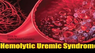
- 1. Done by shaila Robert hameldon
- 2. INTRODUCTION: • Hemolytic uremic syndrome (HUS) is a thrombotic microangiopathy in which microthrombi, consisting primarily of platelet, form and occlude the arterioles and capillaries. • These occlusions result in the simultaneous occurrence of microangiopathic hemolytic anemia, thrombocytopenia, and acute kidney injury (AKI). • HUS predominantly affects children and is caused by bacterial toxins • Thrombotic thrombocytopenic purpura (TTP) and HUS has similar pathophysiology and clinical findings, but different etiologies.
- 3. EPIDEMIOLOGY: • HUS is a rare disease but is more common in children than adults, especially children less than five years of age. • HUS is the leading cause of acute kidney failure in children. • About 200-300 cases of HUS are reported in the United States each year.
- 4. ETIOLOGY: • Bacterial exotoxins • Shiga-like toxin(verotoxin) • From enterohemorrhagic E. coli(EHEC) • Usually transmitted via contaminated foods (e.g., undercooked beef or raw leafy vegetables) • Shiga toxin produced by Shigella dysenteriae • Streptococcus pneumoniae infection. • Complement dysregulation (hereditary or acquired) accounts for approx. 5% of HUS cases with noninfectious etiologies, referred to as atypical HUS(aHUS).
- 5. PATHOPHYSIOLOGY: • Infection with enterohemorrhagic E. coli(EHEC) or another causative organism • Mucosal inflammation facilitates bacterial toxins entering systemic circulation. • Toxins cause endothelial cell damage. • Damaged endothelial cells secrete cytokines that promote vasoconstriction and platelet microthrombus formation at the site of damage (Intravascular coagulopathy → thrombocytopenia (consumption of Platelets) • RBCs are mechanically destroyed as they pass through the platelet microthrombi occluding small blood vessels → hemolysis (schistocytes) and end-organ ischemia and damage, especially in the kidneys → decreased glomerular filtration rate (GFR)
- 6. CLINICAL FEATURES: • A diarrheal illness (usually bloody) for the past 5–10 days precedes the onset of HUS symptoms in many children. The triad of clinical findings occurring in HUS consists of: • Thrombocytopenia • Petechiae, purpura • Mucosal bleeding • Prolonged bleeding after minor cuts • Microangiopathic hemolytic anemia • Fatigue, dyspnea, and pallor • Jaundice • Impaired renal function • Hematuria, • proteinuria • Oliguria, anuria
- 7. DIAGNOSIS & Differential DIAGNOSIS Hemolytic markers • ⬇️ hemoglobin • ⬇️ haptoglobin • ⬆️ indirect bilirubin • ⬆️ reticulocytes • LDH • Schistocytes on blood smear • (Up to 10 percent of rbc’s) • Coagulation profile • ↓ Platelets • Normal/slightly elevated prothrombin time (PT) and activated partial thromboplastin time (aPTT) in contrast to DIC; (see “Differential diagnoses” below) • Normal/slightly elevated Fibrin degradation products and D-dimer levels • ↑ WBC count • Negative Coombs test • Serum chemistry: ↑ BUN and ↑ creatinine (impaired renal function) • Urinalysis: hematuria, proteinuria
- 8. Differential diagnoses HUS TTP Disseminated intravascular coagulation (DIC) Immune thrombocytopenia Purpura (ITP) Pathophysiology Endothelial cell dysfunction due to bacterial toxins ADAMTS13 Deficiency Systematic coagulation activition *Platelet and coagulation factor Consumption Antiplatelet antibodies (Anti – gpllb/llla ) Typical presentation Toddler or preschooler) diarrheal illness * Presents with petechia,jaundice and oliguria Previously healthy Adult , sometimes associated with triggers – infection, surgery, pregnancy Patients with history of serious illness eg(sepsis, trauma, malignancy) Presents with thrombosis, embolism,organ dysfunction Asymptomatic with petechiae, purpura, epistaxis, menorrahgia Peripheral smear +schistocytes ⬇️ platelets + schistocytes ⬇️ platelets +/- schistocytes ⬇️ platelets Normal platelet Morphology ⬇️ platelets PT(INR) and aPTT Normal/ increased Normal/ increased Increased Normal D-dimer,fibrin degradation products Normal/ increased Normal/ increased Increased Normal
- 9. TREATMENT ➡️Avoid antibiotics and antimotility agents; (may increase the likelihood of HUS in suspected infection with EHEC). ➡️Monitor and correct: * Fluid status abnormalities * Electrolyte disturbances * Acid-base abnormalities * Blood pressure * RBC transfusions ➡️Antiepileptic drugs (e.g., diazepam, phenytoin) in patients with seizures *Dialysis (as indicated for AKI): Up to 50% of HUS patients require dialysis. *Plasma exchange therapy: only in refractory cases *Eculizumab ➡️Effective for the treatment of aHUS ➡️May be beneficial in HUS with neurological symptoms
- 10. COMPLICATIONS Heart: ischemia and fluid overload Pancreas: transient or permanent diabetes mellitus Liver: hepatomegaly, transaminase elevations Kidney Hypertension Chronic kidney disease (CKD) End-stage renal disease (ESRD) • CNS • Seizures • Paresis • Stroke • Coma • GI tract • Hemorrhagic colitis • Bowel necrosis, perforation, stricture • Peritonitis • Intussusception HUS can result in microthrombus formation and complications in various organs:
- 11. Prognosis • The prognosis depends primarily on prompt initiation of treatment. Timely treatment can prevent acute complications (AKI, coma, and death) as well as progression to chronic renal failure. • • With treatment, the mortality rate of HUS is low: < 10%. • Long-term renal sequelae occur in 35–55% of children with HUS. • Atypical HUS has a less favorable prognosis and a higher risk of progressing to end-stage renal disease.