Call Girl Surat Madhuri 7001305949 Independent Escort Service Surat
May r.j.-et-al.-2007-clinical-cancer-research
1. Peptide Epitopes from theWilms’ Tumor 1Oncoprotein Stimulate
CD4+
and CD8+
T Cells That Recognize and Kill Human
Malignant Mesothelioma Tumor Cells
RenaJ. May,1
Tao Dao,1
Javier Pinilla-Ibarz,1,2
Tatyana Korontsvit,1
Victoriya Zakhaleva,1
Rong H. Zhang,2
Peter Maslak,2
and David A. Scheinberg1,2
Abstract Purpose:Wilms’ tumor 1protein (WT1), a transcription factor overexpressed in malignant meso-
thelioma, leukemias, and other solid tumors, is an ideal target for immunotherapy. WT1class I
peptide epitopes that were identified and shown to stimulate CD8+
Tcells are being tested as
vaccine candidates in several clinical trials. The induction and maintenance of a robust memory
CD8+
cytotoxicT-cell response requires CD4+
T-cell help.
Experimental Design:Three HLA class II peptide epitopes ofWT1withhigh predictive affinities
to multiple HLA-DRB1molecules were identifiedusing the SYFPEITHIalgorithm. Due to the highly
polymorphic nature of the HLA class II alleles, such reactivity is critical in the development of a
broadly useful therapeutic. One of the WT1CD4+
peptide epitopes, 122-140, comprises a previ-
ously identified CD8+
peptide epitope (126-134). By mutating residue 126 from an arginine to a
tyrosine, we embedded a synthetic immunogenic analogue CD8+
epitope (126-134) inside the
longer peptide (122-140). This analogue was previously designed to improve immunogenicity
and induce a potent CD8+
response.
Results: WT1peptides 328-349 and 423-441are able to stimulate a peptide-specific CD4+
response that can recognize WT1+
tumor cells in multiple HLA-DRB1settings as determined by
IFN-g enzyme-linked immunospot assays. The mutated WT1peptide epitope 122-140 is able to
induce CD4+
and cytotoxic CD8+
WT1-specificT-cell responses that can recognize the native
WT1epitopes on the surface of human WT1+
cancer cells. Cross-priming experiments showed
that antigen-presenting cells pulsed with either mesothelioma or leukemia tumor lysates can
process and present each of the CD4+
peptides identified.
Conclusions: These studies provide the rationale for using the WT1CD4+
peptides in conjunc-
tion with CD8+
peptide epitopes to vaccinate patients withWT1-expressing cancers.
Wilms’ tumor 1 protein (WT1) is a zinc finger transcription
factor that is normally expressed in tissues of the mesodermal
origin during embryogenesis, including the kidney, gonads,
heart, mesothelium, and spleen. In normal adult tissues, WT1
expression is limited to low levels in the nuclei of normal
CD34+
hematopoietic stem cells, myoepithelial progenitor
cells, renal podocytes, and some cells in testis and ovary (1).
Although originally described as a tumor suppressor gene, the
WT1 protein also seems to be involved in tumorigenesis, as
WT1 expression is up-regulated in leukemia cells of all lineages
(2, 3) and in several solid tumors, including malignant
mesothelioma, lung, breast, prostate, and ovarian carcinomas
(4). Therefore, WT1 is an attractive target for immunotherapy.
Malignant mesothelioma is most often seen in patients with
a history of occupational asbestos exposure. Although asbestos
usage has been significantly reduced, incidence of malignant
mesothelioma continues to rise due to the long latency between
exposure and the onset of symptoms (5). To date, there is no
standard curative therapy for mesothelioma, and the prognosis
is poor. Surgical approaches, such as pleurectomy and
extrapleural pneumonectomy, result in high recurrence rates,
and chemotherapy and radiation therapy result in only limited
improvements (6). There are reports describing the relationship
between tumor-infiltrating lymphocytes and prognosis (7) as
well as evidence of immune responsiveness in cases of
spontaneous regression (8), suggesting that some patients can
mount an immune response in vivo against these tumors. New
immunotherapeutic approaches to malignant mesothelioma
Cancer Therapy: Preclinical
Authors’ Affiliations: 1
Molecular Pharmacology and Chemistry Program and
2
Department of Medicine, Memorial Sloan-Kettering Cancer Center, New York,
New York
Received 3/27/07; revised 5/10/07; accepted 5/16/07.
Grant support: NIH grants F32-CA119479A (R.J. May) and PO123766 and RO1
CA55349 (J. Pinilla-Ibarz,T. Korontsvit,V. Zakhaleva,T. Dao, and D.A. Scheinberg),
The Doris Duke Charitable Foundation,The Lymphoma Foundation, and Mr.William
H. Goodwin and Mrs. Alice Goodwin and the Commonwealth Foundation for
Cancer Research and The Experimental Therapeutics Center of Memorial Sloan-
Kettering (R.H. Zhang and P. Maslak). D.A. Scheinberg is a Doris Duke
Distinguished Clinical Scientist.
The costs of publication of this article were defrayed in part by the payment of page
charges.This article must therefore be hereby marked advertisement in accordance
with18 U.S.C. Section1734 solely to indicate this fact.
Note: Current address for J. Pinilla-Ibarz: Malignant Hematology Division, H. Lee
Moffitt Cancer Center,12902 Magnolia Drive,Tampa, FL 33612.
Requests for reprints: David A. Scheinberg, Molecular Pharmacology and
Chemistry Program and Department of Medicine, Memorial Sloan-Kettering
Cancer Center, 1275 York Avenue, NewYork, NY 10021. Phone: 212-639-8635;
Fax: 212-717-3068; E-mail: d-scheinberg@ski.mskcc.org.
F2007 American Association for Cancer Research.
doi:10.1158/1078-0432.CCR-07-0708
www.aacrjournals.org Clin Cancer Res 2007;13(15) August1, 20074547
2. are being explored, including the use of mesothelioma tumor
lysate–pulsed dendritic cells (9) and IFN-h gene therapy via an
adenoviral vector (10). These studies were able to elicit CD8+
T
cells against murine mesothelioma, illustrating the possibilities
of successful immunotherapeutic approaches to this disease.
Expression of WT1 (11) and immunohistochemical detection
of the WT1 protein (12) have been observed in most
mesothelioma cell lines and primary tumor specimens, and
WT1 expression is currently used as a diagnostic marker to
distinguish mesothelioma from similar tumors, such as
pulmonary adenocarcinoma (12). In addition to serving as a
diagnostic marker, the unique overexpression of WT1 may also
serve as a therapeutic target for this disease in particular.
The activation of WT1-specific CD8+
CTLs is critical to
mounting an antitumor response. At least four native peptide
nonamers from human WT1 have been identified and shown
by others to generate a WT1-specific cytotoxic response in the
context of HLA-A0201 and HLA-A2402 that is able to kill
leukemic cell lines and blast cells from patients with acute
myelogenous leukemia or acute lymphoblastic leukemia
(13–18). In an effort to enhance the immunogenicity of these
peptides and overcome possible problems with tolerance to this
self-antigen, we have designed several synthetic analogue HLA
class I peptide epitopes of WT1 via the introduction of specific
amino acid point mutations that induce a more potent
cytotoxic response to tumor cells (19).
Immunization with CD8-specific peptides alone induces a
short-lived antigen-specific CTL response without generating
T-cell memory (20). The need for antigen-specific CD4+
helper
T cells is critical when CTL precursor frequency is low, as in the
case of tumor-specific T cells (21). The presence of significant
amounts of Th1-biased circulating anti-WT1 IgG antibodies in
patients with hematopoietic malignancies compared with
healthy volunteers suggests that the WT1 protein is immuno-
genic and that WT1-specific CD4+
T cells, which are necessary
for immunoglobulin class switching, are activated in these
patients (22). Herein, we report the identification of three HLA
class II peptide epitopes of WT1 and their ability to stimulate
an immune response reactive with human malignant mesothe-
lioma cell lines as well as leukemia cell lines.
Materials and Methods
Synthetic peptides. Each of the peptides used in this study was
purchased and synthesized by Genemed Synthesis, Inc. using fluo-
renylmethoxycarbonyl chemistry and solid-phase synthesis and purified
by high-pressure liquid chromatography. The quality of the peptides
was assessed by high-performance liquid chromatography analysis,
and the expected molecular weight was observed using matrix-assisted
laser desorption mass spectrometry. Peptides were sterile and 70% to
90% pure. The peptides were dissolved in DMSO and diluted in PBS
(pH 7.4) or saline at 5 mg/mL and stored at -80jC.
Amino acid sequences and predicted binding of putative CD4+
epitopes to HLA-DRB1 molecules were identified using the predictive
algorithm of the SYFPEITHI database3
(Table 1; ref. 23). Irrelevant
control peptides used in in vitro experiments were the following: RAS
(TEYKLVVVGAPGVGKSALTIQ) or chronic myelogenous leukemia
(CML) b2a2 (VHSIPLTINKEEALQRPVASDFE) for class II and HIV
pol (ILKEPVHGV) or CML F (YLKALQRPY) for class I.
Cell lines. Human mesothelioma cell lines, including sarcomatoid
(VAMT, H2373, and H28), epithelioid (H2452), and biphasic (JMN,
MSTO, and H-Meso1A) were cultured as described (24, 25).
The cell lines were obtained from the following sources: H-Meso1A
(National Cancer Institute, Bethesda, MD), JMN and VAMT (a gift of
Dr. Sirotnak, Memorial Sloan-Kettering Cancer Center, New York, NY),
H2452 and H2373 (a gift of Dr. Pass, Karmanos Cancer Institute,
Wayne State University, Detroit, MI), and H28 and MSTO (American
Type Culture Collection). Mesothelioma cell lines Meso 11, Meso 34,
Meso 37, Meso 47, and Meso 56 were provided to us by Dr. M. Gregoire
(Institute of Biology, Nantes, France) and cultured as described (26). All
cells were HLA typed by the Department of Cellular Immunology at
Memorial Sloan-Kettering Cancer Center. Melanoma cell line MeWo
(WT1-
, A201+
) was obtained from the American Type Culture
Collection and cultured as directed. SKRC-52 renal cell carcinoma
was obtained from L. Old (Ludwig Institute, New York, NY). Leukemia
cell lines were cultured in RPMI 1640 supplemented with 10% FCS,
penicillin, streptomycin, 2 mmol/L glutamine, and 2-mercaptoethanol
at 37jC/5% CO2. LAMA81, BV173, and 697, Ph+ leukemias that are all
WT1+
, A0201+
were kindly provided by Dr. H.J. Stauss (University
College London, London, United Kingdom). SKLY16 is a human B-cell
lymphoma (WT1-
, A0201+
); K562, RwLeu4, and HL60, all WT1+
leukemias, were obtained from the American Type Culture Collection.
In vitro immunization and human T-cell cultures. After informed
consent on Memorial Sloan-Kettering Cancer Center Institutional
Review Board protocols, peripheral blood mononuclear cells (PBMC)
from HLA-typed healthy donors were obtained by Ficoll density
centrifugation. Monocyte-derived dendritic cells were generated from
PBMCs using a plastic adherence technique. The adherent cells
were cultured for 7 days in RPMI 1640/1% to 5% autologous plasma,
500 units/mL recombinant human interleukin (IL)-4 (R&D Systems),
and 1,000 units/mL recombinant human granulocyte macrophage
3
http://www.syfpeithi.de
Table 1. Peptides from WT1 that are predicted to bind to HLA-DRB molecules
Name Sequence Score*
DRB1*0101 DRB1*0301 DRB1*0401 DRB1*0701 DRB1*1101 DRB1*1501
423 RSDELVRHHNMHQRNMTKL 15 17 20 14 10 24
328 PGCNKRYFKLSHLQMHSRKHTGc 28 11 28 18 25 20
122 SGQARMFPNAPYLPSCLESb 22 18 22 16 16 18
122A1 SGQAYMFPNAPYLPSCLESx 27 17 22 18 16 18
Frequency in Caucasian population (49) 18.5 17.7 23.6 26.2 17.0 19.9
*SYFPEITHI prediction software available at http://www.syfpeithi.de.
cPeptide encompasses the sequence reported by ref. 29 (WT1 amino acids 331-345).
bPeptide encompasses the sequence reported by Kobayashi et al. (WT1 amino acids 124-138; ref. 31).
x Residue Y in bold represents a modification from the native 122 sequence.
Cancer Therapy: Preclinical
www.aacrjournals.orgClin Cancer Res 2007;13(15) August1, 2007 4548
3. colony-stimulating factor (Immunex). On days 2 and 4 of incubation,
fresh medium with IL-4 and granulocyte macrophage colony-stimulat-
ing factor was added. On day 5, 10 Ag/mL peptide was added to the
immature dendritic cells. On day 6, maturation cytokine cocktail was
added [IL-4, granulocyte macrophage colony-stimulating factor, 500
IU/mL IL-1h (R&D Systems), 1,000 IU/mL IL-6 (R&D Systems), 10 ng/
mL tumor necrosis factor-a (R&D Systems), and 1 Ag/mL prostaglandin
E2 (Sigma)]. On day 8, either CD4+
or CD3+
T lymphocytes were
isolated from the same donors using negative selection by depletion
with anti-CD11b, anti-CD56, anti-CD19, and/or anti-CD8 monoclonal
antibodies (Miltenyi) and stimulated at a 10:1 E:T ratio with the
monocyte-derived dendritic cells. The mature dendritic cells expressed
dendritic cell–associated antigens, such as CD80, CD83, CD86, and
HLA class I and class II, on their cell surfaces (data not shown). CD4+
or
CD3+
cells were stimulated for 7 days in the presence of RPMI 1640/5%
autologous plasma, 10 Ag/mL WT1 synthetic peptides, 1 Ag/mL h2-
microglobulin (Sigma), and 10 ng/mL IL-15 (R&D Systems). On day 7,
T cells were restimulated with either a 5:1 E:T ratio of freshly isolated
CD14+
cells or a 50:1 E:T ratio of monocyte-derived dendritic cells. In
some cases, cells were restimulated after another 6 to 7 days in the same
manner. After the second or third stimulation, IFN-g secretion of these
cells was examined by enzyme-linked immunospot (ELISPOT). In some
cases, total PBMCs were stimulated with 10 Ag/mL WT1 synthetic
peptide and 10 ng/mL IL-15. PBMCs were restimulated every 7 days by
spinning down the cells and resuspending in fresh medium containing
10 Ag/mL peptide and 10 ng/mL IL-15. After two or three rounds of
stimulation, the cells were used in an IFN-g ELISPOT assay.
IFN-g ELISPOT. HA-Multiscreen plates (Millipore) were coated
with 100 AL of mouse anti-human IFN-g antibody (10 Ag/mL; clone 1-
D1K; Mabtech) in PBS, incubated overnight at 4jC, washed with PBS to
remove unbound antibody, and blocked with RPMI 1640/10%
autologous plasma for 2 h at 37jC. Purified CD4+
, CD8+
, or CD3+
T
cells (>95% pure) were plated with either autologous CD14+
(5:1 E:T
ratio) or autologous dendritic cells (10:1 E:T ratio). Various test
peptides were added to the wells at 40 Ag/mL. Negative control wells
contained antigen-presenting cells (APC) with or without T cells or
T cells alone plus peptides. Positive control wells contained T cells plus
APC plus 10 Ag/mL phytohemagglutinin (Sigma). All conditions were
done in triplicate. Blocking experiments were done whereby 50 Ag/mL
of anti-HLA-DR monoclonal antibody clone L243 (a gift from Dr.
Denzin, Memorial Sloan-Kettering Cancer Center) were incubated with
1 Â 106
CD14+
target cells for 1 h at 37jC. Microtiter plates were
incubated for 20 h at 37jC and then extensively washed with PBS/
0.05% Tween and 100 AL/well biotinylated detection antibody against
human IFN-g (2 Ag/mL; clone 7-B6-1; Mabtech) was added. Plates were
incubated for an additional 2 h at 37jC and spot development was
done as described (27). Spot numbers were automatically determined
with the use of a computer-assisted video image analyzer with KS
ELISPOT 4.0 software (Carl Zeiss Vision).
Cross-priming experiments. A CD3+
in vitro stimulation was done as
described above. Immature dendritic cells (2 Â 106
) were incubated
with total cellular lysate from 2 Â 106
tumor cells that was previously
prepared by three freeze/thaw cycles. Following an 18-h incubation,
maturation cytokines were added to the dendritic cells as described
above. CD3+
cells were stimulated thrice with these autologous mature
dendritic cells, after which the T cells were tested in an IFN-g ELISPOT
assay, as described above, for reactivity against autologous, mature
dendritic cells that had been previously pulsed with individual CD4+
peptides when in the immature state. These dendritic cells were exposed
to peptide again during the ELISPOT assay as described above.
Chromium-51 cytotoxicity assay. The presence of specific CTLs was
measured in a standard 4-h chromium release assay as described (27).
Briefly, target cells are pulsed with 10 Ag/mL of synthetic peptides
overnight at 37jC, after which they are labeled with 300 ACi of
Na2
51
CrO4 (NEN Life Science Products, Inc.). After extensive washing,
target cells are incubated with T cells at E:T ratios ranging from 100:1 to
10:1. All conditions were done in triplicate. Plates were incubated for
4 h at 37jC in 5% CO2. Supernatant fluids were harvested and
radioactivity was measured in a gamma counter. Percentage specific
lysis was determined from the following formula: 100 Â [(experimental
Table 2. Summary of in vitro stimulation experiment and IFN-g ELISPOT results
Experiment no. WT1DR 328 WT1DR 423 WT1DR 122 WT1DR 122A1
Donor
HLADRB*
Responsec Donor
HLADRB
Response Donor
HLADRB
Response Donor
HLADRB
Response
1 1001/1501 +++ 1001/1501 +++ 1401/15XX — 1401/15XX + (heteroclitic)b
2 301/901 +++ 101/1201 — 1401 + 1401 ++ (heteroclitic)
3 1001/1501 ++ 1001/1501 — 1401 + 1401 + (heteroclitic)
4 407/1302 NAx 407/1302 NA 104/1104 — 104/1104 +++ (heteroclitic)
5 407/1302 +++ 407/1302 + 405/1302 — 301/1601 NA
6 1001/1501 — 1001/1501 — 301/1601 ++ 405/1302 —
7 407/1302 — 407/1302 — 1401/15XX +++ 15XX +++ (heteroclitic)
8 1001/1501 ++ 1001/1501 — 15XX + 422/13XX +++ (heteroclitic)
9 407/1302 ++ 407/1302 + 1401/15XX +++ 422/13XX ++ (heteroclitic)
10 701/1202 ++ 701/1202 — 301/1601 —
11 1401/15XX ++ 1401/15XX — 1401/15XX +
12 407/1302 +++ 422/13XX — 1401/15XX +
13 422/13XX + 422/13XX +++ 15XX +++ (heteroclitic)
14 422/13XX +++ 1001/1501 +++
15 1001/1501 +++
No. donors 6 80%k 6 35.7%k 6 66.6%k 7 76.9%k
*Healthy donor CD3+
/CD4+
T cells were stimulated twice with individual WT1 peptide as described in Materials and Methods. Stimulated T cells
were challenged in an IFN-g ELISPOT.
cELISPOT results comparing T cell reactivity with autologous CD14+
cells pulsed with stimulating peptide versus autologous CD14+
cells pulsed
with irrelevant peptide. -, negative results; +, two times background; ++, three times background; +++, four times background.
bT cells stimulated with WT1DR peptide 122A1 recognized CD14+
cells pulsed with stimulating peptide and CD14+
cells pulsed with WT1DR
peptide 122.
xExperiment failed.
kPercentage of experiments with a significant positive results (P V 0.05).
TCells That Recognize and Kill Tumor Cells
www.aacrjournals.org Clin Cancer Res 2007;13(15) August1, 20074549
4. release - spontaneous release) / (maximum release - spontaneous
release)]. Maximum release was determined by lysis of radiolabeled
targets in 2.5% Triton X-100.
Quantitative reverse transcription-PCR for WT1 transcripts. Total
RNA was isolated from the various cell lines using a phenol/chloroform
extraction method. RNA purity was confirmed by absorbance at 260
nm. The reverse transcription reaction was adapted from protocols
supplied by Applied Biosystems. Beginning with 1 Ag of total RNA,
random hexamers and reverse transcriptase were used to isolate cDNA.
For the PCR, cDNA was mixed with the following WT1 primers and
probe: 5¶-CAGGCTGCAATAAGAGATATTTTAAGCT-3¶ (forward primer,
located on exon 7), 5¶-GAAGTCACACTGGTATGGTTTCTCA-3¶ (reverse
primer, located on exon 8), and 5¶-CTTACAGATGCACAGCAGGAAG-
CACACTG-3¶ (Taqman probe, located on exon 7). The fluorescent WT1
probe 5¶-56-FAM/CTTACAGATGCACAGCAGGAAGCACACTG/
3BHQ_1/-3¶ was labeled with 6-carboxyfluorescein phosphoramide as
reporter dye at the 5¶-end and with the quencher dye carboxytetrame-
thylrhodamine at the 3¶-end (Integrated DNA Technologies). The PCR
has been described (28). Briefly, 2 min at 50jC, 10 min at 95jC
followed by 50 cycles of 15 s at 95jC and 60 s at 62jC. Each reaction
was done in triplicate, and discrepancies >1 Ct in one of the wells were
excluded. The quantitative reverse transcription-PCR and fluorescence
measurements were made on the Applied Biosystems 7500 Real-Time
PCR System. Control ABL primers and probes were 5¶-TGGAGATAA-
CACTCTAAGCATAACTAAAGGT-3¶ (forward primer, located on EnF-
10030), 5¶-GATGTAGTTGCTTGGGACCCA-3¶ (reverse primer, located
on ENR-1063), and 5¶-56-FAM/CCATTTTTGGTTTGGGCTTCACAC-
CATT/3BHQ_1/-3¶ (fluorescent probe, located on ENPr-1043).
Statistics. Statistical analyses were done on StatView software (SAS
Institute) using a two-tailed unpaired t test, with the level of statistical
significance set at 0.05.
Results
Identification of peptides with a high predictive binding to
multiple HLA class II molecules. Due to the permissive nature
of the HLA class II binding pocket, designing a peptide that
displays high affinity to multiple class II molecules, rather than
binding limited to a single DR type, is possible. Such peptides
would therefore be capable of stimulating T cells from a larger
percentage of the target population. The WT1 protein sequence
was screened using the SYFPEITHI algorithm, and three WT1DR
peptides (423, 328, and 122) were identified that had predicted
high-affinity binding to multiple HLA-DRB1 molecules that are
highly expressed in the Caucasian population (Table 1).
Peptide 423 spans WT1 amino acids 423-441, peptide 328
spans WT1 amino acids 328-349, and peptide 122 spans WT1
amino acids 122-140. The maximal scores to each HLA protein
are variable, with HLA-A0201 maximal score being 36. Three
WT1 native 15-mer peptides that were predicted to bind to
HLA-DRB1*0401 (29) and HLA-DP501 (30) and HLA-DR1,
HLA-DR4, and HLA-DR7 (31) have been identified by others.
These peptides were capable of stimulating CD4+
T cells that
were restricted to HLA-DRB1*0401 (29), HLA-DP501 (30), and
HLA-DR53 (31). Peptide 328 is an extended version of the
HLA-DRB1*0401–specific peptide (29), and peptide 122 is
similar to the peptide reported by Kobayashi et al. (31).
WT1DR peptide 122 comprises within it a known CD8+
epitope (126-134; ref. 14) We previously reported the use of
amino acid substitutions at HLA-A0201 binding anchor motifs
to increase the antigenicity and binding affinity of native WT1
9-mer peptides. One such modified peptide, termed WT1A1,
spans the WT1 amino acid residues 126-134 and contains a
modified amino acid at position 126, in which we substituted a
tyrosine for the native arginine. This peptide induces a more
potent cytotoxic CD8+
T-cell response against WT1+
tumors
(19). Due to the location of peptide 126 within the longer
CD4+
122 peptide epitope, we investigated whether the
inclusion of the mutated residue 126 would confer both a
CD4 and a potent CD8+
epitope within the long 122 CD4+
Fig. 1. IFN-g ELISPOTwithWT1DR peptides 328, 423,122, and 1222A1. A, CD3+
Tcells from an HLA-DRB1*1001/1501donor were stimulated twice with either
peptideWT1DR 328 orWT1DR 423 as described in Materials and Methods.
StimulatedTcells were challenged in an IFN-g ELISPOTassay with the following:
unchallenged control (gray columns), CD14+
cells pulsed with stimulating peptide
(either WT1DR 328 or WT1DR 423; black columns), CD14+
cells pulsed with
irrelevant CD4+
peptide epitope (RAS; white columns), and unpulsed CD14+
cells
(hatched columns). *, P <0.05, compared with controls.Yaxis, number of spots per
1 Â105
CD3+
Tcells; X axis, peptide used for T-cell stimulations. B, CD4+
Tcells
from an HLA-DRB1*1001/1501donor were stimulated twice with either peptide
WT1DR 328 orWT1DR 423 as described in Materials and Methods. Stimulated
Tcells were challenged in an IFN-g ELISPOTassay with the following: CD14+
cells
pulsed with stimulating peptide (eitherWT1DR 328 orWT1DR 423; black columns),
CD14+
cells pulsed with stimulating peptide (either WT1DR 328 or WT1DR 423)
andincubated with anti-HLA-DR blocking antibody (gray hatched columns), CD14+
cells pulsed with irrelevant CD4+
peptide epitope (CML b2a2; white columns),
CD14+
cells pulsed with irrelevant CD4+
peptide epitope (CML b2a2) and
incubated with anti-HLA-DR blocking antibody (checkerboard columns), and
unpulsed CD14+
cells (black hatched columns). *, P < 0.04, compared with
irrelevant peptide controls and compared with CD14+
Tcells in the presence of
anti-HLA-DR monoclonal antibody.Y axis, number of spots per1 Â105
CD4+
T
cells; X axis, peptide used forT-cell stimulations. C, CD4+
Tcells from an HLA-
DRB1*1501donor were stimulated twice with either peptideWT1DR122 orWT1DR
122A1as described in Materials and Methods. StimulatedTcells were challenged
in an IFN-g ELISPOTassay with the following: unpulsed CD14+
cells (light gray
columns), CD14+
cells pulsed with irrelevant CD4 peptide epitope (CML b2a2;
white columns), CD14+
cells pulsed withirrelevant CD4 peptide epitope (CML b2a2)
and incubated with anti-HLA-DR blocking antibody (dotted columns), CD14+
cells
pulsed with peptideWT1DR122 (dark gray columns), CD14+
cells pulsed with
peptideWT1DR122 and incubated with anti-HLA-DR blocking antibody (dark gray
hatched columns), CD14+
cells pulsed with peptideWT1DR122A1 (black columns),
and CD14+
cells pulsed with peptideWT1DR122A1and incubated with
anti-HLA-DR blocking antibody (black hatched columns). *, P < 0.05, compared
with irrelevant peptide controls and compared with CD14+
Tcells in the presence
of peptide and anti-HLA-DR monoclonal antibody.Y axis, number of spots per
1 Â105
CD4+
Tcells; X axis, peptide used for T-cell stimulations.
Cancer Therapy: Preclinical
www.aacrjournals.orgClin Cancer Res 2007;13(15) August1, 2007 4550
5. epitope. This mutated peptide, termed 122A1, did not
significantly alter the predictive binding score of the native
WT1DR 122 peptide to the HLA-DRB1 molecules (Table 1) but
offered the possibility of inducing simultaneously a robust
CD4+
and CD8+
WT1-specific T-cell response if the long 122A1
peptide could be processed by the cell into the shorter 126-134
CD8+
epitope.
Induction of a peptide-specific CD4 immune response as
detected by IFN-g assay. Web-based predictive algorithms are
only 50% reliable in identifying peptide epitopes capable of
stimulating T cells (23). Therefore, in vitro stimulation assays
are necessary to confirm those peptides that can stimulate CD4+
T cells. Table 2 lists the results of CD3+
or CD4+
cells from
healthy donors that were stimulated with peptide-loaded
autologous dendritic cells. WT1DR peptide 328 stimulated a
significant peptide-specific CD4+
response in a variety of HLA-
DRB1 settings, a representative sample of which is shown in
Fig. 1A, whereas responses to WT1DR peptide 423 were more
variable. The peptide-specific response seen is HLA-DR restrict-
ed because the addition of anti-HLA-DR antibody blocked the
peptide-specific response (Fig. 1B).
Consistent with previously published results (31), WT1DR
peptide 122 induced a peptide-specific response that was HLA-
DR mediated. CD4+
T cells stimulated with WT1DR peptide
122A1 displayed a more robust heteroclitic T-cell response in
that the stimulated T cells not only recognized the mutated
immunizing peptide 122A1 but also the native 122 peptide
(Fig. 1C, note the scale difference in the Y axis and Table 2).
Here, too, the effect was shown to be HLA-DR mediated. This
cross-reactivity to the native peptide is critical because only
the native sequence would be expressed on the surface of tumor
cells.
These stimulation experiments were reproduced multiple
times in up to seven healthy donors, each with a different HLA-
DRB1 type to assess the efficacy of the peptides in various HLA-
DR settings (Table 2). Up to 15 separate experiments were done
with each WT1DR peptide. Peptide-specific T-cell responses
were seen in 12 of 15 experiments with WT1DR 328, 5 of 14
experiments with WT1DR 423, and 6 of 9 experiments with
WT1DR122. WT1DR 122A1 stimulated T cells that recognized
the immunizing WT1DR 122A1 peptide and the native WT1DR
122 peptide in 10 of 13 experiments. Although the number of
experiments is too small, and the number of HLA-DRB1 alleles
tested is too great to draw any statistically significant
conclusions, there is considerable variation in how the
predicted binding scores correlate to actual efficacy for each
of the HLA-DRB1 alleles.
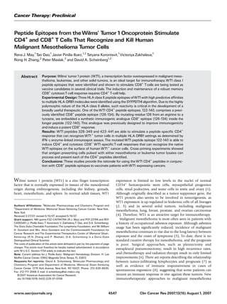
WT1 expression in human mesothelioma cell lines. Whereas
many groups have been focused on developing WT1 peptide
immunotherapy to hematopoietic malignancies, only a few
have studied the feasibility of WT1 vaccine-based therapy of
solid tumors (32, 33). In particular, we were interested in
developing a WT1-based vaccine for patients with malignant
mesothelioma, a disease with a high WT1 expression and poor
prognosis. The WT1 transcript levels in several human meso-
thelioma cell lines (sarcomatoid, epithelioid, and biphasic)
were quantified by reverse transcription-PCR and compared
with various leukemia cell lines with known WT1 expression
(Fig. 2). Although 12 of 12 mesothelioma cell lines expressed
WT1 message, in most cases, the WT1 level was lower in these
cell lines compared with the leukemic cell lines. Melanoma
(MeWo) and lymphoma (SKLY16) cell lines were WT1
negative. Interestingly, SKRC-52, a human renal cell carcinoma
cell line, did not express WT1 despite the low expression of
WT1 in adult renal podocytes (1). Flow cytometry analysis
confirmed that the mesothelioma cell lines express class II
molecules (data not shown), confirming previous studies (34).
However, only mesothelioma cell lines JMN and H2452
expressed class I molecules (data not shown).
Processing and presentation of WT1 epitopes. Peptides that
are presented on the surface of target cells bound to either HLA
class I or class II molecules must first be processed either in the
cytosol for class I peptides or in endocytic vesicles for class II
peptides. Because the peptides we are studying were identified
first by computer algorithms, it was necessary to determine
whether these peptides were properly processed from the WT1
protein and presented in the context of HLA molecules. In
addition, most tumor cells have low MHC II molecule
expression, and it was critical to show that these peptides
derived from dying tumor cells can be processed by APCs and
presented to CD4+
T cells. ‘‘Cross-priming’’ experiments were
done to determine if all the WT1DR peptides under study (423,
328, and 122) were presented and recognized by CD4+
T cells.
Total tumor lysates were prepared from three different cell
lines: 697 (WT1+
, HLA-A0201+
), an e1a2 leukemia cell line;
JMN (WT1+
, HLA-A0201+
), a biphasic mesothelioma cell line;
and as a control, MeWo (WT1-
, HLA-A0201+
), a malignant
melanoma cell line. Dendritic cells from healthy A0201+
Fig. 2. Quantitative reverse transcription-
PCR. RelativeWT1expression levels in a
variety of hematopoietic and mesothelioma
cell lines.WT1levels are shown as relative
values compared with the human leukemia
cell line K562, which is defined as1.0 as
described in ref. 28.
TCells That Recognize and Kill Tumor Cells
www.aacrjournals.org Clin Cancer Res 2007;13(15) August1, 20074551
6. donors were incubated for 18 h with the tumor lysates and then
used to stimulate autologous CD3+
T cells. Following three
stimulations, the T cells were tested for their reactivity to
autologous dendritic cells pulsed with the various WT1
peptides. T cells that had been stimulated with WT1+
tumor
lysates recognized the individual HLA class II peptides (Fig. 3A
and B), whereas T cells stimulated by dendritic cells pulsed with
the WT1-
lysate (MeWo) did not stimulate WT1-specific T cells.
In addition, T cells stimulated with dendritic cells pulsed with
697 tumor lysate recognized the native short class I peptide
WT1A (126-134) and the analogue WT1A1 peptide (Fig. 3A).
These experiments were repeated in five separate donors.
Stimulated T cells could recognize WT1DR peptide 328 and
WT1DR peptide 122A1 in three of five experiments, whereas
stimulated T cells recognized WT1DR 423 in each experiment.
Therefore, despite the low expression of WT1 transcript in the
mesothelioma cell lines, the newly identified WT1 CD4
epitopes seem to be processed and presented in HLA class II
molecules.
The reverse experiment was next conducted to determine if
stimulation with the peptides could result in recognition of the
tumor cells by the T cells, as should occur after vaccination with
the peptides. A sufficient amount of WT1 peptide is presented
on the surface of the WT1+
mesothelioma tumor cell for T cells
stimulated with individual WT1DR peptides to recognize
mesothelioma tumor cells compared with the control WT1-
melanoma cells (Fig. 3C). In another experiment, T cells were
stimulated by the mutated WT1DR 122A1 and challenged with
pulsed and unpulsed adherent cell targets. When control WT1-
target cells are pulsed with additional WT1DR 122A1 peptide,
the amount of IFN-g spots increases. When WT1+
target cells
are pulsed with additional WT1DR 122A1 peptide, spots do not
increase, implying that a maximal response has been achieved
with the native processed peptides (Fig. 3D).
Peptide 122A1 induces a CD4+
and a cytotoxic CD8+
T-cell
response. Although a strong CD8+
epitope is nested within the
longer WT1DR 122 and WT1DR 122A1 peptides, it was critical
to determine if the long peptide could be processed to stimulate
CD8+
T cells. Both WT1DR 122 and WT1DR 122A1 were able
to activate CD8+
T cells against the native short epitope (termed
WT1A, amino acids 126-134) and CD4+
cells against the long
peptide (Fig. 4A and B; ref. 15). The mutated WT1DR 122A1
sequence was a more potent stimulator. WT1DR 122A1, but
not WT1DR 122, stimulated a sufficient amount of CD8+
cells
to be cytotoxic to 697, a WT1+
leukemia cell line that expresses
native WT1 protein. These stimulated CD8+
T cells did not
recognize SKLY16, a WT1-
B-cell lymphoma, unless it had been
pulsed with peptide WT1A (Fig. 4C). T cells stimulated with
WT1DR 122 were unable to kill WT1+
target tumor cell lines
(data not shown). These experiments have been repeated in
four different A0201+
donors, each with a distinct HLA-DRB1
type. In three of four experiments, the long WT1DR 122A1
peptide stimulated CD8+
responses (data not shown). The
magnitude of the response varied, indicating that the potency
of the WT1DR 122A1 peptide is donor dependent.
CD8+
T cells reactive with the WT1A epitope are cytotoxic to
leukemia cell lines (14, 19) that express high levels of WT1.
We have previously shown that T cells stimulated with the
WT1A1 analogue can kill WT1+
leukemia cells (19). In
addition, human T cells stimulated twice with either the
native WT1A or the analogue WT1A1 peptide can kill human
WT1+
mesothelioma cell lines compared with WT1-
control
cell lines (9.2% lysis of MeWo versus 19% lysis of JMN for
WT1A-stimulated T cells; 22.2% lysis of MeWo versus 44.8%
lysis of JMN for WT1A1-stimulated T cells). We were able to
show in this study that CD8+
T cells stimulated with WT1DR
122A1 are cytotoxic to the A0201+
, WT1+
JMN human
mesothelioma cell line and not the A0201+
, WT1-
MeWo
melanoma cell line (Fig. 4D). In contrast, CD4+
cells
Fig. 3. Processing and presentation ofWT1DR peptides. A and B, cross-priming
experiments. A, CD3+
Tcells from an HLA-A0201/301DRB1*1301/1302 healthy
donor were stimulated with autologous dendritic cells (DC) previously incubated
with 697 tumor lysates. 697 is aWT1+
leukemia cell line. StimulatedTcells were
challenged in an IFN-g ELISPOTassay with autologous dendritic cells previously
incubated with either 697 tumor lysate, individualWT1peptides, control peptides,
or unpulsed dendritic cells, as indicated on the X axis. Hatched columns,
background level of spots from autologous dendritic cells incubated in the absence
ofTcells. *, P < 0.05, compared with control peptides.Y axis, number of spots per
1 Â105
CD3+
cells. B, CD3+
Tcells from an HLA-A0201/101DRB1*0301/1601
healthy donor were stimulated with autologous dendritic cells previously incubated
with tumor lysates from eitherJMN, aWT1+
mesothelioma cell line (black columns),
or MeWo, aWT1-
melanoma cell line (white columns). StimulatedTcells were
challenged in an IFN-g ELISPOTassay with autologous dendritic cells previously
incubated with eitherJMN or MeWo tumor lysates, individualWT1DR peptides, or
control class II peptide, as indicated on the X axis. Hatched columns, background
level of spots from autologous dendritic cells incubated in the absence ofTcells.
*, P <0.05, compared with control peptides.Y axis, number of spots per1 Â105
CD3+
cells. C and D, CD3+
IFN-g ELISPOTagainst mesothelioma cell lines. C, total
PBMCs from an HLA-DRB1*13XX donor were stimulated twice with the different
WT1DR peptides as described in Materials and Methods. StimulatedTcells were
challenged in an IFN-g ELISPOTassay with the following: mesothelioma H-Meso1A
cell line (WT1+
, HLA-DRB1*1301; black columns) and control melanoma MeWo cell
line (WT1-
, HLA-DRB1*15XX; gray columns). *, P V 0.01, compared with MeWo
controls.Y axis, number of spots per 2 Â 105
PBMCs; X axis, peptide used for
T-cell stimulation. D, CD3+
Tcells from an HLA-A0201/DRB1*1501donor were
stimulated twice withWT1DR122A1as described in Materials and Methods.
StimulatedTcells were then challenged in an IFN-g ELISPOTassay with the
following target cells: JMN, an A0201/DRB1*1505 WT1+
mesothelioma cell line, or
MeWo, an A0201/DRB1*15XX WT1-
melanoma cell line.The target cells were either
pulsed withWT1DR122A1 (black columns) or not pulsed (gray columns).
*, P <0.05, compared with the unpulsed MeWo target cell.Y axis, number of spots
per1 Â105
CD3+
Tcells; X axis, different cell lines used as target cells.
Cancer Therapy: Preclinical
www.aacrjournals.orgClin Cancer Res 2007;13(15) August1, 2007 4552
7. stimulated with WT1DR 122A1 showed no cytotoxicity to
either WT1+
mesothelioma or WT1-
melanoma cells. However,
we were unable to define the exact CD8+
peptide epitope of
122A1-stimulated CTLs from these experiments. These findings
together provide a rationale to further study WT1-based
immunotherapy for malignant mesothelioma.
Discussion
An approach to improving vaccine potency is to use analogue
peptide epitopes of native tumor antigens, which are created by
introducing amino acid point mutations into certain MHC
anchor motifs (19). These novel peptides, which are homolo-
gous to the native peptide, can generate CTL responses that
recognize the immunizing epitope and the native sequence.
Immunogenic analogue peptides to human CD33 (35), WT1
(19), and bcr-abl (27) and others have been described.
However, the induction and maintenance of a robust memory
CTL response requires CD4+
T-cell help (20). CD4+
T helper
cells recognize peptides ranging from 16 to 19 amino acids that
are bound to the HLA class II molecule. Once activated, CD4+
cells enhance immunity by producing IFN-g and IL-2 and
licensing dendritic cells, thereby maintaining the activation,
proliferation, and survival of potent antitumor activity medi-
ated by CTLs (36, 37). Some in vitro studies have shown that
CTLs at low E:T ratios are not cytotoxic unless CD4+
cells are
added, underscoring the importance of T-cell help at the tumor
site (38). Although some tumor cells are able to process and
present antigens in the context of HLA class I and class II
molecules (34), many tumors typically express low levels of
class II molecules and cannot stimulate antigen-specific CD4+
cells on their own. Therefore, when designing peptide vaccines
for those tumors, it may be valuable to also include CD4+
peptide epitopes. These peptides will be processed and
presented by APCs, which in turn will activate CD4+
T cells
and help induce a potent CD8+
antitumor T-cell response. The
absence of activated CD4+
T-cell help will likely result in the
induction of anergic CTL T-cell clones (39), which may explain
the failure of some vaccination strategies with class I peptides
alone. To overcome this problem, some groups have vaccinated
patients with short tumor-specific CD8+
epitopes along with
universal nonspecific MHC class II–restricted epitopes, such as
keyhole limpet hemocyanin (40) or the promiscuous epitopes
of tetanus toxoid (41). Although a robust CD4+
response to the
peptide is induced, the response to the tumor antigen often
remains limited. An alternative approach to more selectively
enhance the tumor-specific CD8+
response would be to find
CD4+
epitopes directly from the tumor antigen.
Fig. 4. WT1DR peptide122 and122A1stimulate CD8+
T-cell responses. A, CD3+
Tcells from an HLA-A0201/DRB1*1401donor were stimulated twice withWT1DR
122 as described in Materials and Methods. StimulatedTcells were challenged in an
IFN-g ELISPOTassay with autologous CD14+
cells in the presence of different
peptides.Y axis, number of spots per1 Â105
CD3+
cells; X axis, different test
peptides used in the ELISPOT. B, CD3+
Tcells from an HLA-A0201/DRB1*1401
donor were stimulated twice withWT1DR122A1as described in Materials and
Methods. StimulatedTcells were challenged in an IFN-g ELISPOTassay with control
melanoma cell line MeWo (A0201/DRB1*15XX,WT1-
) in the presence of different
peptides. *, P < 0.05, compared with no peptide controls.Y axis, number of spots
per1 Â105
CD3+
cells; X axis, different test peptides used in the ELISPOT. C, CD3+
Tcells from an HLA-A0201/DRB1*0101/15XX donor were stimulated twice with
WT1DR122A1as described in Materials and Methods. After two rounds of
stimulation, CD8+
Tcells were isolated by negative selection and used as effector
cells in a 51
Cr release cytotoxicity assay as described in Materials and Methods.
CD8+
Tcells were incubated with various radiolabeled target cells [pulsed or
unpulsed 697 (A0201+
,WT1+
) or SKLY16 (A0201+
,WT1-
)] at three different E:T
ratios: 100:1 (gray columns), 30:1 (black columns), and10:1 (white columns).
Yaxis, percentage of cytotoxicity; X axis, different target cell conditions. *, P <0.05,
compared with SKLY16 controls at the same E:Tratio. D, CD3+
Tcells from an
HLA-A0201/DRB1*0101/15XX donor were stimulated twice withWT1DR122A1as
described in Materials and Methods. After two rounds of stimulation, CD8+
Tcells
were isolated by negative selection and used as effector cells in a 51
Cr release
cytotoxicity assay as described in Materials and Methods. CD8+
Tcells were
incubated with radiolabeled JMN (A0201+
,WT1+
; black line) or MeWo
(A0201+
,WT1-
, grayline) target cells at four different E:Tratios.Yaxis, percentage of
cytotoxicity; X axis, different E:Tratios. P < 0.001, compared with MeWo controls.
TCells That Recognize and Kill Tumor Cells
www.aacrjournals.org Clin Cancer Res 2007;13(15) August1, 20074553
8. We have identified three WT1 CD4+
epitopes that can
stimulate T cells from a broad group of HLA-DRB types to
recognize target cells pulsed with the stimulating peptide but
not with an irrelevant peptide. Most important, these T cells
also recognized leukemia and mesothelioma cell lines as
assayed by IFN-g ELISPOT.
WT1DR peptide 328 is a modified version of a previously
reported peptide that has high binding affinity to HLA-
DRB1*0401 and has been shown to stimulate CD4+
T cells
that could recognize HLA-DRB1*0401–restricted CML tumor
cells (29). We have added three amino acids to the NH2-
terminal ends and four amino acids to the COOH-terminal
ends, as flanking residues outside the minimal epitope have
been shown to greatly influence processing of the peptide (42)
and increase the predictive binding scores. In addition, because
class II molecules have a more permissive binding pocket,
the elongated 328 peptide was determined to be a more
promiscuous epitope and a potent stimulator of T cells from
donors expressing several HLA-DRB1 alleles.
WT1DR peptide 423 is a new WT1 CD4+
epitope identified
using the SYFPEITHI binding algorithm. A similar peptide from
WT1 (418-432) was recently identified by Rezvani et al. (43)
after screening a WT1 peptide library consisting of overlapping
15-mer peptides and testing the ability of these peptides to
stimulate CD4+
T cells. The cross-priming experiments de-
scribed here confirmed that this peptide is properly processed
and presented by APCs and that the peptide itself could
adequately stimulate T cells to recognize the native processed
protein.
A recent study by Kobayashi (31) identified WT1 peptide
124-138 as a potential CD4+
epitope based on the presence of
HLA-DR1, HLA-DR4, and HLA-DR7 binding motifs. T cells
stimulated with this peptide secreted IFN-g in the presence of
autologous APCs in an HLA-DR53–restricted fashion. In
addition, peptide 124-138 was processed by APC after pulsing
with tumor lysate and it is expressed on the surface of HLA-DR+
tumor cells. Because CD4+
helper T cells play an essential role
in maintenance of CTL antitumor responses, a peptide
encompassing both a CD8+
and CD4+
epitope would be an
advantageous immunogen. However, the reactivity of T cells
stimulated by peptide 124-138 was inhibited by anti-HLA-DR
antibodies, but not anti-HLA class I antibodies, indicating that
CD8+
T cells were not activated (31) despite the presence of the
WT1 CTL epitope (126-134; ref. 14) within the longer peptide.
Therefore, in an attempt to create a peptide that would
simultaneously induce a robust CD4+
and CD8+
T-cell
response, we mutated amino acid 126 from an arginine to a
tyrosine, thereby embedding our previously described (19)
heteroclitic WT1A1 peptide within the longer CD4+
epitope. In
addition, we extended the peptide by several amino acids at the
NH2-terminal and COOH-terminal ends to increase the
likelihood of better processing and increasing the HLA-DR
binding promiscuity. This new long peptide, 122A1, is properly
processed and presented by APC and can stimulate CD4+
and
CD8+
T cells that are cytotoxic to cells expressing the native
WT1 antigen.
Vaccines containing a combination of a CD8+
and CD4+
epitopes result in a more robust CTL response (44). Chimeric
peptides have been made by linking CD4+
and CD8+
epitopes
(45). However, such peptides may not be processed correctly
due to the possible modification of natural protease cleavage
sites, and chimeric peptides contain nonnatural sequences that
are potentially immunogenic. Other investigators have made
peptides in which the CD4+
and CD8+
epitopes overlap and
have shown this elongated peptide to be more effective than
administering separate CD4+
and CD8+
peptides (46, 47). We
describe a different approach wherein a peptide containing a
heteroclitic analogue CD8+
epitope is nested inside a CD4+
epitope. We believe that such a peptide is an ideal vaccine
candidate because it has been shown by others to induce a
CD8+
immune response that is greater in magnitude and
durability than immunizing with the short CD8+
peptide alone
(48). The mutated CD8+
WT1 epitope seems to be a more
potent than its native parent.
This is the first report establishing the in vitro activity of WT1-
based CD4+
and CD8+
T-cell stimulation reactive with human
malignant mesothelioma. The three WT1DR CD4+
epitopes
stimulate T cells that recognize human mesothelioma cell lines,
and WT1-stimulated CTLs are able to kill these tumor cells
despite the lower expression levels of WT1. These studies
provide a rationale for clinical testing of these peptides in
patients with WT1+
cancers. Whether patients with mesotheli-
oma or other solid tumors will have the capacity to respond to
vaccination in a manner similar to that seen in vitro with
healthy donor cells will require a clinical trial. Such a trial is
now open at Memorial Sloan-Kettering Cancer Center.
References
1. Mundlos S, Pelletier J, Darveau A, Bachmann M,
Winterpacht A, Zabel B. Nuclear localization of the
protein encoded by the Wilms’ tumor gene WT1 in
embryonic and adult tissues. Development 1993;119:
1329^41.
2. Inoue K, Ogawa H, SonodaY, et al. Aberrant overex-
pression of the Wilms tumor gene (WT1) in human
leukemia. Blood1997;89:1405^12.
3. Brieger J,Weidmann E, Maurer U, Hoelzer D, Mitrou
PS, Bergmann L. The Wilms’ tumor gene is frequently
expressed in acute myeloblastic leukemias and may
provide a marker for residual blast cells detectable by
PCR. Ann Oncol 1995;6:811^6.
4. OjiY,OgawaH,TamakiH,etal.Expressionof theWilms’
tumorgeneWT1insolidtumorsanditsinvolvementintu-
morcellgrowth. JpnJCancerRes1999;90:194^204.
5. Selikoff IJ, Hammond EC, Seidman H. Latency of as-
bestos disease among insulation workers in theUnited
States and Canada. Cancer1980;46:2736^40.
6. van Ruth S, Baas P, Zoetmulder FA. Surgical treat-
ment of malignant pleural mesothelioma: a review.
Chest 2003;123:551^61.
7. Leigh RA,Webster I. Lymphocytic infiltration of pleu-
ral mesothelioma and its significance for survival. S Afr
Med J1982;61:1007^9.
8. Robinson BW, Robinson C, Lake RA. Localised
spontaneous regression in mesotheliomaöpossible
immunological mechanism. Lung Cancer 2001;32:
197^201.
9. HegmansJP, Hemmes A, Aerts JG, Hoogsteden HC,
Lambrecht BN. Immunotherapy of murine malignant
mesothelioma using tumor lysate-pulsed dendritic
cells. AmJRespir Crit Care Med 2005;171:1168^77.
10. Odaka M, Sterman DH,Wiewrodt R, et al. Eradica-
tion of intraperitoneal and distant tumor by adenovi-
rus-mediated interferon-h gene therapy is attributable
to induction of systemic immunity. Cancer Res 2001;
61:6201^12.
11. Amin KM, Litzky LA, SmytheWR, et al.Wilms’ tumor
1 susceptibility (WT1) gene products are selectively
expressed in malignant mesothelioma. Am J Pathol
1995;146:344^56.
12. Foster MR, JohnsonJE, Olson SJ, Allred DC.Immu-
nohistochemical analysis of nuclear versus cytoplas-
mic staining of WT1in malignant mesotheliomas and
primary pulmonary adenocarcinomas. Arch Pathol
Lab Med 2001;125:1316^20.
13. OkaY, Elisseeva OA,Tsuboi A, et al. Human cytotox-
icT-lymphocyte responses specific for peptides of the
wild-type Wilms’ tumor gene (WT1) product. Immu-
nogenetics 2000;51:99^107.
14. Gao L, Bellantuono I, Elsasser A, et al. Selective
elimination of leukemic CD34(+) progenitor cells by
cytotoxic T lymphocytes specific for WT1. Blood
2000;95:2198^203.
15. Ohminami H,Yasukawa M, Fujita S. HLA class I-re-
stricted lysis of leukemia cells by a CD8(+) cytotoxic
Cancer Therapy: Preclinical
www.aacrjournals.orgClin Cancer Res 2007;13(15) August1, 2007 4554
9. T-lymphocyte clone specific for WT1peptide. Blood
2000;95:286^93.
16. AzumaT, Makita M, Ninomiya K, Fujita S, Harada M,
Yasukawa M. Identification of a novel WT1-derived
peptide which induces human leukocyte antigen-
A24-restricted anti-leukemia cytotoxicT lymphocytes.
BrJHaematol 2002;116:601^3.
17. Bellantuono I, Gao L, Parry S, et al. Two distinct
HLA-A0201-presented epitopes of the Wilms tumor
antigen 1can function as targets for leukemia-reactive
CTL. Blood 2002;100:3835^7.
18. Doubrovina ES, Doubrovin MM, Lee S, et al. In vitro
stimulation with WT1 peptide-loaded Epstein-Barr
virus-positive B cells elicits high frequencies of WT1
peptide-specificTcells with in vitro and in vivo tumor-
icidal activity. Clin Cancer Res 2004;10:7207^19.
19. Pinilla-Ibarz J, May RJ, KorontsvitT, et al. Improved
humanT cell responses against synthetic HLA 0201
analog peptides derived from the WT1 oncoprotein.
Leukemia 2006;11:2025^33.
20. Fernando GJ, Khammanivong V, Leggatt GR, Liu
WJ, Frazer IH. The number of long-lasting functional
memory CD8+
T cells generated depends on the
nature of the initial nonspecific stimulation. Eur J
Immunol 2002;32:1541^9.
21. MinternJD, Davey GM, Belz GT, Carbone FR, Heath
WR. Cutting edge: precursor frequency affects the
helper dependence of cytotoxic T cells. J Immunol
2002;168:977^80.
22. Wu F, Oka Y, Tsuboi A, et al. Th1-biased humoral
immune responses against Wilms tumor gene WT1
product in the patients with hematopoietic malignan-
cies. Leukemia 2005;19:268^74.
23. Rammensee H, Bachmann J, Emmerich NP, Bachor
OA, Stevanovic S. SYFPEITHI: database for MHC
ligands and peptide motifs. Immunogenetics 1999;
50:213^9.
24. Khokhar NZ, Lam AF, Rusch VW, Sirotnak FM.
Despite some expression of folate receptor a in
human mesothelioma cells, internalization of metho-
trexate is predominantly carrier mediated. J Thorac
Cardiovasc Surg 2002;123:862^8.
25. Adusumilli PS, Chan MK, ChunYS, et al. Cisplatin-
induced GADD34 upregulation potentiates oncolytic
viral therapy in the treatment of malignant pleural me-
sothelioma. Cancer BiolTher 2006;5:48^53.
26. Ebstein F, Sapede C, Royer PJ, et al. CytotoxicTcell
responses against mesothelioma by apoptotic cell-
pulsed dendritic cells. Am J Respir Crit Care Med
2004;169:1322^30.
27. Pinilla-Ibarz J, KorontsvitT, ZakhalevaV, Roberts W,
Scheinberg DA. Synthetic peptide analogs derived
from bcr/abl fusion proteins and the induction of
heteroclitic human T-cell responses. Haematologica
2005;90:1324^32.
28. Cilloni D, Gottardi E, De Micheli D, et al. Quantita-
tive assessment of WT1expression by real time quan-
titative PCR may be a useful tool for monitoring
minimal residual disease in acute leukemia patients.
Leukemia 2002;16:2115^21.
29. Muller L, Knights A, Pawelec G. Synthetic peptides
derived from Wilms’ tumor 1protein sensitize human
T lymphocytes to recognize chronic myelogenousleu-
kemia cells. Hematol J 2003;4:57^66.
30. Guo Y, Niiya H, Azuma T, et al. Direct recognition
and lysis of leukemia cells by WT1-specific CD4+
T
lymphocytes in an HLA class II-restricted manner.
Blood 2005;106:1415^8.
31. Kobayashi H, NagatoT, Aoki N, et al. Defining MHC
class II T helper epitopes for WT1 tumor antigen.
Cancer Immunol Immunother 2005;55:850^60.
32. Gillmore R, Xue SA, Holler A, et al. J. Detection of
Wilms’ tumor antigen-specific CTL in tumor-draining
lymph nodes of patients with early breast cancer. Clin
Cancer Res 2006;12:34^42.
33. Tsuboi A, Oka Y, Osaki T, et al. WT1peptide-based
immunotherapy for patients with lung cancer: report
of two cases. Microbiol Immunol 2004;48:175^84.
34. Mutti L,Valle MT, Balbi B, et al. Primary human me-
sothelioma cells express class II MHC, ICAM-1 and
B7-2 and can present recall antigens to autologous
blood lymphocytes. Int JCancer 1998;78:740^9.
35. Bae J, Martinson JA, Klingemann HG. Heteroclitic
CD33 peptide with enhanced anti-acute myeloid leu-
kemic immunogenicity. Clin Cancer Res 2004;10:
7043^52.
36. MarzoAL, Kinnear BF, Lake RA, et al.Tumor-specific
CD4+
Tcells have a major ‘‘post-licensing’’ role in CTL
mediated anti-tumor immunity. J Immunol 2000;165:
6047^55.
37. Hung K, Hayashi R, Lafond-Walker A, Lowenstein
C, Pardoll D, Levitsky H.The central role of CD4(+) T
cells in the antitumor immune response. J Exp Med
1998;188:2357^68.
38. ElsawaSF,RodebergDA,CelisE.T-cellepitopepep-
tide vaccines. Expert Rev Vaccines 2004;3:563^75.
39. Pawelec G, Heinzel S, Kiessling R, Muller L,
Ouyang Q, Zeuthen J. Escape mechanisms in tumor
immunity: a year 2000 update. Crit Rev Oncog
2000;11:97^133.
40. Scheibenbogen C, Schadendorf D, Bechrakis NE,
et al. Effects of granulocyte-macrophage colony-
stimulating factor and foreign helper protein as immu-
nologic adjuvants on theT-cell response to vaccination
with tyrosinase peptides. Int J Cancer 2003;104:
188^94.
41. Panina-Bordignon P,Tan A,Termijtelen A, Demotz S,
Corradin G, Lanzavecchia A. Universally immunogen-
icTcell epitopes: promiscuous binding to human MHC
class II and promiscuous recognition byTcells. Eur J
Immunol 1989;19:2237^42.
42. Eisenlohr LC, Yewdell JW, Bennink JR. Flanking
sequences influence the presentation of an endoge-
nously synthesized peptide to cytotoxic T lympho-
cytes. JExp Med 1992;175:481^7.
43. Rezvani K, Mielke S, Kilical Y, et al. Identification of
novel MHC class I and class II epitopes ofWT1using a
peptide library screen. Atlanta (GA): American Society
of Hematology; 2005.
44. Zwaveling S, Ferreira Mota SC, Nouta J, et al.
Established human papillomavirus type16-expressing
tumors are effectively eradicated following vaccina-
tion with long peptides. JImmunol 2002;169:350^8.
45. Ahlers JD,TakeshitaT, Pendleton CD, BerzofskyJA.
Enhanced immunogenicity of HIV-1vaccine construct
by modification of the native peptide sequence. Proc
Natl Acad Sci U S A1997;94:10856^61.
46. Carreno BM, Turner RV, Biddison WE, Coligan JE.
Overlapping epitopes that are recognized by CD8+
HLA class I-restricted and CD4+
class II-restricted
cytotoxic T lymphocytes are contained within an in-
fluenza nucleoprotein peptide. J Immunol 1992;148:
894^9.
47. Ayyoub M, Merlo A, Hesdorffer CS, et al. Distinct
but overlappingT helper epitopes in the 37-58 region
of SSX-2. Clin Immunol 2005;114:70^8.
48. Knutson KL, Schiffman K, Disis ML. Immunization
with a HER-2/neu helper peptide vaccine generates
HER-2/neu CD8 T-cell immunity in cancer patients.
J Clin Invest 2001;107:477^84.
49. Southwood S, Sidney J, Kondo A, et al. Several
common HLA-DR types share largely overlapping
peptide binding repertoires. J Immunol 1998;160:
3363^73.
TCells That Recognize and Kill Tumor Cells
www.aacrjournals.org Clin Cancer Res 2007;13(15) August1, 20074555