Top Rated Bangalore Call Girls Mg Road ⟟ 8250192130 ⟟ Call Me For Genuine Sex...
CAR T CELL
1. ME67CH06-Ramos ARI 19 August 2015 13:45
R
E
V I E W
S
I
N
A
D V A
N
C
E
CAR-T Cell Therapy for
Lymphoma
Carlos A. Ramos,1,2,3
Helen E. Heslop,1,2,3,4
and Malcolm K. Brenner1,2,3,4
1
Center for Cell and Gene Therapy, Houston Methodist Hospital, Texas Children’s Hospital,
and Baylor College of Medicine, Houston, Texas 77030
2
Dan L. Duncan Cancer Center, 3
Department of Medicine, and 4
Department of Pediatrics,
Baylor College of Medicine, Houston, Texas 77030; email: caramos@bcm.edu,
hheslop@bcm.edu, mbrenner@bcm.edu
Annu. Rev. Med. 2016. 67:6.1–6.19
The Annual Review of Medicine is online at
med.annualreviews.org
This article’s doi:
10.1146/annurev-med-051914-021702
Copyright c
2016 by Annual Reviews.
All rights reserved
Keywords
immunotherapy, adoptive T cell therapy, CD19, CD20, CD30, kappa light
chain
Abstract
Lymphomas arise from clonal expansions of B, T, or NK cells at differ-
ent stages of differentiation. Because they occur in the immunocyte-rich
lymphoid tissues, they are easily accessible to antibodies and cell-based im-
munotherapy. Expressing chimeric antigen receptors (CARs) on T cells is
a means of combining the antigen-binding site of a monoclonal antibody
with the activating machinery of a T cell, enabling antigen recognition in-
dependent of major histocompatibility complex restriction, while retaining
the desirable antitumor properties of a T cell. Here, we discuss the basic
design of CARs and their potential advantages and disadvantages over other
immune therapies for lymphomas. We review current clinical trials in the
field and consider strategies to improve the in vivo function and safety of
immune cells expressing CARs. The ultimate driver of CAR development
and implementation for lymphoma will be the demonstration of their ability
to safely and cost-effectively cure these malignancies.
6.1
Review in Advance first posted online
on August 26, 2015. (Changes may
still occur before final publication
online and in print.)
Changes may still occur before final publication online and in print
Annu.
Rev.
Med.
2016.67.
Downloaded
from
www.annualreviews.org
Access
provided
by
University
of
Sussex
on
09/01/15.
For
personal
use
only.
2. ME67CH06-Ramos ARI 19 August 2015 13:45
HUMAN LYMPHOMAS
Human lymphomas have historically been separated into non-Hodgkin and Hodgkin lymphoma
(NHL and HL). NHL includes a broad group of lymphoid malignancies that arise from clonal
expansions of B, T, or natural killer (NK) cells at various stages of differentiation. B cell lymphomas
are usually derived from germinal center or postgerminal center B cells, whereas T and NK cell
lymphomas may arise at any stage of normal T- or NK-cell lymphopoiesis. Although malignant
cells acquire genetic abnormalities, they also retain many of the phenotypic characteristics of their
normal counterparts. Target antigens for immunotherapy are, therefore, generally expressed on
both lymphoma cells and their nonmalignant counterparts (1).
In contrast, the malignant Reed-Sternberg (RS) cells of HL have an unusual expression of
hematopoietic markers that has no normal counterpart. Although the cell from which HL origi-
nates was long debated, microdissection studies recently showed that RS cells possess clonal heavy-
and light-chain immunoglobulin (Ig) gene rearrangements, and thus HL is likely derived from
crippled germinal center cells (2). HL also differs from NHL in that the malignant RS cells are
relatively rare, and the more prominent, nonmalignant, infiltrating cells in the microenvironment
play an important role in HL biology (2).
SUITABILITY OF NON-HODGKIN AND HODGKIN LYMPHOMA
FOR IMMUNOTHERAPY
Despite their biological differences, both HL and NHL have proven to be good targets for im-
munotherapy. Both lymphomas occur in the immune-rich lymphoid tissues and are therefore
easily accessible for antibodies and cell-based immunotherapy. Moreover, T cells targeted to
tumor-associated antigens expressed by B cell lymphomas are likely to receive the costimulation
they require if they are to pass through immune checkpoints, as B cells are excellent antigen-
presenting cells. In addition, lymphomas express both lineage-restricted (e.g., CD19) and unique
(e.g., Ig idiotype) tumor antigens that can be targeted by antibodies and/or effector cells (3). Finally,
35–45% of all human lymphomas are associated with persistent infection with Epstein-Barr virus
(EBV), so that antigens associated with viral latency can be detected in many patients with HL or
with many NHL subtypes, including Burkitt lymphoma, NK/T cell lymphomas, and diffuse large
B cell lymphoma (DLBCL). These viral antigens can be successfully targeted with EBV-specific
T cells (4, 5).
Although the majority of target antigens on human lymphomas include lineage-restricted
antigens that are also present on normal B cells and some T cells, eradication of normal as well
as malignant lymphocytes may be considered an acceptable toxicity. Hence, antibodies to CD20
are included in most B cell lymphoma treatment regimens, and the expression of CD30 both on
the RS cells of HL and on a subpopulation of activated T and NK cells has not prevented the use
of CD30 antibody to treat HL (6).
HOW CAN CAR-T CELLS BUILD UPON LYMPHOMA
IMMUNOTHERAPY?
Chimeric antigen receptors (CARs) were first developed in the mid-1980s (7). In 1993, the Eshhar
group (8) modified the concept to use (a) an extracellular domain (ectodomain) from a single-
chain variable fragment (scFv), composed of the antigen-binding regions of both heavy and light
chains of a monoclonal antibody (mAb); (b) a transmembrane domain; and (c) an intracellular
domain (endodomain) with a cell-signaling component derived from the T cell receptor (TCR)
6.2 Ramos ·Heslop ·Brenner
Changes may still occur before final publication online and in print
Annu.
Rev.
Med.
2016.67.
Downloaded
from
www.annualreviews.org
Access
provided
by
University
of
Sussex
on
09/01/15.
For
personal
use
only.
3. ME67CH06-Ramos ARI 19 August 2015 13:45
Monoclonal
antibody
α β
TCR complex
γ ε ε δ
ζ ζ
Tumor
antigen
T cell
T
T c
cell
ell
Intracytoplasmic
Tumor
vL
vH
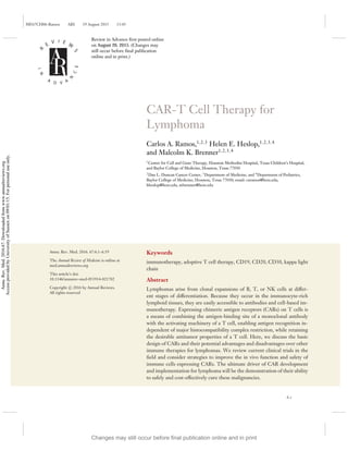
Figure 1
The basic structure of first-generation chimeric antigen receptors (CARs). The most common CARs combine the extracellular
antigen-recognition site of a monoclonal antibody and the intracellular domains of a T cell receptor (TCR) complex molecule.
Clustering of CARs induced by antigen binding on the surface of tumor cells initiates signal transduction that leads to T cell activation
and killing of tumor cells.
ζ chain (Figures 1 and 2). Most subsequent CARs have followed this same structural pattern,
with incorporation of accessory or costimulatory signaling components (Figure 2). In contrast to
conventional T cells, which rely on their native TCRs for tumor antigen recognition, CAR-T cells
recognize unprocessed antigen and therefore kill tumor cells independently of their expression of
major histocompatibility complex (MHC) antigens.
Design of CAR Ectodomains
To date, all CARs for lymphoma have been based on scFvs (Table 1), whose effectiveness depends
in part on the affinity of the CAR itself and on the properties of the antigenic epitope recognized.
For instance, CARs containing high-affinity scFvs for ROR1 confer greater effector function to
T cells than those containing low-affinity scFvs (9). However, the location of the recognized
epitope on the antigen also affects CAR function (10, 11). For example, T cells expressing a CAR
with an scFv that recognizes a membrane-proximal epitope on CD22 have greater antileukemic
activity than CAR-T cells recognizing a distal epitope (10).
Other components of the ectodomain may also influence CAR effectiveness, such as the pres-
ence of flexible linker sequences in the scFv and the type of elements connecting the ecto- to
the endodomain (hinge and transmembrane regions). The hinge and transmembrane regions can
affect CAR-T cell function profoundly by modifying the length and flexibility of the resulting
CAR, its cell surface density, its tendency to self-aggregate and produce T cell exhaustion by
www.annualreviews.org • CAR-T Cell Therapy for Lymphoma 6.3
Changes may still occur before final publication online and in print
Annu.
Rev.
Med.
2016.67.
Downloaded
from
www.annualreviews.org
Access
provided
by
University
of
Sussex
on
09/01/15.
For
personal
use
only.
4. ME67CH06-Ramos ARI 19 August 2015 13:45
Spacer
Linker
scFv
ζ (zeta)
ζ
CD28
Ectodomain
Transmembrane
Endodomain
4-1BB
CD28
ζ
First
generation
Second
generation
Third
generation
Figure 2
Three generations of chimeric antigen receptors (CARs). First-generation CARs include an extracellular
domain (ectodomain), usually derived from a single-chain variable fragment (scFv), composed of the antigen-
binding regions of both heavy and light chains of a monoclonal antibody; a transmembrane domain; and an
intracellular domain (endodomain) with a cell-signaling component derived from the T cell receptor, usually
the ζ chain. Most subsequent CARs have followed this same structural pattern, with incorporation of one
(second-generation CARs) or more (third-generation CARs) accessory or costimulatory signaling
components, such as CD28, CD137 (4-1BB), and CD134 (OX40). These additional costimulatory
endodomains improve T cell activation and proliferation, and thus may promote killing of target tumor cells.
tonic signaling, and its potential binding to molecules other than the intended target antigen. For
example, a CD19-specific CAR with a CD3-ζ transmembrane domain is less stable over time on
the cell surface of T cells than a CD19-specific CAR with the same scFv but a CD28 transmem-
brane domain (12) (G. Dotti, B. Savoldo, unpublished data). Also, for instance, CD19-CARs with
a hinge derived from the IgG4 CH2–CH3 region are functional in vitro but may have impaired
Table 1 Chimeric antigen receptor (CAR) targets in development or in use for lymphomas
Antigen Malignancy lineage CAR ectodomain Clinical trial
BCMA B cell scFv Unavailablea
CD19 B cell scFv See Table 2
CD20 B cell scFv See Table 2
CD22 B cell scFv Ongoing (90)
CD30 B cell and T cell scFv See Table 2
CD70 B cell and T cell CD70 ligand Unavailable
ROR1 B cell scFv Ongoing (91)
κ B cell scFv See Table 2
a
Clinical trial available only for multiple myeloma.
6.4 Ramos ·Heslop ·Brenner
Changes may still occur before final publication online and in print
Annu.
Rev.
Med.
2016.67.
Downloaded
from
www.annualreviews.org
Access
provided
by
University
of
Sussex
on
09/01/15.
For
personal
use
only.
5. ME67CH06-Ramos ARI 19 August 2015 13:45
antitumor activity in vivo due to interaction between the Fc domain within the hinge and Fc
receptor-bearing myeloid cells (13).
Design of CAR Endodomains
Upon antigen recognition, CAR endodomains transmit activation and costimulatory signals to
T cells. T cell activation relies on the phosphorylation of immunoreceptor tyrosine-based acti-
vation motifs (ITAMs) present in the cytoplasmic CD3-ζ domain of the TCR complex (14). In
addition to this stimulus from TCR engagement (signal 1), T cells require costimulation (sig-
nal 2) for sustained growth and function, as TCR stimulation without costimulation induces T
cell anergy. The endodomain of early CARs contained only the CD3-ζ signaling component
(Figure 1). To improve CAR-T cell function and persistence, costimulatory endodomains (such
as CD28, OX40, or 4-1BB) were incorporated into so-called second- (and even third-) generation
CARs (Figure 2) to ensure the transgenic T cells are fully activated after engaging their specific
target (15–19).
The superiority of second- over first-generation CAR-transduced T cells was decisively demon-
strated in a study comparing the two constructs head to head (12). This phase I trial treated subjects
with refractory or relapsed B cell lymphomas, mostly DLBCL, who were simultaneously infused
with two autologous T cell products. Both products were retrovirally transduced with a CD19-
CAR, but one CAR encoded both the CD28 and ζ endodomains whereas the other included only
the ζ endodomain. This strategy allowed direct measurement of the consequences of adding a
CD28 costimulatory endodomain to CAR-redirected T cells in the same subject and established
that T cells bearing a second-generation CAR that contains the CD28 endodomain have enhanced
in vivo proliferation and survival compared to T cells expressing a first-generation CAR lacking
CD28. The contribution of costimulatory domains is discussed in detail in the section on CD19-
CAR trials. All the subsequent studies described in the “Lymphoma Antigens Targeted in Current
Clinical Trials” section used second-generation CARs, except where indicated.
Advantages and Disadvantages of Using CARs
Even though mAbs such as CD20 have been highly successful in the treatment of lymphomas,
T cells expressing CARs directed at the same or related antigens additionally offer the poten-
tial benefits of active trafficking to tumor sites, in vivo expansion, and long-term persistence.
Moreover, because CAR-T cells have MHC-unrestricted activity, they can circumvent some of
the major mechanisms by which tumors avoid MHC-restricted T cell recognition, such as the
downregulation of human leukocyte antigen (HLA) class I molecules and defective antigen pro-
cessing (20–23). Finally, CARs can target nonprotein antigens, which allows them to recognize
lymphoma tumor antigens derived from carbohydrates or glycolipids, which are not detectable by
conventional TCRs.
T CELL EXPRESSION OF CARS
Although it is possible to transiently express CARs in T cells by transfecting them with naked
DNA plasmids or mRNA (for instance, to more safely test them for toxicity, since any adverse
effects should reverse once the transgene is eliminated), in most instances the goal is to obtain
sustained expression to ensure durability of response (24). Replication-defective retroviruses (gam-
maretroviruses or lentiviruses) can integrate a reverse-transcribed sequence into the host cell DNA
through the action of a viral integrase, thus becoming part of that cell’s genome (25). Because
www.annualreviews.org • CAR-T Cell Therapy for Lymphoma 6.5
Changes may still occur before final publication online and in print
Annu.
Rev.
Med.
2016.67.
Downloaded
from
www.annualreviews.org
Access
provided
by
University
of
Sussex
on
09/01/15.
For
personal
use
only.
6. ME67CH06-Ramos ARI 19 August 2015 13:45
retroviral production under good manufacturing practices is time consuming and expensive, some
groups have adopted nonviral methods for permanent transduction, specifically transposon-based
systems, including Sleeping Beauty (26) and PiggyBac (27), which lead to stable integration of the
transgene, although these systems may be less effective overall.
Ideally, CAR molecules should be grafted onto a subset of T cells that can traffic to tumor
sites, receive appropriate costimulation, and expand and persist in vivo. Lymphoma studies have
focused on αβ-TCR+
T cells, and investigators are now trying to use phenotypic profiling to
identify the optimal subset within this population (28, 29). T cells with a memory-associated
phenotype may be optimal for in vivo persistence, and investigators have used positively selected
cells expressing a central memory–associated marker (CD62L) to express CD19-directed CARs
in a clinical study at the Fred Hutchinson Cancer Research Center. More recently, a T cell subset
has been identified with even greater proliferative potential and longer survival in vivo (28). These
T stem cell memory populations can differentiate into memory and effector populations, but their
ultimate value in human CAR studies has not yet been established.
An alternative to T cell selection based on surface phenotype is to physiologically select T cells
that have an established capacity to act as effector T cells, enter the memory pool, and re-expand
on re-exposure to antigens in vivo. Virus-specific T cells (VSTs) have these abilities (5, 30). VSTs
also express chemokine receptors that should allow them to traffic to sites of lymphoma (31).
Moreover, the ability of CAR-VSTs to interact through their native TCR with viral antigens on
professional antigen-presenting cells may provide a range of costimulatory signals that enhance
their persistence after adoptive transfer and that increase their activity against tumor targets, which
is mediated through their transgenic CAR (32).
In addition to CAR gene expression in αβ T cells, T cells with γδ-TCR may also be amenable
to transduction with a CAR and provide additional functionality (33). Similarly, other lymphocyte
populations may offer specific advantages and can also be transduced using the same methods. For
instance, invariant-chain TCR T cells (or NK-T cells) may have preferential tumor trafficking
properties and inherent activity against tumor-associated macrophages, while NK cells may have
additional intrinsic antitumor lytic potential (34). None of these alternative cell sources has been
used yet for CAR therapy of lymphoma.
CHOICE OF LYMPHOMA ANTIGEN FOR CAR TARGETING
Unlike the native TCR, the majority of ScFv-based CARs only recognize intact target antigens
expressed directly on the cell surface, rather than peptide fragments from processed proteins that
are presented in association with MHC molecules. This limited recognition hinders CAR-T cells’
ability to detect most truly tumor-specific antigenic epitopes (since these are usually derived from
internal mutant oncogenes and translocations). Unfortunately, normal B or T cells also express
the majority of lymphoma target antigens suitable for CAR recognition.
CAR-T cells can, for example, be used to target a highly and consistently expressed lineage-
specific antigen (e.g., CD19, CD20, CD22) resulting in elimination of malignant B cells. However,
because these antigens are also expressed by their normal counterparts, B cell ablation is a frequent
side effect, albeit relatively benign because replacement therapy using intravenous Ig is feasible.
In general, however, it might be preferable to target more restricted lineage-associated antigens,
such as BCMA (B cell maturation antigen), which is predominantly expressed by plasma cells and
subsets of mature B cells (35). As another example, in many B cell malignancies it is possible to
target either the κ or the λ light chain associated with all cells of the (clonal) malignancy, and this
is desirable to spare normal (polyclonal) B cells expressing the reciprocal light chain and maintain
immune function (18).
6.6 Ramos ·Heslop ·Brenner
Changes may still occur before final publication online and in print
Annu.
Rev.
Med.
2016.67.
Downloaded
from
www.annualreviews.org
Access
provided
by
University
of
Sussex
on
09/01/15.
For
personal
use
only.
7. ME67CH06-Ramos ARI 19 August 2015 13:45
The argument in favor of targeting an entire lineage is less acceptable for T cell lymphomas
because T cell function is less amenable to replacement therapies than that of B cells. Nonethe-
less, as discussed below, a more selectively expressed T lineage marker, such as CD30, may be
acceptable, particularly if expression is high on the tumor cells.
Targeting single antigens carries the inherent risk of immune escape (36–38), which can be
reduced by targeting multiple antigens. Expressing multiple CARs in T cells also has the potential
to increase safety by generating T cells that recognize a unique antigen pattern that is only present
on tumor cells or their associated stroma (39).
LYMPHOMA ANTIGENS TARGETED IN CURRENT CLINICAL TRIALS
CD19
With the important exceptions of hematopoietic stem cells and plasma cells, CD19 is expressed
during all stages of B cell differentiation and is maintained on the vast majority of cells that
have undergone neoplastic transformation (40), such as in B cell NHL and chronic lymphocytic
leukemia (CLL). Most of the initial CAR-T cell trials in B cell malignancies, including NHL and
acute lymphoblastic leukemia (ALL), targeted this antigen. Results in NHL, albeit impressive,
have not been as striking as in ALL, for which complete remission rates of ∼90% have been
described (41, 42).
CAR ζ-chain signaling is insufficient for CAR T cell persistence. Early experience treating
B cell malignancies with CD19-CAR T cells demonstrated the feasibility of the approach but also
its lack of objective antitumor effects. All of these trials used first-generation CARs with a single
signaling domain (derived from the ζ chain of the TCR complex) (43). In one of these studies,
two patients with refractory follicular lymphoma received T cells expressing a CD19-CAR after
undergoing treatment with lymphodepleting doses of fludarabine. The T cells had undergone
polyclonal activation with a CD3 antibody (OKT3), plasmid electroporation, and hygromycin
selection (for which the plasmid also encoded a resistance gene). After CAR-T cell infusion,
patients received low-dose subcutaneous interleukin (IL)-2 injections. Transferred T cells were
detectable by polymerase chain reaction (PCR) for fewer than seven days. As expected given the
cells’ limited persistence, neither clinical responses nor overt toxicities were observed. Of note,
cellular antitransgene immune rejection responses were documented in both patients, although
whether this activity was directed at the CAR or the hygromycin resistance gene is unknown (43).
Results from such trials using first-generation CAR-T cells demonstrated that a single stim-
ulatory domain was insufficient to fully activate the chimeric T cells and confirmed that host
lymphopenia also facilitates expansion of adoptively transferred T cells. Lymphopenia creates
“space” for the oncoming adoptively transferred cells and enhances their homeostatic expan-
sion while also depleting the endogenous regulatory T cells, which normally secrete inhibitory
cytokines [e.g., transforming growth factor β (TGFβ) and IL-10] that limit effector T cell ex-
pansion (44). Additionally, T cell growth homeostatic cytokines, such as IL-7 and IL-15, which
ordinarily exist in limiting amounts, may become readily available owing to less competition and
increased production by lymphopoietic stromal cells (45).
An early report from a trial using a second-generation, CD28-containing CD19-CAR described
one patient with advanced follicular lymphoma, who was treated with a preparative chemotherapy
regimen followed by autologous T cells retrovirally modified to express the CAR. The patient’s
tumor underwent partial regression, and B cells were absent from circulation for at least 39 weeks
after T cell infusion, despite recovery of other blood series. The CD19-CAR transgene was
www.annualreviews.org • CAR-T Cell Therapy for Lymphoma 6.7
Changes may still occur before final publication online and in print
Annu.
Rev.
Med.
2016.67.
Downloaded
from
www.annualreviews.org
Access
provided
by
University
of
Sussex
on
09/01/15.
For
personal
use
only.
8. ME67CH06-Ramos ARI 19 August 2015 13:45
detected in the peripheral blood up to 27 weeks after infusion (46). Nonetheless, in vivo expansion
of these second-generation CAR T cells may still be modest, and clinical responses are limited.
In another trial of six patients with DLBCL, only two had transient stable disease and four had
disease progression. The inadequate activity suggests that alternative costimulatory domains (or
a different CAR design) might be necessary for more potent activation of chimeric T cells.
Later-acting costimulatory domains may be more efficacious than CD28. Although co-
stimulatory signals from CD28 seemed to improve expansion and persistence, a trial using a
second-generation CAR incorporating 4-1BB (CD137) as an alternative costimulatory domain
(47) reported the most dramatic expansion and clinical activity in indolent B cell malignancies.
CD28 costimulation is usually provided physiologically by professional antigen-presenting cells
and represents an “early” costimulatory signal, but “late” costimulatory molecules, including mem-
bers of the tumor necrosis factor receptor (TNFR) family such as OX40 (also known as CD134)
and 4-1BB (CD137), also play crucial roles. After binding to their specific ligands, these molecules
recruit TNFR-associated-factor (TRAF) adapter proteins, which represent an entirely distinct ac-
tivation pathway from CD28 costimulation and may be associated with more potent activation of
T cells (48), at least in certain disease settings.
The first three patients reported from this second-generation, 4-1BB-containing CD19-CAR
trial had large-burden, relapsed B cell CLL. They were infused with autologous CAR-T cells
after receiving lymphodepleting chemotherapy (47, 49). In contrast to other trials, a lentivirus
was used to transfect T cells. These CAR-T cells had a 1,000-fold expansion in vivo, trafficked
to bone marrow, and continued to express functional CARs at high levels for at least six months.
Despite large tumor burdens, results were impressive: two long-term complete remissions and one
prolonged partial remission were seen in the three CLL patients treated. Each infused CAR-T
cell was calculated to have eradicated at least 1,000 CLL cells on average. Significant adverse
effects were noted, however, including an acute systemic inflammatory response syndrome (fever
with hypotension, respiratory distress, or tumor lysis syndrome) as well as late on-target, off-
tumor toxicities, namely B cell aplasia associated with decreased numbers of plasma cells and
hypogammaglobulinemia (see the section on toxicities below).
Nonetheless, the issue of whether late costimulatory domains are always better than early ones
is far from being settled. CD28-containing CD19-CARs have continued to show encouraging
activity in other trials, reports from which have reinforced the need for lymphodepletion prior
to CAR-T cell infusion, at least in the autologous setting. For example, the outcomes of seven
additional patients were described in an update (50) to the single-patient report (46) mentioned
in the previous section and, more recently, the same group published results in 13 other NHL
patients (51). Nine patients achieved complete remission, which lasted up to 23 months. Adverse
events included long-term depletion of normal B cells and prominent elevations in serum levels
of inflammatory cytokines, which appeared to correlate with the severity of acute toxicities (fever
and hypotension). In addition, central nervous system toxicity, of unclear etiology, was observed
in some patients.
A similar second-generation, CD28-containing CAR was used in another trial in which eight
CLL patients (and one ALL patient) were treated (52). All patients tolerated the CAR-T cell
infusions well, but one patient had rapid clinical deterioration and died less than 48 h after CAR-
T cell infusion (see section on toxicities below). Some of the other patients developed fever with or
without hypotension a few days after T cell infusion. One of the patients with CLL had a partial
response, and none developed B cell aplasia. Persistence of infused CAR-T cells was inversely
proportionaltothetumorburdenbutenhancedbypriorcyclophosphamideadministration,further
favoring the use of lymphodepleting chemotherapy before CAR-T cell infusion.
6.8 Ramos ·Heslop ·Brenner
Changes may still occur before final publication online and in print
Annu.
Rev.
Med.
2016.67.
Downloaded
from
www.annualreviews.org
Access
provided
by
University
of
Sussex
on
09/01/15.
For
personal
use
only.
9. ME67CH06-Ramos ARI 19 August 2015 13:45
All the CD19-CAR trials described above used cell products generated from autologous T
cells. Two additional protocols, summarized in Table 2, employed allogeneic cells (53, 54).
CD20
A first-generation CAR targeting CD20 has been used in a few studies. In one of these, seven
patients with follicular or mantle cell lymphomas received CD20-specific CAR-modified T cell
infusions, with minimal toxicities. T cells were subjected to polyclonal activation, plasmid elec-
troporation, and neomycin selection. The modified T cells persisted in vivo up to nine weeks in
patients, who also received low-dose subcutaneous IL-2 injections. Two patients had continued
complete responses, one achieved a partial response, and four had stable disease (55). In another
study, two patients with recurrent DLBCL were treated with cloned CD8+
T cells expressing
another first-generation CD20-CAR (and neomycin resistance) after autologous hematopoietic
stem cell transplantation. Neither clinical responses nor overt toxicities were observed. In that
trial, the transferred T cells were detectable by PCR for fewer than seven days (43).
CD30
Almost all HLs and some NHLs express the CD30 antigen both at diagnosis and relapse, and mAbs
targeting CD30 produce objective antitumor responses. The effects of mAbs, however, appear to
be limited in duration, encouraging the substitution of CD30-CARs on longer-lived T cells. A
phase I dose escalation study of activated autologous CAR-CD30-T cells treated nine patients with
relapsed/refractory EBV1/N
CD30+
HL or NHL (seven with HL and two with CD30+
anaplastic
large cell lymphoma). Eight of these patients had either relapsed or progressed after treatment
with the CD30 mAb brentuximab. CAR-T cell infusions produced no attributable adverse events;
in particular, the frequency of T cell precursors targeting cytomegalovirus, EBV, adenovirus, and
influenza virus remained unchanged by treatment (assuaging an important concern, since CD30 is
upregulated on some activated T cells). The molecular signal from the CAR-T cells peaked at one
week following infusion, but decreased by four weeks. Of eight evaluable patients, six weeks after
treatment, four patients had stable disease, one had a complete response, and one had a partial
response, while three had disease progression (56; C. Ramos, G. Dotti, B. Savoldo, unpublished
data).
κ Light Chain
As mentioned above, by taking advantage of the clonal restriction of mature B cell malignancies,
which express either a κ or λ light Ig chain, it may be feasible to target B cell malignancies
more selectively. A CAR specific for the κ light chain, for example, should selectively target
κ+
lymphoma cells and spare the normal B cells expressing the nontargeted λ light chain, thus
minimizing damage to humoral immunity. To assess the value of this approach, nine NHL/CLL
patients were treated with a κ-directed CAR (57). Infusions were well tolerated without side effects.
A CAR-κ-specific Q-PCR assay showed that molecular signals peaked 1–2 weeks after infusion
and remained detectable for at least six weeks and up to nine months. Of the seven patients with
relapsed NHL, two entered complete remission (after two and three infusions), one had a partial
response, and four progressed; and both patients with CLL progressed before or shortly after the
six-week evaluation. These data indicate that infusion of CAR-κ-T cells is safe and can be effective
in patients with κ+
lymphoma (57). We are currently preparing studies with modified light-chain
CARs and with increased conditioning.
www.annualreviews.org • CAR-T Cell Therapy for Lymphoma 6.9
Changes may still occur before final publication online and in print
Annu.
Rev.
Med.
2016.67.
Downloaded
from
www.annualreviews.org
Access
provided
by
University
of
Sussex
on
09/01/15.
For
personal
use
only.
10. ME67CH06-Ramos ARI 19 August 2015 13:45
Table
2
Published
results
from
clinical
trials
of
CAR-T
cells
targeting
non-Hodgkin
and
Hodgkin
lymphoma
Antigen
targeted
Diseases
N
Construct
CAR
gene
transfer
T
cell
origin
Auxiliary
therapy
SAEs
Outcomes
CD19
(43)
FL
2
scFv
+
CH
2
CH
3
+
CD4
TM
+
CD3ζ
(first
generation)
Electroporation
(hygromycin
selection)
Autologous
FLU
(post
T-cell
infusion)
and
IL-2
None
2
NR
CD19
(46)
FL
1
scFv
+
CD28
+
CD3ζ
(second
generation)
Retroviral
Autologous
Lymphodepletion
(CTX/FLU)
and
IL-2
None
1
PR
CD19
(12)
DLBCL,
transformed
FL
6
scFv
+
CH
2
CH
3
±
CD28
+
CD3ζ
(first
and
second
generation)
Retroviral
Autologous
None
None
2
SD,
4
NR
CD19
(47,
49)
CLL/SLL
3
scFv
+
CD8
TM
+
4-1BB
+
CD3ζ
(second
generation)
Lentiviral
Autologous
Lymphodepletion
(BEN
or
CTX/PTS)
TLS,
CRS,
BC
aplasia
2
CR,
1
PR
CD19
(52)
CLL/SLL
a
8
scFv
+
CD28
+
CD3ζ
(second
generation)
Retroviral
Autologous
None
or
lymphodepletion
(CTX)
Fever,
death
1
PR,
2
SD,
4
NR,
1
death
CD19
(50)
FL,
CLL/SLL,
SMZL
8
scFv
+
CD28
+
CD3ζ
(second
generation)
Retroviral
Autologous
Lymphodepletion
(CTX/FLU)
and
IL-2
Mild
CRS,
BC
aplasia
1
CR,
5
PR,
1
SD,
1
NE
CD19
(51)
DLBCL,
PMBCL,
CLL/SLL,
SMZL
15
scFv
+
CD28
+
CD3ζ
(second
generation)
Retroviral
Autologous
Lymphodepletion
(CTX/FLU)
TLS,
CRS,
CNS
toxicity,
death
8
CR,
4
PR,
1
SD,
1
death,
1
NE
CD19
(92)
CLL/SLL,
transformed
CLL/SLL
a
4
scFv
+
CH
2
CH
3
+
CD28
+
CD3ζ
(second
generation)
Retroviral
Allogeneic
Allo-HSCT
preparative
regimen;
none
immediately
before
T-cell
infusion
None
1
PR,
1
SD,
2
NR
6.10 Ramos ·Heslop ·Brenner
Changes may still occur before final publication online and in print
Annu.
Rev.
Med.
2016.67.
Downloaded
from
www.annualreviews.org
Access
provided
by
University
of
Sussex
on
09/01/15.
For
personal
use
only.
11. ME67CH06-Ramos ARI 19 August 2015 13:45
CD19
(93)
CLL/SLL,
DLBCL,
MCL
10
scFv
+
CD28
+
CD3ζ
(second
generation)
Retroviral
Allogeneic
Allo-HSCT
preparative
regimen,
DLI;
none
immediately
before
T-cell
infusion
TLS,
CRS,
fever
1
CR,
1
PR,
6
SD,
2
NR
CD20
(43)
DLBCL
2
scFv
+
CH
2
CH
3
+
CD4
TM
+
CD3ζ
(first
generation)
Electroporation
(G418
selection)
Autologous
Post-ASCT
None
2
cCR
CD20
(55)
FL,
MCL
7
scFv
+
CH
2
CH
3
+
CD4
TM
+
CD3ζ
(first
generation)
Electroporation
(hygromycin
selection)
Autologous
None
or
IL-2
None
2
cCR,
1
PR,
4
SD
CD30
(56)
HL,
ALCL
9
scFv
+
CH
2
CH
3
+
CD28
+
CD3ζ
(second
generation)
Retroviral
Autologous
None
or
post-ASCT
None
1
CR,
4
SD,
3
NR,
1
NE
κ
(57)
DLBCL,
transformed
FL,
CLL/SLL,
LPL,
MCL
a
7
scFv
+
CH
2
CH
3
+
CD28
+
CD3ζ
(second
generation)
Retroviral
Autologous
None
or
low
dose
CTX
None
2
CR,
1
PR,
4
NR
a
Trial
included
patients
with
acute
lymphoblastic
leukemia
or
multiple
myeloma
but
these
are
excluded
from
this
table.
Abbreviations:
ALCL,
anaplastic
large
cell
lymphoma;
FL:
follicular
lymphoma,
DLBCL:
diffuse
large
B-cell
lymphoma,
CLL/SLL:
chronic
lymphocytic
leukemia/small
lymphocytic
lymphoma;
SMZL:
splenic
marginal
zone
lymphoma,
PMBCL:
primary
mediastinal
B-cell
lymphoma,
LPL:
lymphoplasmacytic
lymphoma,
scFv:
single-chain
variable
fragment:
patient,
TM:
transmembrane
segment,
EBV:
Epstein-Barr
virus,
LCL:
lymphoblastoid
cell
line,
CMV:
cytomegalovirus,
AdV:
adenovirus,
Mon:
monocytes,
CTX:
cyclophosphamide,
FLU:
fludarabine,
BEN:
bendamustine,
PTS:
pentostatin,
Allo-HSCT:
allogeneic
hematopoietic
stem
cell
transplantation,
DLI:
donor
lymphocyte
infusion,
TLS:
tumor
lysis
syndrome,
CRS:
cytokine
release
syndrome,
BC:
B
cell,
CNS:
central
nervous
system,
NR:
no
response,
SD:
stable
disease,
PR:
partial
response,
cCR:
continued
complete
response
(i.e.,
patient
had
no
evidence
of
disease
before
and
after
infusion),
CR:
complete
response,
NE:
not
evaluable.
www.annualreviews.org • CAR-T Cell Therapy for Lymphoma 6.11
Changes may still occur before final publication online and in print
Annu.
Rev.
Med.
2016.67.
Downloaded
from
www.annualreviews.org
Access
provided
by
University
of
Sussex
on
09/01/15.
For
personal
use
only.
12. ME67CH06-Ramos ARI 19 August 2015 13:45
OVERALL SAFETY OF CAR-T CELLS FOR LYMPHOMA
Although the results of several of the above studies confirm the promise of CAR-T cell therapy for
lymphoma, they also reveal two concerns. The first is that significant, even fatal, treatment-related
toxicity may occur, and the second is that the effectiveness of the approach for lymphoma appears
lower than for acute B cell leukemia. In order for the therapy to succeed, both the safety and
efficacy of CAR-T cells will need to be improved.
Toxicities
The most striking acute safety concern is an example of an on-target toxicity, namely systemic
inflammatory response syndrome (SIRS) or cytokine release syndrome (CRS). This toxicity is
attributable to rapid and extensive activation of infused CAR-T cells upon antigen engagement,
with general perturbation of the immune system, and the associated release of high levels of
proinflammatory cytokines, such as TNFα and IL-6 (49, 58). To reduce the likelihood or severity
of CRS, investigators are modifying T cell dose escalation and have introduced the prompt use of
antibodies blocking the effects of TNFα and IL-6.
Longer-term on-target toxicities are attributable to the consequences of destruction of the
normal tissues expressing the targeted antigen, such as B cell aplasia and ultimately hypogam-
maglobulinemia (41, 47, 49, 55, 58–60). On-target toxicity may be reduced by targeting antigens
that are more restricted in their expression, such as the κ light chain of Ig as described above
(57), or by targeting multiple antigens when their combination occurs only in tumor cells. Al-
ternatively, patients may receive T cells with only transient expression of the CAR, for example
after electroporation of mRNA encoding the receptor (61–63). Unlike T cells transduced with a
genome-integrating vector, in which each daughter cell contains the same transgene, translated
to the same level, mRNA-transduced T cells express the transgene for a finite period of time
(depending on the stability of the mRNA and the translated protein); moreover, levels of expres-
sion diminish as the cells divide, and the transcripts become progressively diluted. However, since
CAR-T cells may expand 1,000–10,000-fold over 7–10 days, this dilutional effect may take place
too quickly for the therapy to be effective.
There are also concerns about toxicity from the gene delivery system, in particular the in-
sertional mutagenesis induced by gamma-retroviral vectors (47, 49, 58, 64) that has led to T cell
leukemias following retroviral vector–mediated gene transfer to CD34+
hematopoietic progenitor
cells (65). Alternatively, there may be transduction of circulating tumor cells during preparation of
the CAR-T cells, and it is conceivable that such inadvertent “marking experiments” would intro-
duce a new driver mutation (66). Insertional mutagenesis leading to uncontrolled proliferation of
T lymphocytes (including CAR-T cells) has not yet occurred, perhaps because integration is oc-
curring into more differentiated cells with fewer developmental pathways open to disruption by in-
tegration events. Although oncogenicity from retroviruses is currently only a hypothetical concern
for CAR-T cells, there is considerable interest in developing vector systems that retain significant
genomic integration capacity but are based on DNA plasmids such as the transposon/transposase
systems, which may be less likely to integrate in critical sites in the genome (67, 68).
Safety Switches
The specific measures outlined above may all be beneficial, but the inherent potential of T cells to
persist and expand means that the associated toxicities may show corresponding persistence and
worsen with time. Thus, there is a strong incentive to use engineered T cells that also express
a suicide or safety switch along with the CAR. These cells retain their long-term capacity for
6.12 Ramos ·Heslop ·Brenner
Changes may still occur before final publication online and in print
Annu.
Rev.
Med.
2016.67.
Downloaded
from
www.annualreviews.org
Access
provided
by
University
of
Sussex
on
09/01/15.
For
personal
use
only.
13. ME67CH06-Ramos ARI 19 August 2015 13:45
engraftment, expansion, and expression, but can be eliminated quickly by the activation of the
suicide gene in the event of toxicity. Investigators have developed a safety switch based on the
caspase-9 molecule that rapidly induces apoptosis in the cell upon exposure to an otherwise bioinert
small molecule (chemical inducer of dimerization) (69, 70). Other studies use transfer of the
herpes thymidine kinase enzyme that phosphorylates a prodrug such as ganciclovir to an inhibitory
nucleoside, or transduce the T cells with a surface-expressed protein that can be targeted in vivo
by a lytic mAb, but these may act more slowly or less completely than the caspase-based system.
INCREASING EFFICACY OF CARS
Malignant cells and their supporting stroma have developed a multiplicity of means to evade or sub-
vert the immune system. Many tumors, including most lymphomas, secrete immunosuppressive
cytokines, attract immunosuppressive cells, inhibit dendritic cell maturation, express molecules on
the cell surface that suppress immune cells, and create a metabolic environment that is immuno-
suppressive (Figure 3). Although T cell costimulation mediated by CD28 or 4-1BB endodomains
in CAR molecules may mitigate some of the above inhibitory effects, other causes of T cell anergy
are more difficult to overcome (71).
Three broad approaches have been adopted as countermeasures to overcome tumor immuno-
suppression: (a) increasing the level of CAR-T cell activation or decreasing physiological downreg-
ulation by checkpoint molecules; (b) engineering the CAR-T cells to be resistant to the inhibitory
cytokines used by the tumor; and (c) targeting the cellular components of tumor stroma. Any one
countermeasure may affect more than one mechanism of tumor immunosuppression (Figure 3).
Overcoming Checkpoint Inhibition
Inhibiting downregulatory signals with mAbs enables CAR-T cells to overcome the “false” check-
point signals presented by many lymphomas. Antibodies that block the cytotoxic T lymphocyte–
associated antigen 4 (CTLA-4), the programmed death 1 (PD-1) receptor, or the PD-1 ligand
(PD-L1) in the tumor microenvironment have produced encouraging clinical results as single
agents, for example in HL (72). Several investigators are now combining these agents with CAR-
T cells in patients with lymphoma.
As an alternative or complementary solution, it is possible to supply immunostimulatory signals
that reduce the inhibitory effects of the checkpoint signals on the CAR-T cells. For example, IL-15
is mainly produced by monocytes, macrophages, and dendritic cells. IL-15 promotes the prolifer-
ation of T lymphocytes and also prevents apoptosis and exhaustion (73, 74), reverses anergy (73),
stimulates long-lasting antigen-experienced memory cells (75), and overcomes Treg-mediated in-
hibition (76–79). IL-15 can be used either as a growth factor for the ex vivo expansion of CAR-T
cells, where it may “imprint” long-lasting resistance to Tregs (80, 81), or as a recombinant protein
in vivo to support T-cell expansion after adoptive transfer (80), thereby enhancing the antitumor
activity of adoptively transferred T cells in animal models. CAR-T cells can be genetically modified
to produce their own IL-15 and achieve the hoped-for benefits at the tumor site while avoiding
the toxicity associated with systemic administration of the cytokine (76, 78). Local production of
cytokines such as IL-7 and IL-12 may be equally beneficial.
Overcoming Inhibitory Cytokines
CAR-T cells can be engineered to be resistant to inhibitory cytokines, such as IL-4 and TGFβ,
which are widely used by tumors as an immune evasion strategy. For example, TGFβ promotes
www.annualreviews.org • CAR-T Cell Therapy for Lymphoma 6.13
Changes may still occur before final publication online and in print
Annu.
Rev.
Med.
2016.67.
Downloaded
from
www.annualreviews.org
Access
provided
by
University
of
Sussex
on
09/01/15.
For
personal
use
only.
14. ME67CH06-Ramos ARI 19 August 2015 13:45
Lactate
IL-4 TGF-β
VEGF
Fas-L
Arginase
Arginase
IDO
PD-L1
IDO
IDO
Cancer
cell
CAR-T cell
CAR-T cell
MDSC
Endothelium
CAF
Immature
DC
Treg
Figure 3
Tumor strategies for immune evasion. Malignant cells and their supporting stroma secrete
immunosuppressive cytokines, such as transforming growth factor β (TGF—β); attract immunosuppressive
cells, such T regulatory cells (Tregs) and myeloid-derived suppressive cells (MDSCs); inhibit dendritic cell
(DC) maturation; express immunosuppressive molecules on their surface, including Fas ligand and PD-L1
(programmed death 1 ligand); and create a metabolic environment that is immunosuppressive, including
high lactate levels, low tryptophan levels, and high kynurenine levels [through the activity of indoleamine
2,3-dioxygenase (IDO) in tumor cells and immature DCs], as well as low arginine levels (through the activity
of arginase in MDSCs). Malignant cells and stroma also secrete vascular endothelial growth factor (VEGF),
which promotes tumor vascularization and growth via recruitment of endothelial cells. Possible
countermeasure strategies include increasing the level of CAR-T cell activation or decreasing physiological
downregulation (e.g., by autocrine production of IL-15 or inclusion of additional costimulatory domains);
engineering the CAR-T cells to be resistant to tumor immune evasion strategies (such as expressing a
dominant negative receptor for TGF-β in CAR-T cells); and targeting the cellular components of tumor
stroma [cancer-associated fibroblasts (CAFs) and endothelial cells] using an additional CAR.
tumor growth and limits effector T cell function through SMAD-mediated pathways, resulting
in decreased expression of cytolytic gene products such as perforin, decreased cell proliferation,
and increased apoptosis (82, 83). These detrimental effects can be negated by modifying T cells
to express a dominant-negative TGFβ receptor type II (TGFβ-DNR), which lacks most of the
cytoplasmic component including the kinase domain (84, 85). DNR expression interferes with
TGFβ signaling, thereby blocking TGFβ-induced SMAD2 phosphorylation so that T cell effector
function is sustained even in the presence of TGFβ. This approach has shown benefit in patients
with relapsed/resistant EBV-associated HL/NHL who were treated with TGFβ-resistant T cells
specific for EBV antigens. Clinical benefit was observed, including complete responses (85), even
in patients who had failed treatment with EBV-specific T cells expressing only wild-type TGFβ
receptor type II. Other lymphoma studies in which the TGFβ-DNR is expressed in CAR-T cells
are now being planned.
6.14 Ramos ·Heslop ·Brenner
Changes may still occur before final publication online and in print
Annu.
Rev.
Med.
2016.67.
Downloaded
from
www.annualreviews.org
Access
provided
by
University
of
Sussex
on
09/01/15.
For
personal
use
only.
15. ME67CH06-Ramos ARI 19 August 2015 13:45
Targeting the Cellular Components of Tumor Stroma
Lymphomas have a stromal compartment that supports tumor growth directly through paracrine
secretion of growth factors and provision of nutrients, and that also contributes to tumor-induced
immunosuppression (86–89). This compartment may be a suitable target for CAR-T cell therapy,
but as yet no clinical trials have been reported.
HOW WILL CAR-T CELL THERAPY DEVELOP?
CAR-T cells will require a new approach to drug development and distribution. The conventional
pharmaceutical model was created for items that have low manufacturing costs, are sold at high
prices, and ameliorate rather than eradicate diseases. Unlike most pharmaceuticals, CAR-T cells
have the potential to be “one and done” therapy, meaning a single treatment could prove curative.
CAR-T cells are also expensive to manufacture. Moreover, we will likely need to combine CAR
therapy with other expensive targeted therapies such as checkpoint antibodies for optimal results,
making it difficult to know how to price and pay for these agents.
The complexity of CAR therapies also implies that their development will require multiple
small-scale iterations of clinical trials followed by modifications and further clinical trials—akin
to the beta testing and version upgrades of the software industry rather than the pattern for
conventional therapeutics, in which interruption of progression through phases I–III tends to be
a terminal event for the agent rather than an opportunity for an upgrade. The ultimate driver of
the development and implementation of CAR-T cells in lymphoma will be demonstrating their
increasing success in safely and cost-effectively curing disease.
DISCLOSURE STATEMENT
M.K.B. is a cofounder of Adcyte, serves on the scientific advisory boards of Blue Bird Bio,
Conkwest, PLC, TESSA Therapeutics, and Harvard Medical School, and serves on data safety
monitoring boards for Boston Children’s Hospital and Great Ormond Street Hospital. H.E.H.
is a cofounder of Viracyte, has a licensing agreement with Cell Medica, and serves on an advisory
board for Chimerix; her center has a collaborative research agreement with Celgene.
ACKNOWLEDGMENTS
This work was supported in part by grants from the Leukemia and Lymphoma Society Specialized
Center of Research (grant 7018) and the National Institutes of Health National Cancer Institute
(grant 3P50CA126752).
LITERATURE CITED
1. Weinstock DM, Dalla-Favera R, Gascoyne RD, et al. 2015. A roadmap for discovery and translation in
lymphoma. Blood 125:2175–77
2. Kuppers R, Engert A, Hansmann ML. 2012. Hodgkin lymphoma. J. Clin. Invest. 122:3439–47
3. Schuster SJ, Neelapu SS, Gause BL, et al. 2011. Vaccination with patient-specific tumor-derived antigen
in first remission improves disease-free survival in follicular lymphoma. J. Clin. Oncol. 29:2787–94
4. Bollard CM, Gottschalk S, Torrano V, et al. 2014. Sustained complete responses in patients with lym-
phoma receiving autologous cytotoxic T lymphocytes targeting Epstein-Barr virus latent membrane pro-
teins. J. Clin. Oncol. 32:798–808
5. Heslop HE, Slobod KS, Pule MA, et al. 2010. Long-term outcome of EBV-specific T-cell infusions to
prevent or treat EBV-related lymphoproliferative disease in transplant recipients. Blood 115:925–35
www.annualreviews.org • CAR-T Cell Therapy for Lymphoma 6.15
Changes may still occur before final publication online and in print
Annu.
Rev.
Med.
2016.67.
Downloaded
from
www.annualreviews.org
Access
provided
by
University
of
Sussex
on
09/01/15.
For
personal
use
only.
16. ME67CH06-Ramos ARI 19 August 2015 13:45
6. Moskowitz CH, Nademanee A, Masszi T, et al. 2015. Brentuximab vedotin as consolidation therapy after
autologous stem-cell transplantation in patients with Hodgkin’s lymphoma at risk of relapse or progression
(AETHERA): a randomised, double-blind, placebo-controlled, phase 3 trial. Lancet 385:1853–62
7. Becker ML, Near R, Mudgett-Hunter M, et al. 1989. Expression of a hybrid immunoglobulin-T cell
receptor protein in transgenic mice. Cell 58:911–21
8. Stancovski I, Schindler DG, Waks T, et al. 1993. Targeting of T lymphocytes to Neu/HER2-expressing
cells using chimeric single chain Fv receptors. J. Immunol. 151:6577–82
9. Hudecek M, Lupo-Stanghellini MT, Kosasih PL, et al. 2013. Receptor affinity and extracellular domain
modifications affect tumor recognition by ROR1-specific chimeric antigen receptor T cells. Clin. Cancer
Res. 19:3153–64
10. Haso W, Lee DW, Shah NN, et al. 2013. Anti-CD22-chimeric antigen receptors targeting B-cell pre-
cursor acute lymphoblastic leukemia. Blood 121:1165–74
11. Hombach AA, Schildgen V, Heuser C, et al. 2007. T cell activation by antibody-like immunoreceptors:
the position of the binding epitope within the target molecule determines the efficiency of activation of
redirected T cells. J. Immunol. 178:4650–57
12. Savoldo B, Ramos CA, Liu E, et al. 2011. CD28 costimulation improves expansion and persistence of
chimeric antigen receptor-modified T cells in lymphoma patients. J. Clin. Investig. 121:1822–26
13. Hudecek M, Sommermeyer D, Kosasih PL, et al. 2015. The nonsignaling extracellular spacer domain of
chimeric antigen receptors is decisive for in vivo antitumor activity. Cancer Immunol. Res. 3:125–35
14. Irving BA, Weiss A. 1991. The cytoplasmic domain of the T cell receptor zeta chain is sufficient to couple
to receptor-associated signal transduction pathways. Cell 64:891–901
15. Krause A, Guo HF, Latouche JB, et al. 1998. Antigen-dependent CD28 signaling selectively enhances
survival and proliferation in genetically modified activated human primary T lymphocytes. J. Exp. Med.
188:619–26
16. Finney HM, Lawson AD, Bebbington CR, Weir AN. 1998. Chimeric receptors providing both primary
and costimulatory signaling in T cells from a single gene product. J. Immunol. 161:2791–97
17. Pule MA, Straathof KC, Dotti G, et al. 2005. A chimeric T cell antigen receptor that augments cytokine
release and supports clonal expansion of primary human T cells. Mol. Ther. 12:933–41
18. Vera J, Savoldo B, Vigouroux S, et al. 2006. T lymphocytes redirected against the kappa light chain of
human immunoglobulin efficiently kill mature B lymphocyte-derived malignant cells. Blood 108:3890–97
19. Imai C, Mihara K, Andreansky M, et al. 2004. Chimeric receptors with 4-1BB signaling capacity provoke
potent cytotoxicity against acute lymphoblastic leukemia. Leukemia 18:676–84
20. Jakobsen MK, Restifo NP, Cohen PA, et al. 1995. Defective major histocompatibility complex class
I expression in a sarcomatoid renal cell carcinoma cell line. J. Immunother. Emphasis Tumor Immunol.
17:222–28
21. Lou Y, Basha G, Seipp RP, et al. 2008. Combining the antigen processing components TAP and Tapasin
elicits enhanced tumor-free survival. Clin. Cancer Res. 14:1494–501
22. Singh R, Paterson Y. 2007. Immunoediting sculpts tumor epitopes during immunotherapy. Cancer Res.
67:1887–92
23. Vago L, Perna SK, Zanussi M, et al. 2009. Loss of mismatched HLA in leukemia after stem-cell trans-
plantation. N. Engl. J. Med. 361:478–88
24. Rosenberg SA, Aebersold P, Cornetta K, et al. 1990. Gene transfer into humans—immunotherapy of pa-
tients with advanced melanoma, using tumor-infiltrating lymphocytes modified by retroviral gene trans-
duction. N. Engl. J. Med. 323:570–78
25. Varmus H. 1988. Retroviruses. Science 240:1427–35
26. Geurts AM, Yang Y, Clark KJ, et al. 2003. Gene transfer into genomes of human cells by the sleeping
beauty transposon system. Mol. Ther. 8:108–17
27. Wilson MH, Coates CJ, George AL Jr. 2007. PiggyBac transposon-mediated gene transfer in human
cells. Mol. Ther. 15:139–45
28. Gattinoni L, Klebanoff CA, Restifo NP. 2012. Paths to stemness: building the ultimate antitumour T cell.
Nat. Rev. Cancer 12:671–84
29. Restifo NP, Dudley ME, Rosenberg SA. 2012. Adoptive immunotherapy for cancer: harnessing the T
cell response. Nat. Rev. Immunol. 12:269–81
6.16 Ramos ·Heslop ·Brenner
Changes may still occur before final publication online and in print
Annu.
Rev.
Med.
2016.67.
Downloaded
from
www.annualreviews.org
Access
provided
by
University
of
Sussex
on
09/01/15.
For
personal
use
only.
17. ME67CH06-Ramos ARI 19 August 2015 13:45
30. Rooney CM, Smith CA, Ng CY, et al. 1998. Infusion of cytotoxic T cells for the prevention and treatment
of Epstein-Barr virus-induced lymphoma in allogeneic transplant recipients. Blood 92:1549–55
31. Hislop AD, Taylor GS, Sauce D, Rickinson AB. 2007. Cellular responses to viral infection in humans:
lessons from Epstein-Barr virus. Annu. Rev. Immunol. 25:587–617
32. Pule MA, Savoldo B, Myers GD, et al. 2008. Virus-specific T cells engineered to coexpress tumor-specific
receptors: persistence and antitumor activity in individuals with neuroblastoma. Nat. Med. 14:1264–70
33. Rischer M, Pscherer S, Duwe S, et al. 2004. Human gammadelta T cells as mediators of chimaeric-receptor
redirected anti-tumour immunity. Br. J. Haematol. 126:583–92
34. Imai C, Iwamoto S, Campana D. 2005. Genetic modification of primary natural killer cells overcomes
inhibitory signals and induces specific killing of leukemic cells. Blood 106:376–83
35. Carpenter RO, Evbuomwan MO, Pittaluga S, et al. 2013. B-cell maturation antigen is a promising target
for adoptive T-cell therapy of multiple myeloma. Clin. Cancer Res. 19:2048–60
36. Gottschalk S, Ng CY, Perez M, et al. 2001. An Epstein-Barr virus deletion mutant associated with fatal
lymphoproliferative disease unresponsive to therapy with virus-specific CTLs. Blood 97:835–43
37. Yee C, Thompson JA, Byrd D, et al. 2002. Adoptive T cell therapy using antigen-specific CD8+ T
cell clones for the treatment of patients with metastatic melanoma: in vivo persistence, migration, and
antitumor effect of transferred T cells. PNAS 99:16168–73
38. Dunn GP, Old LJ, Schreiber RD. 2004. The three Es of cancer immunoediting. Annu. Rev. Immunol.
22:329–60
39. Hegde M, Corder A, Chow KK, et al. 2013. Combinational targeting offsets antigen escape and enhances
effector functions of adoptively transferred T cells in glioblastoma. Mol. Ther. 21:2087–101
40. Scheuermann RH, Racila E. 1995. CD19 antigen in leukemia and lymphoma diagnosis and immunother-
apy. Leuk. Lymphoma 18:385–97
41. Davila ML, Riviere I, Wang X, et al. 2014. Efficacy and toxicity management of 19-28z CAR T cell
therapy in B cell acute lymphoblastic leukemia. Sci. Transl. Med. 6:224ra25
42. Maude SL, Frey N, Shaw PA, et al. 2014. Chimeric antigen receptor T cells for sustained remissions in
leukemia. N. Engl. J. Med. 371:1507–17
43. Jensen MC, Popplewell L, Cooper LJ, et al. 2010. Antitransgene rejection responses contribute to atten-
uated persistence of adoptively transferred CD20/CD19-specific chimeric antigen receptor redirected T
cells in humans. Biol. Blood Marrow Transplant. 16:1245–56
44. Antony PA, Piccirillo CA, Akpinarli A, et al. 2005. CD8+ T cell immunity against a tumor/self-antigen is
augmented by CD4+ T helper cells and hindered by naturally occurring T regulatory cells. J. Immunol.
174:2591–601
45. Gattinoni L, Finkelstein SE, Klebanoff CA, et al. 2005. Removal of homeostatic cytokine sinks by lym-
phodepletion enhances the efficacy of adoptively transferred tumor-specific CD8+ T cells. J. Exp. Med.
202:907–12
46. Kochenderfer JN, Wilson WH, Janik JE, et al. 2010. Eradication of B-lineage cells and regression of
lymphoma in a patient treated with autologous T cells genetically engineered to recognize CD19. Blood
116:4099–102
47. Porter DL, Levine BL, Kalos M, et al. 2011. Chimeric antigen receptor-modified T cells in chronic
lymphoid leukemia. N. Engl. J. Med. 365:725–33
48. Williams KM, Hakim FT, Gress RE. 2007. T cell immune reconstitution following lymphodepletion.
Semin. Immunol. 19:318–30
49. Kalos M, Levine BL, Porter DL, et al. 2011. T cells with chimeric antigen receptors have potent antitumor
effects and can establish memory in patients with advanced leukemia. Sci. Transl. Med. 3:95ra73
50. Kochenderfer J, Dudley M, Feldman S, et al. 2012. B-cell depletion and remissions of malignancy along
with cytokine-associated toxicity in a clinical trial of anti-CD19 chimeric-antigen-receptor-transduced T
cells. Blood 119:2709–20
51. Kochenderfer JN, Dudley ME, Kassim SH, et al. 2015. Chemotherapy-refractory diffuse large B-cell
lymphoma and indolent B-cell malignancies can be effectively treated with autologous T cells expressing
an anti-CD19 chimeric antigen receptor. J. Clin. Oncol. 33:540–49
www.annualreviews.org • CAR-T Cell Therapy for Lymphoma 6.17
Changes may still occur before final publication online and in print
Annu.
Rev.
Med.
2016.67.
Downloaded
from
www.annualreviews.org
Access
provided
by
University
of
Sussex
on
09/01/15.
For
personal
use
only.
18. ME67CH06-Ramos ARI 19 August 2015 13:45
52. Brentjens R, Rivière I, Park J, et al. 2011. Safety and persistence of adoptively transferred autologous
CD19-targeted T cells in patients with relapsed or chemotherapy refractory B-cell leukemias. Blood
118:4817–28
53. Cruz CR, Micklethwaite KP, Savoldo B, et al. 2013. Infusion of donor-derived CD19-redirected virus-
specific T cells for B-cell malignancies relapsed after allogeneic stem cell transplant: a phase 1 study. Blood
122:2965–73
54. Kochenderfer JN, Dudley ME, Carpenter RO, et al. 2013. Donor-derived CD19-targeted T cells cause re-
gression of malignancy persisting after allogeneic hematopoietic stem cell transplantation. Blood 122:4129–
39
55. Till BG, Jensen MC, Wang J, et al. 2008. Adoptive immunotherapy for indolent non-Hodgkin lymphoma
and mantle cell lymphoma using genetically modified autologous CD20-specific T cells. Blood 112:2261–
71
56. Ramos C, Ballard B, Liu E, et al. 2015. Chimeric T cells for therapy of CD30+ Hodgkin and non-Hodgkin
lymphomas (HL NHL). ASCGT Annu. Meet. Abstr. Mol. Ther. 23:Abstr. C-9
57. Ramos CA, Savoldo B, Liu E, et al. 2013. Clinical responses in patients infused with T lymphocytes
redirected to target κ-light immunoglobulin chain. ASH Annu. Meet. Abstr. Blood 122:506
58. Grupp SA, Kalos M, Barrett D, et al. 2013. Chimeric antigen receptor-modified T cells for acute lymphoid
leukemia. N. Engl. J. Med. 368:1509–18
59. Brentjens RJ, Riviere I, Park JH, et al. 2011. Safety and persistence of adoptively transferred autolo-
gous CD19-targeted T cells in patients with relapsed or chemotherapy refractory B-cell leukemias. Blood
118:4817–28
60. Kochenderfer JN, Dudley ME, Feldman SA, et al. 2012. B-cell depletion and remissions of malig-
nancy along with cytokine-associated toxicity in a clinical trial of anti-CD19 chimeric-antigen-receptor-
transduced T cells. Blood 119:2709–20
61. Zhao Y, Moon E, Carpenito C, et al. 2010. Multiple injections of electroporated autologous T cells
expressing a chimeric antigen receptor mediate regression of human disseminated tumor. Cancer Res.
70:9053–61
62. Barrett DM, Zhao Y, Liu X, et al. 2011. Treatment of advanced leukemia in mice with mRNA engineered
T cells. Hum. Gene Ther. 22:1575–86
63. Almasbak H, Rian E, Hoel HJ, et al. 2011. Transiently redirected T cells for adoptive transfer. Cytotherapy
13:629–40
64. Bear AS, Morgan RA, Cornetta K, et al. 2012. Replication-competent retroviruses in gene-modified T
cells used in clinical trials: Is it time to revise the testing requirements? Mol. Ther. 20:246–49
65. Cavazzana-Calvo M, Fischer A, Hacein-Bey-Abina S, Aiuti A. 2012. Gene therapy for primary immun-
odeficiencies: part 1. Curr. Opin. Immunol. 24:580–84
66. Brenner MK, Rill DR, Moen RC, et al. 1993. Gene-marking to trace origin of relapse after autologous
bone-marrow transplantation. Lancet 341:85–86
67. Hackett PB, Largaespada DA, Switzer KC, Cooper LJ. 2013. Evaluating risks of insertional mutagenesis
by DNA transposons in gene therapy. Transl. Res. 161:265–83
68. Nakazawa Y, Huye LE, Salsman VS, et al. 2011. PiggyBac-mediated cancer immunotherapy using EBV-
specific cytotoxic T-cells expressing HER2-specific chimeric antigen receptor. Mol. Ther. 19:2133–43
69. Di Stasi A, Tey SK, Dotti G, et al. 2011. Inducible apoptosis as a safety switch for adoptive cell therapy.
N. Engl. J. Med. 365:1673–83
70. Zhou X, Di Stasi A, Tey SK, et al. 2014. Long-term outcome after haploidentical stem cell transplant and
infusion of T cells expressing the inducible caspase 9 safety transgene. Blood 123:3895–905
71. Ninomiya S, Narala N, Huye L, et al. 2015. Tumor indoleamine 2,3-dioxygenase (IDO) inhibits CD19-
CAR T cells and is downregulated by lymphodepleting drugs. Blood 125:3905–16
72. Ansell SM, Lesokhin AM, Borrello I, et al. 2015. PD-1 blockade with nivolumab in relapsed or refractory
Hodgkin’s lymphoma. N. Engl. J. Med. 372:311–19
73. Li XC, Demirci G, Ferrari-Lacraz S, et al. 2001. IL-15 and IL-2: a matter of life and death for T cells in
vivo. Nat. Med. 7:114–18
74. Mueller K, Schweier O, Pircher H. 2008. Efficacy of IL-2- versus IL-15-stimulated CD8 T cells in
adoptive immunotherapy. Eur. J. Immunol. 38:2874–85
6.18 Ramos ·Heslop ·Brenner
Changes may still occur before final publication online and in print
Annu.
Rev.
Med.
2016.67.
Downloaded
from
www.annualreviews.org
Access
provided
by
University
of
Sussex
on
09/01/15.
For
personal
use
only.
19. ME67CH06-Ramos ARI 19 August 2015 13:45
75. Ochoa MC, Mazzolini G, Hervas-Stubbs S, et al. 2013. Interleukin-15 in gene therapy of cancer. Curr.
Gene Ther. 13:15–30
76. Hoyos V, Savoldo B, Quintarelli C, et al. 2010. Engineering CD19-specific T lymphocytes with
interleukin-15 and a suicide gene to enhance their anti-lymphoma/leukemia effects and safety. Leukemia
24:1160–70
77. Hsu C, Hughes MS, Zheng Z, et al. 2005. Primary human T lymphocytes engineered with a codon-
optimized IL-15 gene resist cytokine withdrawal-induced apoptosis and persist long-term in the absence
of exogenous cytokine. J. Immunol. 175:7226–34
78. Perna SK, De Angelis B, Pagliara D, et al. 2013. Interleukin 15 provides relief to CTLs from regulatory
T cell-mediated inhibition: implications for adoptive T cell-based therapies for lymphoma. Clin. Cancer
Res. 19:106–17
79. Quintarelli C, Vera JF, Savoldo B, et al. 2007. Co-expression of cytokine and suicide genes to enhance
the activity and safety of tumor-specific cytotoxic T lymphocytes. Blood 110:2793–802
80. Brentjens RJ, Latouche JB, Santos E, et al. 2003. Eradication of systemic B-cell tumors by genetically
targeted human T lymphocytes co-stimulated by CD80 and interleukin-15. Nat. Med. 9:279–86
81. Klebanoff CA, Finkelstein SE, Surman DR, et al. 2004. IL-15 enhances the in vivo antitumor activity of
tumor-reactive CD8+ T cells. PNAS 101:1969–74
82. Akhurst RJ. 2004. TGF beta signaling in health and disease. Nat. Genet. 36:790–92
83. Thomas DA, Massague J. 2005. TGF-beta directly targets cytotoxic T cell functions during tumor evasion
of immune surveillance. Cancer Cell 8:369–80
84. Bollard CM, Rossig C, Calonge MJ, et al. 2002. Adapting a transforming growth factor beta-related tumor
protection strategy to enhance antitumor immunity. Blood 99:3179–87
85. Bollard CM, Dotti G, Gottschalk S, et al. 2010. Administration of tumor-specific cytotoxic T lymphocytes
engineered to resist TGF-β to patients with EBV-associated lymphomas. ASH Annu. Meet. Abstr. Blood
116:560
86. Cirri P, Chiarugi P. 2011. Cancer associated fibroblasts: the dark side of the coin. Am. J. Cancer Res.
1:482–97
87. Kakarla S, Song XT, Gottschalk S. 2012. Cancer-associated fibroblasts as targets for immunotherapy.
Immunotherapy 4:1129–38
88. Marx J. 2008. Cancer biology. All in the stroma: cancer’s Cosa Nostra. Science 320:38–41
89. Mueller MM, Fusenig NE. 2004. Friends or foes—bipolar effects of the tumour stroma in cancer. Nat.
Rev. Cancer 4:839–49
90. National Cancer Institute. 2014. Anti-CD22 chimeric receptor T cells in pediatric and young adults with
recurrent or refractory CD22-expressing B cell malignancies. ClinicalTrials. gov, Bethesda, MD, Natl.
Libr. Med. http://clinicaltrials.gov/show/NCT02315612
91. MD Anderson Cancer Center, CLL Global Research Foundation Alliance. 2014. Autologous ROR1R-
CAR-T cells for chronic lymphocytic leukemia (CLL). ClinicalTrials. gov, Bethesda, MD, Natl. Libr.
Med. http://clinicaltrials.gov/show/NCT02194374
92. Cruz CR, Micklethwaite KP, Savoldo B, et al. 2013. Infusion of donor-derived CD19-redirected-virus-
specific T cells for B-cell malignancies relapsed after allogeneic stem cell transplant: a phase I study. Blood
122:2965–73
93. Kochenderfer J, Dudley M, Carpenter R, et al. 2013. Donor-derived CD19-targeted T cells cause regres-
sion of malignancy persisting after allogeneic hematopoietic stem cell transplantation. Blood 122:4129–39
www.annualreviews.org • CAR-T Cell Therapy for Lymphoma 6.19
Changes may still occur before final publication online and in print
Annu.
Rev.
Med.
2016.67.
Downloaded
from
www.annualreviews.org
Access
provided
by
University
of
Sussex
on
09/01/15.
For
personal
use
only.