1. ORIGINAL ARTICLE
Improved human T-cell responses against synthetic HLA-0201 analog peptides derived
from the WT1 oncoprotein
J Pinilla-Ibarz, RJ May, T Korontsvit, M Gomez, B Kappel, V Zakhaleva, RH Zhang and DA Scheinberg
Molecular Pharmacology and Chemistry Program, Memorial Sloan-Kettering Cancer Center, New York, NY, USA
Wilms tumor protein 1 (WT1) is a transcription factor over-
expressed in several types of leukemia and solid tumors. For
this reason, WT1 is an attractive target for immunotherapy.
Four peptide nonamers from WT1 have been identified by
others to generate a WT1-specific cytotoxic response in the
context of human leukocyte antigen (HLA)-A0201 and A2402.
However, as WT1 is a self-antigen, breaking tolerance is a
potential obstacle to vaccination. Here, we use a strategy to
circumvent tolerance by designing synthetic immunogenic
analog peptides that could crossreact to the native peptides
(a heteroclitic response). A number of synthetic peptides
derived from nonamer sequences of the WT1 protein were
designed in which single amino-acid substitutions were
introduced at HLA-A0201 major histocompatibility complex
(MHC)-binding positions. Several of new peptides could
stabilize MHC class I A0201 molecules better than native
sequences. Some analogs were also able to elicit WT1-specific
T-cell recognition and cytotoxic T-cell lymphocytes more
effectively than native sequences. Importantly, T cells stimu-
lated with the new analogs crossreacted with the native WT1
peptide sequence and were able to kill HLA-matched chronic
myeloid leukemia cell lines. In conclusion, analog heteroclitic
WT1 peptides with increased immunogenicity can be synthe-
sized and are potential cancer vaccine candidates.
Leukemia (2006) 20, 2025–2033. doi:10.1038/sj.leu.2404380;
published online 31 August 2006
Keywords: WT1 peptides; cancer vaccine; peptide epitope;
cytolytic T cells
Introduction
Wilms tumor protein 1 (WT1) is a zinc-finger transcription factor
expressed during normal ontogenesis (fetal kidney, testis and
ovary).1–3
In adults, WT1 expression is limited to low levels in
the nuclei of normal CD34 þ hematopoietic stem cells,
myoepithelial progenitor cells, renal podocytes and some cells
in testis and ovary.4–6
The WT1 gene is overexpressed in acute
and chronic myelogenous leukemia and myelodysplastic
syndromes.7–9
Solid tumors such as breast, lung, thyroid,
testicular and ovarian carcinomas and melanomas also express
the WT1 gene.10
The WT1 gene seems to be involved in the
process of leukemogenesis and its expression may be essential
to maintain the uncontrolled proliferation and defective
differentiation of leukemic cells.11,12
Thus, WT1 is an attractive
target for immunotherapy.
WT1-specific antibodies directed against the N-terminus
portion of the WT1 protein were found in the sera of 15–30%
of patients with acute myeloid leukemia (AML), but in only 2%
of healthy donors.13,14
This implies that WT1-specific CD4T
helper responses should be present in these patients. WT1 class
II peptide candidates specific for human leukocyte antigen
(HLA) DRB1*0401, DP-5 and DR53 (DRB4*0101) have been
identified.15–18
In addition, class I immunogenic peptides
derived from WT1 protein have been described. At least four
peptide nonamers from WT1 have been identified by others to
generate a WT1-specific cytotoxic response in the context of
HLA-A0201 and A2402. T-cell lines or T-cell clones expanded
from single healthy donors, but not from patients with
leukemias, were able to lyse WT1 þ , HLA-A0201 þ or
2402 þ leukemic cell lines and in some cases, blast cells from
HLA-matched patients with AML or acute lymphoblastic
leukemia (ALL).19–24
Animal models also have been used to show the immuno-
genicity of WT1 protein. Mice immunized with either the WT1
peptide or DNA encoding WT1 elicited specific cytotoxic T
lymphocytes (CTL) and rejected a challenge from WT1-
expressing tumor cells.25,26
Importantly, histopathologic studies
performed in immunized animals did not show any evidence of
autoimmunity. Finally, nonobese diabetic-severe combined
immunodeficient (NOD-SCID) mouse models have been used
to demonstrate the activity of human T-cell lines or clones in the
eradication of clonogenic cells capable of transferring leuke-
mia.24,27
Immunological tolerance to self-proteins can prevent the
development of immune responses to many self-cancer anti-
gens. In the present study, we use a new strategy to circumvent
tolerance by designing synthetic immunogenic analog peptides
to generate T-cell responses that could crossreact with the native
peptides (a heteroclitic response). We report the use of amino-
acid substitutions at HLA-A0201-binding anchor positions in
native nine-mer WT1 peptides to increase the binding affinity of
the modified nine-mer WT1 peptides to HLA-A0201 major
histocompatibility complex (MHC) molecules. These analog
peptides could more efficiently induce WT1-specific HLA-
A0201-restricted CTL than the natural nine-mer WT1 peptides.
Materials and methods
Synthetic peptides
Each of the peptides utilized in this study was purchased and
synthesized by Genemed Synthesis Inc. (San Francisco, CA,
USA) using fluorenylmethoxycarbonyl chemistry, solid phase
synthesis and purified by high-pressure liquid chromatography.
The quality of the peptides was assessed by high-performance
liquid chromatography analysis, and the expected molecular
weight was observed using matrix-assisted laser desorption mass
Received 13 June 2006; revised 24 July 2006; accepted 26 July 2006;
published online 31 August 2006
Correspondence: Professor Dr DA Scheinberg, Molecular Pharmaco-
logy and Chemistry Program, Memorial Sloan-Kettering Cancer
Center, 1275 York Avenue, New York, NY 10021, USA.
E-mail: d-scheinberg@ski.mskcc.org or Dr J Pinilla-Ibarz, H Lee Moffit
Cancer Center, 12902 Magnolia drive SRB-4, Tampa, FL, USA.
E-mail: pinillji@moffitt.usf.edu
Leukemia (2006) 20, 2025–2033
& 2006 Nature Publishing Group All rights reserved 0887-6924/06 $30.00
www.nature.com/leu
2. spectrometry. Peptides were sterile and 70–90% pure. The
peptides were dissolved in dimethyl sulfoxide (DMSO) and
diluted in phosphate-buffered saline (PBS; pH 7.4) or saline to
give a concentration of 5 mg/ml and were stored at À801C.
The amino-acid sequences and predicted score for binding to
HLA-A0201, generated by two online available databases
(BIMAS and SYFPEITHI), are shown (Table 1).28
For in vitro
experiments, we also used an irrelevant control peptide, HLA-
A24 consensus.
Cell lines
Cell lines were cultured in Rosewell Park Memorial Institute
(RPMI) 1640 medium supplemented with 5% fetal calf serum
(FCS), penicillin, streptomycin, 2 mM glutamine and 2-mercap-
toethanol at 371C in humidifier air containing 5% CO2.
LAMA81 is a cell line expressing b3a2 translocation, WT1
positive and HLA-A0201, BV173 was established from periph-
eral blood of a male patient with chronic myeloid leukemia
(CML) in blast crisis, b2a2, WT1 positive and HLA-A0201, cell
line 697 was established from the bone marrow of a child with
ALL, is e1a2, WT1 positive and HLA-A0201 (these cell lines
were kindly provided by Hans J Stauss). SKLY-16 is a human
B-cell lymphoma expressing HLA-A0201, but WT1 negative. T2
is an HLA-A0201 human cell line lacking TAP1 and TAP2 and
therefore unable to present peptides derived from cytosolic
proteins.
T2 assay for peptide binding and stabilization of HLA-
A0201
T2 cells (TAPÀ, HLA-A0201 þ ) were incubated overnight at 27
or 371C at 1 Â 106
cells/ml in FCS-free RPMI medium supple-
mented with 5 mg/ml human b2m (Sigma, St Louis, MO, USA) in
the absence (negative control) or presence of either a positive
reference tyrosinase peptide or test peptides at various final
concentrations (50, 10, 1 and 0.1 mg/ml). Following a 4-h
incubation with 5 mg/ml brefeldin A (Sigma), T2 cells were
labeled for 30 min at 41C with a saturating concentration of anti-
HLA-A2.1 (BB7.2) mAb, then washed twice. The cells were then
incubated for 30 min at 41C with a saturating concentration of
fluorescein isothiocyanate-conjugated goat immunoglobulin
(Ig)G F(ab0
)2 anti-mouse Ig (Caltag, South San Francisco, CA,
USA), washed twice, fixed in PBS/1% paraformaldehyde and
analyzed using a FACS Calibur cytofluorometer (Becton Dick-
inson, Immunocytometry systems, San Jose, CA, USA).
The mean intensity of fluorescence (MIF) observed for each
peptide concentration (after division of the MIF observed
without peptide) was used as an estimate of peptide binding
and expressed as a fluorescence index. Stabilization assays were
performed similarly. Following initial evaluation of peptide
binding at time 0, cells were washed in RPMI complete medium
to remove free peptides and incubated in the continuous
presence of 0.5 mg/ml brefeldin-A for 2, 4, 6 and 8 h. The
amount of stable peptide–HLA-A0201 complexes was estimated
as described above by indirect immunofluorescence analysis.
The half-time of complexes is an estimate of the time required
for a 50% reduction of the time 0 MIF value.
In vitro immunization and human T-cell cultures
After Institutional Research Board approved informed consent,
peripheral blood mononuclear cells (PBMCs) from HLA-A0201-
positive healthy donors were obtained by Ficoll-density
centrifugation. Peripheral blood dendritic cells (DCs) were
generated as follows: monocyte-enriched PBMC fractions were
isolated, using a plastic adherence technique, from total PBMCs.
The plastic-adherent cells were cultured further in RPMI 1640
medium supplemented with 1–5% autologous plasma, 1000 U/
ml recombinant human interleukin (IL)-4 (Shering-Plough,
Kenilworth, NJ, USA) and 1000 U/ml recombinant human
granulocyte–macrophage colony-stimulating factor (GM-CSF)
(Immunex, Seattle, WA, USA). On days 2 and 4 of incubation,
part of the medium was exchanged for fresh culture medium
supplemented with IL-4 and GM-CSF, and culture was
continued. On day 6, half of the medium was exchanged for
culture medium supplemented with IL-4, GM-CSF and 10 ng/ml
recombinant human tumor necrosis factor-a (R&D Systems,
Minneapolis, MN, USA) and 500 ng/ml of trimeric soluble
CD40L (Immunex, Seattle). On day 9, the cells were harvested
Table 1 HLA A0201 native peptides and synthetic analogues
Name Sequence Predicted half-lifea
Scoreb
WT1 A RMFPNAPYL 126–134 313c
22
WT1 A1 YMFPNAPYL 1444 24
WT1 B SLGEQQYSV 187–195 285c
27
WT1 B1 YLGEQQYSV 1311 27
WT1 C ALLPAVPSL 10–19 181 33
WT1 C1 YLLPAVPSL 836 33
WT1 D NLGATLKGV 242–250 159c
24
WT1 D1 YLGATLKGV 735 26
WT1E DLNALLPAV 7–15 11 27
WT1E1 YLNALLPAV 735 30
WT1F GVFRGIQDV 292–300 51 22
WT1F1 GLRRGIQDV 12 27
WT1G KRYFKLSHL 332–340 1 16
WT1G1 KLYFKLSHL 550 26
WT1H ALLLRTPYS 214–222 1 17
WT1H1 ALLLRTPYV 1415 27
WT1J CMTWNQMNL 235–243 15c
17
WT1J1 YMTWNQMNL 70 19
Abbreviations: HLA, human leukocyte antigen; WT1, Wilms tumor protein 1.
Residues in bold represent modifications from the native sequence.
a
BIMAS prediction software available at http://bimas.dcrt.nih.gov/cgi-bin/molbio/ken_parker_comboform.
b
SYFPEITHI prediction software available at http://syfpeithi.bmi-heidelberg.com/.
c
Peptides described previously in the literature as immunogenic sequences derived from WT1.
Improved human T-cell responses against synthetic HLA-0201 analog peptides
J Pinilla-Ibarz et al
2026
Leukemia
3. and used as monocyte-derived DCs for antigen stimulation. The
cells generated expressed DC-associated antigens, such as
CD80, CD83, CD86, and HLA class I and class II on their cell
surfaces (data not shown). T lymphocytes were isolated from the
same donors using negative selection by depletion with an anti-
CD11b, anti-CD56 and CD19 MoAb (Miltenyi, CA, USA). A
total of 1 Â 106
pure T lymphocytes were cultured with 1 Â 105
autologous DCs in RPMI 1640 medium supplemented with 5%
heat-inactivated human autologous plasma with WT1 synthetic
peptides at a concentration of 10 mg/ml and b2 microglobulin at
2 mg/ml in 24-well plates in the presence of 5–10 ng/ml
recombinant human IL-7 (Genzyme) and 0.1 ng/ml of IL-12.
After culture for 3 days 20 U/ml of IL-2 was added. After 10
days, 1 Â 106
cells were stimulated again by adding 2 Â 105
autologous magnetically isolated CD14 þ monocytes together
with 10 ng/ml of IL-7 and 20 U/ml of IL-2 and peptide at a
concentration of 10 mg/ml. In some cases, after culture for
another 7 days, the cells were stimulated a third time, in the
same manner. After the second or third stimulation, interferon
(IFN)-g secretion of these cells was then examined by ELISPOT.
T cells were then again stimulated with autologous B cells
expanded in the presence of CD40L and IL-4 or with autologous
transformed EBV-B cell lines. After several additional stimula-
tions, cells were tested for cytotoxicity using a Chromium-51
assay.
IFN-g ELISPOT
HA-Multiscreen plates (Millipore, Burlington, MA, USA) were
coated with 100 ml of mouse anti-human IFN-g antibody (10 mg/
ml; clone 1-D1K, Mabtech, Sweden) in PBS, incubated over-
night at 41C, washed with PBS to remove unbound antibody and
blocked with RPMI/autologous plasma for 1 h at 371C. Purified
CD8 þ T cells (more than 95% pure) were plated at a
concentration of 1 Â 105
/well. T cells were stimulated with
1 Â 104
T2 cells per well pulsed with 10 mg/ml of b2-
microglobulin (Sigma, St Louis, MO, USA) and either 50 mg/ml
of test peptide positive control influenza matrix peptide, or
irrelevant control peptide at a final volume of 100–200 ml/well.
Control wells contained T2 cells with or without CD8 þ cells.
Additional controls included medium or CD8 þ alone plus PBS/
5% DMSO diluted according to the concentrations of peptides
used for pulsing T2 cells. After incubation for 20 h at 371C,
plates were extensively washed with PBS/0.05% Tween and
100 ml/well biotinylated detection antibody against human IFN-g
(2 mg/ml; clone 7-B6-1, Mabtech, Sweden) was added. Plates
were incubated for an additional 2 h at 371C and spot
development was performed as described.29
Spot numbers were
automatically determined with the use of a computer-assisted
video image analyzer with KS ELISPOT 4.0 software (Carl Zeiss
Vision, Germany).
Chromium 51 cytotoxicity assay
The presence of specific CTLs was measured in a standard 4-h
chromium release assay as follows: 4 Â 106
targets were labeled
with 300 mCi of Na2
51
CrO4 (NEN Life Science Products Inc.,
Boston, MA, USA) for 1 h at 371C. After washing, cells at
2 Â 106
/ml were incubated with or without synthetic peptides
at a concentration of 10 mg/ml for 2 h at 201C in the presence of
b2-microglobulin at 3 mg/ml. After washing by centrifugation,
target cells were resuspended in complete media at 5 Â 104
cells/ml and plated in a 96-well U-bottom plate (Becton
Dickinson NY) at 5 Â 103
cells/well with effector cells at effector
to target ratios (E/T) ranging from 100:1 to 10:1. Plates were
incubated for 4 h at 371C in 5% CO2. Supernatant fluids were
harvested and radioactivity was measured in a gamma counter.
Percent specific lysis was determined from the following
formula: 100 Â ((experimental releaseÀspontaneous release)/
(maximum releaseÀspontaneous release)). Maximum release
was determined by lysis of targets in 2.5% Triton X-100.
Quantitative RT-PCR for WT-1 transcripts
Total RNA was isolated from the various cell lines using a
phenol/chloroform extraction method. RNA purity was con-
firmed by absorbance at 260 nm. The reverse transcriptase (RT)
reaction was adapted from protocols supplied by Applied
Biosystems (Foster City, CA, USA). Beginning with 1 mg of total
RNA, random hexamers and RT were used to isolate cDNA. For
the polymerase chain reaction (PCR) reaction, cDNA was mixed
with the following WT1 primers and probe: forward primer
(located on exon 7): 50
-CAGGCTGCAATAAGAGATATTT
TAAGCT-30
; and reverse primer (located on exon 8): 50
-
GAAGTCACACTGGTATGGTTTCTCA-30
; Taqman probe (lo-
cated on exon 7) 50
-CTTACAGATGCACAGCAGGAAGCA
CACTG-30
. The fluorescent probe was labeled with 6-carboxy-
fluorscein phosphoramide (FAM) as repoter dye at the 50
-end
and with the quencher dye carboxy-tetramethyl-rhodamine at
the 30
-end (Integrated DNA Technologies, Coralville, IA, USA).
The PCR reaction30
began with a 2 min step at 501C to activate
the UNG enzyme, followed by 10 min at 951C to inactivate the
0.5 0.5
1
1
1.5
1.5
2
2
2.5
2.5
3
Fluorescenceindex
Fluorescenceindex
WT1-B1
WT1-B
WT1-A1
WT1-A
WT1-D1
WT1-D
WT1-C1
WT1-C
50 µgr 10 µgr 1 µgr 0.1 µgr 50 µgr 10 µgr 1 µgr 0.1 µgr
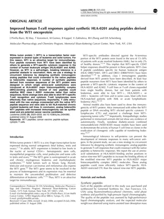
Figure 1 T2 stabilization assay using HLA-A0201 peptides derived from WT1 protein. The assay was conducted as described in Materials and
methods. Sequences of the peptides are shown in Table 1. Fluorescence index is the ratio between the median fluorescence with the peptide tested
divided by median fluorescence with no peptide. The X axis shows different concentrations per well of the peptide tested.
Improved human T-cell responses against synthetic HLA-0201 analog peptides
J Pinilla-Ibarz et al
2027
Leukemia
4. UGN enzyme and to provide a ‘Hot start’ activation of the
AmpliTaq polymerase. Subsequently, 50 cycles of denaturation
at 951C for 15 s followed by annealing/extention at 621C for 60 s
were performed. Each reaction was performed in triplicate, and
discrepancies 41 Ct in one of the wells were excluded. The
Q-RT-PCR reaction and fluorescence measurements were made
on the Applied Biosystems 7500 Real Time PCR System. Control
ABL primers and probes were as follows: forward 50
-TGGAGA
TAACACTCTAAGCATAACTAAAGGT-30
; reverse 50
-GATGTAG
TTGCTTGGGACCCA-30
; fluorescent probe 50
-/56 FAM/CCAT
TTTTGGTTTGGGCTTCACACCATT /3BHQ_1/-30
.
Results
Identification and generation of peptides with a high
probability to bind to HLA-A0201
Peptides potentially reactive with CTL can be predicted by
means of a peptide library-based scoring system for MHC class
I-binding peptides. Amino-acid sequences of the full human
WT-1 protein were scanned for peptides with a potential
binding capacity for HLA-A0201 (about 40% of the Caucasian
population). By using the software of the Bioinformatics and
Molecular Analysis Section (National Institutes of Health,
Washington, DC, USA) available at http://bimas.dcrt.nih.gov/
cgi-bin/molbio/ ken_parker_comboform, which ranks nine-mer
peptides on a predicted half-time dissociation coefficient from
common HLA class I molecules, we initially selected four
peptides (A, B, C and D) from the WT1 protein with
comparatively high binding scores for HLA-A0201. A second
set of five peptides (E, F, G, H and J) was selected with lower
binding scores with the idea of choosing peptides with less
probability of having their T-cell receptors (TCRs) deleted
peripherally by thymic selection. In six out of nine peptides
derived from HLA-A0201, which had already preferred residues
at position two and nine (primary anchor motifs), we looked in
silico for further improvement in the ability of these peptides to
bind to HLA-A0201 molecules. Previous studies have shown
that modification of the secondary anchor motif at position one
by an aromatic amino acid, tyrosine (Y), substitution increases
the binding and affinity of the peptides.31,32
After this alteration,
the computer algorithm predicted half-lives up to 70 times that
of the native peptide sequence for the six peptides (Table 1). The
other three peptides derived from the prediction of binding to
HLA-0201 molecules were improved by a single acid substitu-
tion introduced at HLA-A0201 primary preferred residues, two
of them by a leucine substitution at position two and one by
valine substitutions at position nine. Once again, these changes
improved the predicted half-life of the peptides up to thousand
times that of the native sequence (Table 1).28
Binding of HLA-A0201 by selected peptides derived
from WT1 protein
The immunogenicity of MHC class I-restricted peptides requires
the capacity to bind and stabilize MHC class I molecules on the
live cell surface. Moreover, the computer prediction models
above have only 60–80% predictive accuracy; so we next
sought direct measurement of the strength of the interaction
between the peptides and the HLA-A0201 molecules using a
conventional binding and stabilization assay that uses the
antigen-transporting-deficient (TAP2 negative) HLA-A0201 hu-
man T2 cells. T2 cells lack TAP function and consequently are
defective in properly loading class I molecules with antigenic
peptides generated in the cytosol. The association of exogen-
ously added peptides with thermolabile, empty HLA-A0201
molecules stabilizes them and results in an increase in the level
of surface HLA-A0201 recognizable by specific anti-HLA-A0201
mAb such as BB7.2.
The first set of peptides was analyzed (Figure 1). These four
peptides had well-recognized binding motifs and a tyrosine
0
1
2
3
4
5
FluorescenceIndexFluorescenceIndexFluorescenceIndex
WT1-F1
WT1-F
WT1-E1
WT1-E
0.5
1
1.5
2
2.5
3
3.5
WT1-J1
WT1-J
0
1
2
3
4
5
6
WT1-H1
WT1-H
WT1-G1
WT1-G
100µgr 50 µgr 10µgr 1µgr
100 µgr 50 µgr 10 µgr 1µgr
100 µgr 50 µgr 10 µgr 1µgr
Figure 2 T2 stabilization assay using HLA-A0201 peptides derived
from WT1 protein. The assay was conducted as described in Materials
and methods. Sequences of the peptides are shown in Table 1.
Fluorescence index is the ratio between the median fluorescence with
the peptide tested divided by median fluorescence with no peptide.
The X axis represents different concentrations per well of the peptide
tested.
Improved human T-cell responses against synthetic HLA-0201 analog peptides
J Pinilla-Ibarz et al
2028
Leukemia
5. substitution in position one was introduced to increase the
stability of the peptides in the HLA groove. Three of the native
peptides (A, B and D) from which the analogs were derived have
been shown to be immunogenic by other groups.20,23
For
peptides B and D, the amino-acid substitution (sequences B1
and D1) improved the measured binding of the peptide to the
HLA-0201 molecules. For peptide A, a well-recognized
immunogenic peptide,21
the analog peptide showed similar
behavior in the binding assay. The peptide C analog showed
worse binding ability after the tyrosine substitution.
A second set of five pairs of peptides was evaluated in which
the native peptides had lower binding predictions. These
peptides were chosen based on the rationale that the reactive
T cells may not have been deleted from the peripheral TCR
repertoire. As expected, it was far easier to improve these
peptides of initial poor affinity. Only one of the native peptides
(J) from which the analog was derived had been shown to
be immunogenic by others.23
The tyrosine substitution improved
the binding of the synthetic peptide E1. Peptide F1, an analog
with demonstrated immunogenicity (see below), was pre-
dicted to have no better binding than the native sequence
by BIMAS analysis and somewhat better binding by SYFPEITHI.
In the in vitro binding assay, the new synthetic peptide (F1)
did not show any improvement. Peptides G and H had
poor predicted binding. After substitutions of a lysine in position
two for peptide G and a valine in position nine for peptide
H, the new synthetic peptides G1 and H1 improved their
predicted binding 500–1000 times. These modifications
corresponded with an improved binding in the in vitro T2
assay. Finally, peptide J, the only peptide known to be
immunogenic in the second group of peptides,23
was predicted
to be improved further by a tyrosine substitution in position one.
However, the binding assay demonstrated the change to be
deleterious and peptide J1 had a lower binding than its native
pair (Figure 2).
Early and more potent induction of CD8 immune
response against new synthetic peptides detected by
IFN-g assay
Although affinity for MHC molecules is necessary for peptide
immunogenicity, there is also a requirement for presence of
reactive precursor T cells with appropriate TCRs. Using an
optimized T-cell expansion system, with monocyte-derived DC,
0
100
200
300
400
500
600
0
50
100
150
200
250
300
0
200
400
600
800
0
50
100
150
200
WT1 H WT1 H1 WT1 J WT1 J1
WT1 A WT1 A1 WT1 B WT1 B1
Figure 3 CD8 þ /CD3 þ IFN-g ELISPOT from different healthy HLA-A0201 donors. The assay was conducted as described in Materials and
methods. T cells were stimulated in vitro with the peptides WT1A, -A1, -B, -B1, -H, -H1, -J and -J1. Black bars: CD8 þ plus APC T2; gray bars:
CD8 þ plus T2 pulsed with analog peptide; light gray bars: CD8 þ plus T2 pulsed with native peptide; white bars: CD8 þ plus T2 pulsed with
negative peptide. Upper left: T cells after two rounds of stimulation with WT1A or -A1. Upper right: T cells after two rounds of stimulation with
WT1B or -B1. Lower left: T cells after two rounds of stimulation with WT1H or -H1. Lower right: T cells after two rounds of stimulation with WT1J
or -J1. The Y axis represents the number of spots per 1 Â 105
CD8 þ /CD3 þ T cells. The X axis shows the different peptides used for stimulations.
Experiments were performed in triplicate and confirmed three to five times.
Improved human T-cell responses against synthetic HLA-0201 analog peptides
J Pinilla-Ibarz et al
2029
Leukemia
6. CD14 þ antigen presenting cells, and purified CD3 þ T cells,
we investigated whether the new synthetic WT1 analogs could
stimulate peptide-specific CTLs. Ten healthy HLA-A0201 donors
were studied.
After two or three in vitro stimulations, cells of individuals
responded, generating T cells that secreted IFN-g when
challenged with different peptide-pulsed T2 cells as targets.
Peptides A1, B1, C1, H1 and J1 revealed the best profiles. When
compared with native peptides, these sequences were able to
generate a better response, which was up to 100 times larger in
some donors. The spot numbers were consistently higher with
peptides that bound with higher affinity to HLA-A0201
molecules, as determined by the T2 assay. More importantly,
T cells generated in the presence of the new synthetic analogs
were able to recognize the native sequences. T cells stimulated
with mutant sequences WT1A1, -B1, -H1 and -J1 were able to
stimulate T cells to recognize their respective native sequences
with a similar level of response (Figure 3). Peptide WT1A, the
native sequence from WT1A1, is a well-recognized HLA-
A0201-binding peptide described in the literature to have
immunogenic properties in autologous and allogenic systems
and there is indirect evidence that it can be naturally expressed
on the surface of leukemic blasts.24
After only two rounds of
stimulations, WT1A1 was able to generate a robust immune
response, whereas the WT1A native sequence was still near the
limit of detection by the sensitive IFN-g ELISPOT technique. The
WT1-C1 sequence also generated a strong immune response but
did not crossreact with the native sequence. In some donors,
WT1A, -C and -D native sequences were able to generate an
immune response, but not comparable with the mutant
sequence. No immune response could be generated against
the D1 and E1 peptides, despite attempts using different donors.
Analog peptides derived from WT1 sequence kill pulsed
targets cell as well as leukemic cell lines
Positive results in an IFN-g ELISPOT assay are not always
associated with functional killing. Therefore, we tested the
T-cell lines, and in some cases T-cell clones, obtained after
multiple stimulations with the analog peptide in a chromium-51
release assay using peptide-pulsed target cell lines. T cells
generated in vitro in the presence of WT1A1, -B1, -F1, -G1 and
-J1 were able to kill T2 cells pulsed with specific peptides, but
not T2 cells without peptide or T2 cells pulsed with a control
peptide. Therefore, these peptides were able to generate a
cytolytic response by cytotoxic T cells. Importantly, T cells
stimulated with these mutant peptides were able to recognize
the native sequences, confirming the necessary heteroclitic
response (Figure 4). Peptide F1 generated positive cytotoxicity
with pulsed target cells with some donors, but not others.
Experiments were also performed using HLA-matched CML cell
lines expressing WT1 protein as targets (Figure 5). We were only
0
20
40
60
80
0
20
40
60
80
%cytotoxicity%cytotoxicity
T2-neg peptide
T2-WT1A1
T2-WTA
T2-no peptide
T2-neg peptide
T2-WT1B1
T2-WT1B
T2-no peptide
20
30
40
50
60
70
80
90
T2-neg peptide
T2-WT1G1
T2-WT1G
T2-no peptide
0
20
40
60
80
100
T2-neg peptide
T2-WT1F1
T2-WT1F
T2-no peptide
100 30 10
100 30 10 100 30 10
100 30 10
Figure 4 Cytotoxicity assay with T cells from a healthy HLA-A0201 donor after multiple stimulations in vitro. The assay was conducted as
described in Materials and methods. T cells were stimulated in vitro following the protocol described in Material and methods with the peptides
WT1A1, -B1, -F1 and -G1. Target cells used were the T2 cell line pulsed with the respective peptides. The Y axis shows percentage of cytotoxicity
and the X axis shows different ratios between T cells to target cells. Experiments were confirmed two to five times.
Improved human T-cell responses against synthetic HLA-0201 analog peptides
J Pinilla-Ibarz et al
2030
Leukemia
7. able to generate T-cell-mediated specific cytotoxicity when
using T cells stimulated with peptides WT1A1 (Figure 5). This
raises the possibility that the different native peptides were
differently processed from the full-length WT1 protein or not
sufficiently expressed on the surface of the leukemic cells, as has
been postulated by others. Considerable differences in WT1
expression were confirmed in the cell lines by RT-PCR (Figure 6).
The cytotoxicity against HLA-matched CML blasts from patient
samples was low (range ¼ 10–30%) with some donors and not
significantly different from controls.
Discussion
The WT1 gene was initially defined as a tumor suppressor gene,
but much evidence suggests that in leukemias this gene behaves
more as an oncogene rather than a tumor suppressor gene.33,34
The overexpression of WT1 in leukemias and various solid
tumors, the growth inhibition by WT1 antisense oligomers and
the promotion of growth in hematopoietic progenitors cells
transfected with the wild-type WT1 gene are strong arguments
for its oncogenic function.35,36
The selective expression of WT1
in adult tissues also makes WT1 an excellent target for
immunotherapy.
Immunological tolerance to self-protein can prevent the
development of immune responses to many cancer self-
antigens. Recent reports have shown low, but detectable
frequencies of WT1 IFN-g-secreting T cells in peripheral blood
of 50% of patients with AML.37
Other groups using a more
sensitive method were able to detect IFN-g mRNA in T cells
stimulated with WT1 peptides in more than 50% of healthy
individuals and in 60% of patients with CML.38,39
WT1-specific
HLA-A0201 tetramers were found in a high percentage of
patients with CML and from a small group of healthy subjects.40
However, only low-avidity CD8 þ T-cell responses were seen
likely secondary to tolerance mechanisms. Furthermore, there is
no evidence that the specific T cells detected were able to lyse
leukemic blasts or normal cell in vivo. Therefore, the expression
of WT1 in normal and tumor cells would be expected to cause
partial or complete tolerance of high-avidity CTLs.
Although WT1-specific T cells can be generated in healthy
donors,19–24
it is likely that peptide immunization of patients
with large numbers of leukemia cells (100–1000 billion) each
expressing high levels of WT1 protein would be difficult.
However, a successful phase I trial with an HLA-A2402 WT1
peptide vaccination in leukemias, myelodysplastic syndromes,
lung and breast cancer has been recently published.41
The
vaccination of 26 patients with different doses of a natural or
modified A2401-WT1 peptide with Montanide as adjuvant
yielded tolerable toxicity. No abnormal renal function was
observed. Clinical improvements were seen in 12 of 20 patients,
which included decreased metastasis size or lowered tumor
markers and levels of WT1 by RT-PCR. Immune responses
assessed by tetramer staining, but not with functional cytoplas-
mic IFN-g, correlated with clinical changes. A clinical response
in a patient with a recurrent AML after vaccination with an
A0201-WT1 peptide with KLH and GM-CSF as adjuvant also
0
10
20
30
40
50
%cytotoxicity%cytotoxicity
697+WT1A1
697
LAMA81+WT1A1
LAMA81
BV173+WT1A1
BV173
SKLY
0
25
50
75
100
LAMA81
BV173SKLY
100 30 10
Clone C5 Clone G3 Clone G5 Clone F4
a
b
Figure 5 (a) Cytotoxicity assay with T-cell lines from a healthy HLA-
A0201 donor after six stimulations in vitro. T cells were stimulated in
vitro following the protocol described in ‘Material and methods’ with
the peptide WT1A1. Target cells used were SKLY16 cells (WT1À,
HLA-A0201 þ ), BV173 cells (WT1 þ , HLA-A0201 þ ), LAMA81 cells
(WT1 þ , HLA-A0201 þ ) and 697 cells (WT1 þ , HLA-A0201 þ ),
unused or pulsed with the WT1A1 peptide. Y axis: percentage of
cytotoxicity. X axis: different ratios between effector T cells and
targets. (b) Cytotoxicity assay with T-cell clones generated from a
healthy HLA-A0201 donor after multiple stimulations in vitro. T cells
were generated in vitro following the protocol described in Materials
and methods with the peptide WT1A1. Target cells used were SKLY16,
BV173 and LAMA81 cell lines; cells were used unpulsed. Y axis:
percentage of cytotoxicity. X axis is clone number.
K562 SKLY LAMA 697 BV173
6
5
4
3
2
1
0
relativeexpressionlevel
Figure 6 WT1 expression levels in target leukemia lines as
determined by quantitative RT- PCR assay (see Materials and
methods). K562 was used as the standard and the levels in the other
lines listed on the X axis (SKLY16, LAMA, 697, BV173) are shown.
Improved human T-cell responses against synthetic HLA-0201 analog peptides
J Pinilla-Ibarz et al
2031
Leukemia
8. has been reported.42
Larger trials are necessary to confirm these
observations.
In this paper, we described a way to bypass tolerance creating
several new synthetic analog peptides derived from native
sequences from WT1 protein. Furthermore, we showed that
these peptides were able to generate a stronger immune
response as measured by IFN-g ELISPOT assay. Some of the
new sequences generated cytotoxic responses and crossreacted
against the native sequences. Finally, in some cases, the
synthetic peptides described were able to recognize naturally
presented WT1 peptides on the surface of the leukemia cell
lines. We have generated these potent immunogenic sequences
based on epitopes already described by others (A1, B1 and
J1).20,23
In addition, we described new peptides not previously
recognized (C1, F1 and G1). However, direct cytotoxicity
against CML cell lines was only shown with the A1 (Figure 5)
and F1 peptides. High levels of direct cytotoxicity on fresh CML
blasts were difficult to demonstrate. This reduced activity against
fresh blasts may be due to the lower expression of WT1 by the
target cells, to the heterogeneity of the CD34 þ blast popula-
tions in the preparations or to inefficient processing of WT1.
Other groups using high-avidity CTLs only were able to kill fresh
leukemic blasts at low level, arguing that only a subpopulation
of CD34 þ leukemic cells express adequate amounts of WT1
protein for cytolysis.21
A correlation between antigenicity and MHC-binding affinity
and/or stability of MHC–peptide complexes for class I epitopes
has been demonstrated. Previous reports have demonstrated that
peptides from viral- and tumor-derived proteins that bound with
higher affinity to HLA class I molecules elicited stronger CTL
immune responses than those that bound with lower affinity to
these molecules.43,44
Enhancement of the antigenicity of
epitopes with low to intermediate MHC affinity may therefore
be achieved by improvement of their binding affinity and/or
MHC/peptide complex stability, resulting in the induction of a
strong CTL-specific response for the epitopes.45–48
Sugiyama’s group22
previously described a modified
WT1A2401 peptide (methionine by tyrosine in position two)
with better binding and immunogenic characteristics assessed
by cytotoxicity. This peptide also has been tested in a phase I
clinical trial, although no correlation was found between the use
of the modified peptide and the clinical activity in comparison
with the native peptide.41
Stauss et al. circumvented the problem of tolerance by
developing an allo-restricted CTL approach. T cells from
HLA-A0201À donors are stimulated with HLA-A0201 þ APCs
in the presence of peptides. This technique allowed the
generation of high-avidity allo-restricted CTLs against an HLA-
A0201 WT-1-derived peptide. The CTLs generated were able to
specifically kill WT1 þ , HLA-A0201 þ leukemia cell lines and
blasts. The high-avidity CTL eliminated the progenitors of CFU-
GM and BFU-GM from CD34 þ CML samples, whereas low-
avidity CTL had no effect. In an LTC-IC assay, the authors also
showed the absence of inhibition of normal CD34 þ progeni-
tors/stem cells.21,23
These T cells were able to selectively
deplete clonogenic cells capable of transferring leukemia in
NOD-SCID mice.27
However, the isolation of peptide-specific
allogenic CTL was unsuccessful in many donors, as allogenic
stimulator cells often provoked dominant CTL responses against
allogenic epitopes unrelated to the A2 peptide presented WT1
peptide. Unfortunately, this approach is difficult to apply
clinically and the same authors have recently developed a
method to transfect the specific TCR responsible for this
response in autologous T lymphocytes for possible future
clinical applications.49
In conclusion, new heteroclitic peptides from the WT1
protein could offer alternative sequences for studies leading to
clinical trials for vaccination of patients with myeloid malig-
nancies.
Acknowledgements
We thank Hans Stauss for providing some of the cell lines used in
this paper. This work was supported by NIH PO1 23766, RO1
55349, F32-CA119479A, The Doris Duke Charitable Foundation,
The Lymphoma Foundation, The William and Alice Goodwin
Commonwealth Foundation for Cancer Research. JPI was an
ASCO Young Investigator 03-04 and DAS is a Doris Duke
Distinguished Clinical Scientist.
References
1 Call KM, Glaser T, Ito CY, Buckler AJ, Pelletier J, Haber DA et al.
Isolation and characterization of a zinc finger polypeptide gene at
the human chromosome 11 Wilms’ tumor locus. Cell 1990; 60:
509–520.
2 Pritchard-Jones K, Fleming S, Davidson D, Bickmore W, Porteous
D, Gosden C et al. The candidate Wilms’ tumour gene is involved
in genitourinary development. Nature 1990; 346: 194–197.
3 Huang A, Campbell CE, Bonetta L, McAndrews-Hill MS, Chilton-
MacNeill S, Coppes MJ et al. Tissue, developmental, and tumor-
specific expression of divergent transcripts in Wilms tumor.
Science 1990; 250: 991–994.
4 Maurer U, Brieger J, Weidmann E, Mitrou PS, Hoelzer D,
Bergmann L. The Wilms’ tumor gene is expressed in a subset of
CD34+ progenitors and downregulated early in the course of
differentiation in vitro. Exp Hematol 1997; 25: 945–950.
5 Park S, Schalling M, Bernard A, Maheswaran S, Shipley GC,
Roberts D et al. The Wilms tumour gene WT1 is expressed in
murine mesoderm-derived tissues and mutated in a human
mesothelioma. Nat Genet 1993; 4: 415–420.
6 Pelletier J, Schalling M, Buckler AJ, Rogers A, Haber DA, Housman
D. Expression of the Wilms’ tumor gene WT1 in the murine
urogenital system. Genes Dev 1991; 5: 1345–1356.
7 Inoue K, Ogawa H, Sonoda Y, Kimura T, Sakabe H, Oka Y et al.
Aberrant overexpression of the Wilms tumor gene (WT1) in human
leukemia. Blood 1997; 89: 1405–1412.
8 Brieger J, Weidmann E, Maurer U, Hoelzer D, Mitrou PS,
Bergmann L. The Wilms’ tumor gene is frequently expressed in
acute myeloblastic leukemias and may provide a marker for
residual blast cells detectable by PCR. Ann Oncol 1995; 6: 811–
816.
9 Cilloni D, Gottardi E, Messa F, Fava M, Scaravaglio P, Bertini M
et al. Significant correlation between the degree of WT1 expression
and the International Prognostic Scoring System Score in patients
with myelodysplastic syndromes. J Clin Oncol 2003; 21: 1988–
1995.
10 Oji Y, Ogawa H, Tamaki H, Oka Y, Tsuboi A, Kim EH et al.
Expression of the Wilms’ tumor gene WT1 in solid tumors and
its involvement in tumor cell growth. Jpn J Cancer Res 1999; 90:
194–204.
11 Inoue K, Tamaki H, Ogawa H, Oka Y, Soma T, Tatekawa T et al.
Wilms’ tumor gene (WT1) competes with differentiation-inducing
signal in hematopoietic progenitor cells. Blood 1998; 91: 2969–
2976.
12 Svedberg H, Chylicki K, Baldetorp B, Rauscher III FJ, Gullberg U.
Constitutive expression of the Wilms’ tumor gene (WT1) in the
leukemic cell line U937 blocks parts of the differentiation
program. Oncogene 1998; 16: 925–932.
13 Gaiger A, Carter L, Greinix H, Carter D, McNeill PD, Houghton RL
et al. WT1-specific serum antibodies in patients with leukemia.
Clin Cancer Res 2001; 7 (3 Suppl): 761s–765s.
14 Elisseeva OA, Oka Y, Tsuboi A, Ogata K, Wu F, Kim EH et al.
Humoral immune responses against Wilms tumor gene WT1
product in patients with hematopoietic malignancies. Blood 2002;
99: 3272–3279.
Improved human T-cell responses against synthetic HLA-0201 analog peptides
J Pinilla-Ibarz et al
2032
Leukemia
9. 15 Knights AJ, Zaniou A, Rees RC, Pawelec G, Muller L. Prediction of
an HLA-DR-binding peptide derived from Wilms’ tumour 1 protein
and demonstration of in vitro immunogenicity of WT1(124–138)-
pulsed dendritic cells generated according to an optimised
protocol. Cancer Immunol Immunother 2002; 51: 271–281.
16 Muller L, Knights A, Pawelec G. Synthetic peptides derived from
the Wilms’ tumor 1 protein sensitize human T lymphocytes to
recognize chronic myelogenous leukemia cells. Hematol J 2003;
4: 57–66.
17 Guo Y, Niiya H, Azuma T, Uchida N, Yakushijin Y, Sakai I et al.
Direct recognition and lysis of leukemia cells by WT1-specific
CD4+ T lymphocytes in an HLA class II-restricted manner. Blood
2005; 106: 1415–1418.
18 Kobayashi H, Nagato T, Aoki N, Sato K, Kimura S, Tateno M et al.
Defining MHC class II T helper epitopes for WT1 tumor antigen.
Cancer Immunol Immunother 2006; 55: 850–860.
19 Ohminami H, Yasukawa M, Fujita S. HLA class I-restricted lysis of
leukemia cells by a CD8(+) cytotoxic T-lymphocyte clone specific
for WT1 peptide. Blood 2000; 95: 286–293.
20 Oka Y, Elisseeva OA, Tsuboi A, Ogawa H, Tamaki H, Li H et al.
Human cytotoxic T-lymphocyte responses specific for peptides of
the wild-type Wilms’ tumor gene (WT1 ) product. Immunogenetics
2000; 51: 99–107.
21 Gao L, Bellantuono I, Elsasser A, Marley SB, Gordon MY,
Goldman JM et al. Selective elimination of leukemic CD34(+)
progenitor cells by cytotoxic T lymphocytes specific for WT1.
Blood 2000; 95: 2198–2203.
22 Tsuboi A, Oka Y, Udaka K, Murakami M, Masuda T, Nakano A
et al. Enhanced induction of human WT1-specific cytotoxic T
lymphocytes with a 9-mer WT1 peptide modified at HLA-A*2402-
binding residues. Cancer Immunol Immunother 2002; 51: 614–
620.
23 Bellantuono I, Gao L, Parry S, Marley S, Dazzi F, Apperley J et al.
Two distinct HLA-A0201-presented epitopes of the Wilms tumor
antigen 1 can function as targets for leukemia-reactive CTL. Blood
2002; 100: 3835–3837.
24 Doubrovina ES, Doubrovin MM, Lee S, Shieh JH, Heller G, Pamer
E et al. In vitro stimulation with WT1 peptide-loaded Epstein–Barr
virus-positive B cells elicits high frequencies of WT1 peptide-
specific T cells with in vitro and in vivo tumoricidal activity. Clin
Cancer Res 2004; 10: 7207–7219.
25 Nakajima H, Kawasaki K, Oka Y, Tsuboi A, Kawakami M, Ikegame
K et al. WT1 peptide vaccination combined with BCG-CWS is
more efficient for tumor eradication than WT1 peptide vaccination
alone. Cancer Immunol Immunother 2004; 53: 617–624.
26 Tsuboi A, Oka Y, Ogawa H, Elisseeva OA, Li H, Kawasaki K et al.
Cytotoxic T-lymphocyte responses elicited to Wilms’ tumor gene
WT1 product by DNA vaccination. J Clin Immunol 2000; 20:
195–202.
27 Gao L, Xue SA, Hasserjian R, Cotter F, Kaeda J, Goldman JM et al.
Human cytotoxic T lymphocytes specific for Wilms’ tumor
antigen-1 inhibit engraftment of leukemia-initiating stem cells in
non-obese diabetic-severe combined immunodeficient recipients.
Transplantation 2003; 75: 1429–1436.
28 Gomez-Nunez M, Pinilla-Ibarz J, Dao T, May RJ, Pao M, Jaggi JS
et al. Peptide binding motif predictive algorithms correspond
with experimental binding of leukemia vaccine candidate peptides
to HLA-A*0201 molecules. Leuk Res 2006; March 11 [Epub ahead
of print].
29 Herr W, Linn B, Leister N, Wandel E, Meyer zum Buschenfelde
KH, Wolfel T. The use of computer-assisted video image analysis
for the quantification of CD8+ T lymphocytes producing tumor
necrosis factor alpha spots in response to peptide antigens.
J Immunol Methods 1997; 203: 141–152.
30 Cilloni D, Gottardi E, De Micheli D, Serra A, Volpe G, Messa F
et al. Quantitative assessment of WT1 expression by real time
quantitative PCR may be a useful tool for monitoring minimal
residual disease in acute leukemia patients. Leukemia 2002; 16:
2115–2121.
31 Ruppert J, Sidney J, Celis E, Kubo RT, Grey HM, Sette A. Prominent
role of secondary anchor residues in peptide binding to HLA-A2.1
molecules. Cell 1993; 74: 929–937.
32 Tourdot S, Scardino A, Saloustrou E, Gross DA, Pascolo S,
Cordopatis P et al. A general strategy to enhance immunogenicity
of low-affinity HLA-A2.1-associated peptides: implication in the
identification of cryptic tumor epitopes. Eur J Immunol 2000; 30:
3411–3421.
33 Sugiyama H. Wilms’ tumor gene WT1: its oncogenic function and
clinical application. Int J Hematol 2001; 73: 177–187.
34 Sugiyama H. Wilms tumor gene WT1 as a tumor marker for
leukemic blast cells and its role in leukemogenesis. Methods Mol
Med 2002; 68: 223–237.
35 Tsuboi A, Oka Y, Ogawa H, Elisseeva OA, Tamaki H, Oji Y et al.
Constitutive expression of the Wilms’ tumor gene WT1 inhibits the
differentiation of myeloid progenitor cells but promotes their
proliferation in response to granulocyte-colony stimulating factor
(G-CSF). Leuk Res 1999; 23: 499–505.
36 Yamagami T, Sugiyama H, Inoue K, Ogawa H, Tatekawa T, Hirata
M et al. Growth inhibition of human leukemic cells by WT1
(Wilms tumor gene) antisense oligodeoxynucleotides: implications
for the involvement of WT1 in leukemogenesis. Blood 1996; 87:
2878–2884.
37 Scheibenbogen C, Letsch A, Thiel E, Schmittel A, Mailaender V,
Baerwolf S et al. CD8 T-cell responses to Wilms tumor gene
product WT1 and proteinase 3 in patients with acute myeloid
leukemia. Blood 2002; 100: 2132–2137.
38 Rezvani K, Grube M, Brenchley JM, Sconocchia G, Fujiwara H,
Price DA et al. Functional leukemia-associated antigen-specific
memory CD8+ T cells exist in healthy individuals and in patients
with chronic myelogenous leukemia before and after stem cell
transplantation. Blood 2003; 102: 2892–2900.
39 Rezvani K, Brenchley JM, Price DA, Kilical Y, Gostick E, Sewell AK
et al. T-cell responses directed against multiple HLA-A*0201-
restricted epitopes derived from Wilms’ tumor 1 protein in patients
with leukemia and healthy donors: identification, quantification,
and characterization. Clin Cancer Res 2005; 11 (24 Part 1):
8799–8807.
40 Gannage M, Abel M, Michallet AS, Delluc S, Lambert M,
Giraudier S et al. Ex vivo characterization of multiepitopic
tumor-specific CD8T cells in patients with chronic myeloid
leukemia: implications for vaccine development and adoptive
cellular immunotherapy. J Immunol 2005; 174: 8210–8218.
41 Oka Y, Tsuboi A, Taguchi T, Osaki T, Kyo T, Nakajima H et al.
Induction of WT1 (Wilms’ tumor gene)-specific cytotoxic T
lymphocytes by WT1 peptide vaccine and the resultant cancer
regression. Proc Natl Acad Sci USA 2004; 101: 13885–13890.
42 Mailander V, Scheibenbogen C, Thiel E, Letsch A, Blau IW,
Keilholz U. Complete remission in a patient with recurrent acute
myeloid leukemia induced by vaccination with WT1 peptide in the
absence of hematological or renal toxicity. Leukemia 2004; 18:
165–166.
43 Sette A, Vitiello A, Reherman B, Fowler P, Nayersina R, Kast WM
et al. The relationship between class I binding affinity and
immunogenicity of potential cytotoxic T cell epitopes. J Immunol
1994; 153: 5586–5592.
44 van der Burg SH, Visseren MJ, Brandt RM, Kast WM, Melief CJ.
Immunogenicity of peptides bound to MHC class I molecules
depends on the MHC-peptide complex stability. J Immunol 1996;
156: 3308–3314.
45 Clay TM, Custer MC, McKee MD, Parkhurst M, Robbins PF,
Kerstann K et al. Changes in the fine specificity of gp100(209–
217)-reactive T cells in patients following vaccination with a
peptide modified at an HLA-A2.1 anchor residue. J Immunol 1999;
162: 1749–1755.
46 Parkhurst MR, Salgaller ML, Southwood S, Robbins PF, Sette A,
Rosenberg SA et al. Improved induction of melanoma-reactive CTL
with peptides from the melanoma antigen gp100 modified at HLA-
A*0201-binding residues. J Immunol 1996; 157: 2539–2548.
47 Rosenberg SA, Yang JC, Schwartzentruber DJ, Hwu P,
Marincola FM, Topalian SL et al. Immunologic and therapeutic
evaluation of a synthetic peptide vaccine for the treatment of
patients with metastatic melanoma [see comments]. Nat Med
1998; 4: 321–327.
48 Valmori D, Fonteneau JF, Valitutti S, Gervois N, Dunbar R, Lienard
D et al. Optimal activation of tumor-reactive T cells by selected
antigenic peptide analogues. Int Immunol 1999; 11: 1971–1980.
49 Xue SA, Gao L, Hart D, Gillmore R, Qasim W, Thrasher A et al.
Elimination of human leukemia cells in NOD/SCID mice by
WT1-TCR gene-transduced human T cells. Blood 2005; 106:
3062–3067.
Improved human T-cell responses against synthetic HLA-0201 analog peptides
J Pinilla-Ibarz et al
2033
Leukemia