1. M U S C L E D E V E L O P M E N T
MuSK is a BMP co-receptor that shapes BMP
responses and calcium signaling in muscle cells
Atilgan Yilmaz,1,2
* Chandramohan Kattamuri,3
Rana N. Ozdeslik,4
Carolyn Schmiedel,1
Sarah Mentzer,1
Christoph Schorl,2
Elena Oancea,4
Thomas B. Thompson,3
Justin R. Fallon1†
Bone morphogenetic proteins (BMPs) function in most tissues but have cell type–specific effects. Giv-
en the relatively small number of BMP receptors, this exquisite signaling specificity requires additional
molecules to regulate this pathway’s output. The receptor tyrosine kinase MuSK (muscle-specific ki-
nase) is critical for neuromuscular junction formation and maintenance. Here, we show that MuSK also
promotes BMP signaling in muscle cells. MuSK bound to BMP4 and related BMPs with low nanomolar
affinity in vitro and to the type I BMP receptors ALK3 and ALK6 in a ligand-independent manner both in
vitro and in cultured myotubes. High-affinity binding to BMPs required the third, alternatively spliced
MuSK immunoglobulin-like domain. In myoblasts, endogenous MuSK promoted BMP4-dependent
phosphorylation of SMADs and transcription of Id1, which encodes a transcription factor involved in
muscle differentiation. Gene expression profiling showed that MuSK was required for the BMP4-induced
expression of a subset of genes in myoblasts, including regulator of G protein signaling 4 (Rgs4). In
myotubes, MuSK enhanced the BMP4-induced expression of a distinct set of genes, including transcripts
characteristic of slow muscle. MuSK-mediated stimulation of BMP signaling required type I BMP receptor
activity but was independent of MuSK tyrosine kinase activity. MuSK-dependent expression of Rgs4 re-
sulted in the inhibition of Ca2+
signaling induced by the muscarinic acetylcholine receptor in myoblasts.
These findings establish that MuSK has dual roles in muscle cells, acting both as a tyrosine kinase–
dependent synaptic organizing molecule and as a BMP co-receptor that shapes BMP transcriptional
output and cholinergic signaling.
INTRODUCTION
Bone morphogenetic proteins (BMPs) function in virtually all tissues in
developing and mature organisms, and cell type–specific output of BMP
signaling is essential for proper tissue function and differentiation. How-
ever, the large BMP family is served by only a handful of BMP receptors.
Thus, signaling specificity in distinct cellular and developmental contexts
requires additional molecules to modulate pathway output. Here, we iden-
tify a muscle-specific kinase (MuSK) as a co-receptor that potentiates
BMP signaling in myogenic cells.
BMPs are a large subfamily of conserved signaling molecules within
the transforming growth factor–b (TGFb) superfamily. Two different
classes of receptors, type I (ALK2, ALK3, or ALK6) and type II (BMPRII,
ActRIIB, and ActRII), bind BMPs on the cell surface (1, 2). After ligand
binding, the type I receptor is phosphorylated by the constitutively active
type II receptor (2). The activated type I receptor phosphorylates small
mothers against decapentaplegic 1 (SMAD1), SMAD5, or SMAD8, which
then associates with SMAD4 (3). This complex translocates to the nucleus
where it can function as part of the transcriptional activator or repressor
complexes in a cell type–specific fashion (4, 5). For example, BMP4 in-
duces osteoblast differentiation but inhibits myoblast differentiation (6, 7).
The canonical BMP signaling pathway is modulated at several levels.
The best understood mechanism is the control of ligand availability by
either secreted or cell surface–associated BMP-binding molecules. For ex-
ample, the secreted proteins noggin, chordin, and members of the DAN
(differential screening-selected gene in neuroblastoma) family sequester
BMPs and prevent their association with signaling receptors (8). The
glycophosphatidylinositol-anchored RGM/Dragon family co-receptors
bind BMPs and positively regulate signaling, whereas the transmembrane
protein BAMBI (BMP and activin membrane-bound inhibitor) acts as a
type I pseudoreceptor to inhibit BMP signaling (9). BMP pathway modula-
tors can also act in SMAD-dependent and SMAD-independent manners
(10, 11). Finally, receptor tyrosine kinases (RTKs) such as c-KIT and ROR-
2 can modulate signaling by binding TGFß family ligands and receptors (12).
MuSK is an RTK that is highly abundant at the postsynaptic membrane
of the neuromuscular junction (NMJ) and is well established as the master
regulator of the formation and maintenance of this synapse (13–15). Proper
signaling in this context requires neuronal agrin, low-density lipoprotein
receptor-related protein 4 (LRP4), docking protein 7 (DOK7), and MuSK
tyrosine kinase activity (13). MuSK is also present outside the NMJ, nota-
bly in intact slow-twitch muscle, denervated fast-twitch muscle, and brain
(16–18). However, MuSK function in these nonsynaptic contexts is poorly
understood.
Here, we report that MuSK binds BMPs and influences the BMP4-
mediated gene expression signature in muscle cells. MuSK promoted the
BMP4-induced phosphorylation of SMAD1/5/8 and expression of Id1,
which encodes a transcriptional coactivator. In myoblasts, MuSK was re-
quired for the BMP4-induced expression of a subset of genes, including
1
Department of Neuroscience, Brown University, Providence, RI 02912,
USA. 2
Department of Molecular Biology, Cell Biology, and Biochemistry,
Brown University, Providence, RI 02912, USA. 3
Department of Molecular Ge-
netics, Biochemistry, and Microbiology, University of Cincinnati, Medical
Sciences Building, Cincinnati, OH 45267, USA. 4
Department of Molecular
Pharmacology, Physiology and Biotechnology, Brown University, Provi-
dence, RI 02912, USA.
*Present address: The Azrieli Center for Stem Cells and Genetic Research,
Department of Genetics, Silberman Institute of Life Sciences, The Hebrew
University, Jerusalem 91904, Israel.
†Corresponding author. Email: justin_fallon@brown.edu
R E S E A R C H A R T I C L E
www.SCIENCESIGNALING.org 6 September 2016 Vol 9 Issue 444 ra87 1
onSeptember8,2016http://stke.sciencemag.org/Downloadedfrom
2. the guanosine triphosphatase (GTP)–activating protein (GAP) Rgs4. This
MuSK-dependent, BMP4-responsive GAP regulated muscarinic acetylcho-
line receptor (mAChR)–mediated Ca2+
signaling in these cells. In myotubes,
MuSK stimulated the BMP4-induced expression of a large set of genes,
including the slow muscle–enriched genes myosin heavy chain 15 (Myh15)
and carbonic anhydrase 3 (Car3). MuSK bound to the type I BMP recep-
tors ALK3 and ALK6 and the type I activin receptor ALK4 in a ligand-
independent manner. Type I BMP receptor activity was necessary for the
regulation of MuSK-dependent transcripts, but MuSK tyrosine kinase ac-
tivity was dispensable. We propose that MuSK acts as a BMP co-receptor
to confer cell type–specific signaling in muscle. These findings also es-
tablish that MuSK has dual roles in muscle cells, acting as both a tyrosine
kinase–dependent synaptic organizing molecule and a BMP co-receptor
that shapes transcriptional output and Ca2+
signaling in myogenic cells.
RESULTS
MuSK binds to BMPs
Because both BMP4 and MuSK are important for muscle differentiation
(19), we tested whether MuSK and BMP interact in vitro. We preincu-
bated purified BMP4 with the His-tagged MuSK ectodomain (Fig. 1A)
or a control protein (His-tagged tobacco etch virus protease). After deple-
tion of the tagged MuSK or control protein with nickel beads, we assessed
BMP4 activity in the supernatants using a cell line harboring a luciferase
reporter gene under the control of the BMP-responsive elements of the Id1
promoter [C2C12BRA (20)]. Luciferase activity in the supernatants was
reduced about 50%, indicating that BMP4 had coprecipitated with MuSK
on the nickel beads (Fig. 1B). We observed no significant inhibition of
BMP4 activity with the control protein. Enzyme-linked immunosorbent
assay (ELISA) analysis of the bead-containing pellets confirmed the spe-
cific pull-down of BMP4 by MuSK (Fig. 1C). Thus, BMP4 and MuSK
bind in solution.
To determine the kinetics and binding affinity of the MuSK-BMP4
interaction, we used surface plasmon resonance (SPR). BMP4 was immo-
bilized as the ligand, and the MuSK ectodomain was used as the analyte.
A kinetic analysis using a heterogeneous ligand model revealed high-
affinity binding between these molecules, with a dissociation constant (Kd)
of 6.1 nM, similar in magnitude to the high-affinity interaction of BMP2
with its type I receptors (Fig. 1D and table S1) (21). We also used SPR
to determine whether the MuSK ectodomain binds to the closely related
family members BMP2 and BMP7 (22, 23). These proteins bound MuSK
with comparable affinity to BMP4 (5.6 and 11.8 nM for BMP2 and
BMP7, respectively) (fig. S1 and table S1). Thus, the MuSK ectodomain
binds with high affinity to a closely related set of the BMP family mem-
bers that includes BMP2, BMP4, and BMP7.
As a further test of the interaction between MuSK and BMP4, we treated
C2C12BRA cells either with BMP4 alone or with purified recombinant
MuSK ectodomain. Coincubation with 50 or 100 nM MuSK ectodomain
inhibited BMP4-induced luciferase activity by 50% (fig. S2). In contrast,
no inhibition was observed with the His-tagged control protein (200 nM;
fig. S2). Thus, the MuSK ectodomain inhibits the BMP4-induced activation
of the Id1 reporter in this cell line, possibly by sequestering BMP4.
The Ig3 domain of MuSK is required for high-affinity
BMP4 binding
We next determined which regions of the MuSK extracellular domain
were required for binding to BMP4. One candidate domain was suggested
by MuSK alternative splicing. A major splice isoform of MuSK lacks the
Ig3 domain in the extracellular portion of the molecule. The biological
importance of this DIg3 form of MuSK has been unclear because the nat-
urally occurring splice form with an intact cytoplasmic domain is sufficient
for forming postsynaptic specializations in muscle cells in culture and in
vivo (24). To test the potential role of this domain in MuSK-BMP4 binding,
we generated constructs encoding Fc-fusion ectodomain proteins either
containing or lacking the Ig3 domain (FL and DIg3, respectively; Fig.
1E) and tested their binding to soluble BMP4 over a range of concentra-
tions. BMP4 displayed saturable, high-affinity binding to FL-ecto-MuSK
with a half maximum of ~30 nM (Fig. 1F). In contrast, only low, nonsa-
turable BMP4 binding was observed with DIg3-ecto-MuSK.
We considered the possibility that the failure to observe specific bind-
ing of BMP4 to Fc-DIg3-ecto-MuSK might be due to artifactual misfolding
of the ectodomain. To control for such potential misfolding, we assessed
the binding of both proteins to biglycan, which associates with MuSK in a
manner that is independent of the Ig3 domains (25). Biglycan bound
equivalent amounts of both the FL-ecto-MuSK and DIg3-ecto-MuSK pro-
teins (fig. S3). Together, these data indicate that the alternatively spliced
Ig3 domain is necessary for MuSK binding to BMP4.
MuSK affects the expression of distinct sets of
BMP4-induced genes in myoblasts
BMPs are potent and selective regulators of gene transcription. We there-
fore determined whether MuSK modulates BMP-induced gene tran-
scription in myogenic cells. As a first test of this possibility, we treated
either wild-type or MuSK−/−
myoblasts with BMP4 and analyzed their
gene expression profiles using microarrays. We identified 120 transcripts
that increased in abundance in wild-type but not in MuSK−/−
cells after
BMP4 treatment [≥1.5-fold; false discovery rate (FDR)–corrected P < 0.05].
Thus, these BMP-regulated transcripts are MuSK-dependent (Fig. 2A and
table S2). In addition, 67 transcripts increased in abundance both in wild-
type and MuSK−/−
myoblasts and, therefore, were not qualitatively regulated
by MuSK (Fig. 2A and table S3). Notably, many of the genes induced in
both genotypes encoded proteins active in the canonical BMP pathway
including Id1, Id2, and Id3 and Smad6, Smad7, and Smad9. Finally, 42
transcripts increased abundance only in MuSK−/−
myoblasts (Fig. 2A and
table S4).
Because the arbitrary fold change and statistical significance cutoffs
might lead to false-positive hits in gene array studies, we validated
the responses revealed by the microarrays for a subset of transcripts. Using
quantitative reverse transcription polymerase chain reaction (qRT-PCR),
the relative amount of transcripts of these genes was measured and nor-
malized to 18S ribosomal RNA (rRNA). Fabp7, a previously unidentified
BMP4 target, was prominent among the 67 MuSK-independent tran-
scripts. qRT-PCR analysis showed that Fabp7 transcript abundance
increased more than 15-fold between untreated and treated wild-type myo-
blasts, as well as between untreated and treated MuSK−/−
myoblasts, after
an 8-hour BMP4 treatment (Fig. 2B). Next, we validated a group of
MuSK-dependent genes. BMP4 selectively increased the expression of
Ptgs2 and Ptger4 in wild-type as compared to MuSK−/−
myoblasts (Fig.
2, C and D). Ptger2 encodes cyclooxygenase-2 (COX2), the key enzyme
in the prostaglandin pathway, and Ptger4 encodes a prostaglandin re-
ceptor. BMP4 also induced an eightfold increase in the expression of
Rgs4, a regulator of heterotrimeric guanine nucleotide–binding protein
(G protein) signaling, in wild-type cells, but this transcript did not
change in abundance in MuSK−/−
cells (Fig. 2E). Finally, Rgs4 also
showed a strict MuSK dependence when cells were stimulated with
BMP4 for a shorter period (2 hours; fig. S4). Together, these results
confirm the microarray results and demonstrate that MuSK-dependent
transcripts show robust and selective responses to BMP stimulation in
myoblasts.
R E S E A R C H A R T I C L E
www.SCIENCESIGNALING.org 6 September 2016 Vol 9 Issue 444 ra87 2
onSeptember8,2016http://stke.sciencemag.org/Downloadedfrom
3. MuSK affects the expression of distinct sets of genes in
myoblasts and myotubes
MuSK is highly abundant at the NMJ in differentiated muscle, where its
role as a synapse-organizing molecule is well established. We next de-
termined whether MuSK also modulates BMP-induced gene transcription
in differentiated muscle cells. We stimulated wild-type or MuSK−/−
myo-
tubes with BMP4 and the profiled gene expression in these cells by mi-
croarray. BMP4 induced the up-regulation of 134 and 202 transcripts in
wild-type and MuSK−/−
myotubes, respectively (≥1.5-fold increase; FDR,
<0.05). There were 72 transcripts that increased in abundance only in
His6
Ig1 Ig2 Ig3 CRD
Relativeluciferaseunits
0
0.2
0.4
0.6
0.8
1
1.2
BMP4 (45 pM):
His-tagged ecto-MuSK (nM):
His-tagged TEV protease (nM):
+ + +
– 100 –
– – 100
6
8
14
16
18
0
2
4
10
12
BMP4inpellet(pmoles×10–3
)
***
BMP4 (45 pM):
His-tagged ecto-MuSK (nM):
++
+–
–+
**
A
D
CB
Full-length ectodomain
∆Ig3 ectodomain
Ig1 Ig2 Ig3
Ig1 Ig2
CRD
CRD
Fc
Fc
E
dnuob4PMB
)mn054(ecnabrosbA±MES
0
0.5
1
1.5
BMP4 incubated (nM)
0 50 100 150 200
MuSK ecto-Fc fusion
MuSK ecto-∆Ig3-Fc fusion
Response(RU)
20
40
60
-
-
-
-
-
-
-
Time (s)
0 200 400 600 800
F
ns
MuSK ectodomain
His-tagged TEV protease (nM):
20
Fig. 1. MuSK ectodomain binds to BMP4. (A) Schematic representation of the MuSK
ectodomain fusion protein used in the reporter and the solution-binding assays. The
fusion protein contains the three immunoglobulin (Ig)–like domains (Ig1, Ig2, and Ig3)
and the cysteine-rich domain (CRD) of the extracellular portion of MuSK fused to a
His-tag at the C terminus. (B) BMP4 depletion. Soluble BMP4 was co-incubated
with His-tagged MuSK ectodomain or a control protein [His-tagged tobacco etch
virus (TEV) protease] followed by precipitation with nickel beads. Residual soluble
BMP4 activity was measured using the C2C12BRA reporter cell line. The average
value of untreated cells was set as 100% activity. Data are means ± SD from three
independent experiments with eight replicates in each [n = 3; **P < 0.01; ns, not
significant versus BMP4-only treatment, Bonferroni-adjusted one-way analysis of
variance (ANOVA)]. (C) BMP4 coprecipitates with the MuSK ectodomain. The
amount of BMP4 in eluates from the bead pellets was analyzed by ELISA. Data
are representative of three independent experiments. Values indicate the average
from eight replicates and are means ± SD (***P < 0.0001, unpaired, two-sided
Student’s t test). RU, response units. (D) SPR binding analysis of MuSK with
BMP4. Representative SPR profiles are shown for various concentrations of MuSK
binding to BMP4. Sensorgrams were normalized for MuSK binding to a mock-
coupled flow cell. The black lines show the experimental measurements of a
twofold serial dilution over the concentration range 2 mM to 1.96 nM of each sen-
sorgram, and the red lines correspond to global fits of the data to a 1:1 model
using a heterogeneous surface model with the program EVILFIT. (E) Schematics
of Fc-fusion of full length (FL) and Ig3-lacking (DIg3) MuSK ectodomain proteins
used in (F). (F) The MuSK Ig3 domain is required for BMP4 binding. Immobilized
FL or DIg3 MuSK ectodomain Fc-fusions were incubated with BMP4 (0 to 200 nM).
Data are means ± SD from three independent experiments with four technical re-
plicates in each (n = 3).
R E S E A R C H A R T I C L E
www.SCIENCESIGNALING.org 6 September 2016 Vol 9 Issue 444 ra87 3
onSeptember8,2016http://stke.sciencemag.org/Downloadedfrom
4. wild-type myotubes, whereas 62 transcripts increased in abundance in
both genotypes (Fig. 3A and tables S5 and S6). Further 140 transcripts
increased abundance only in MuSK−/−
myotubes (Fig. 3A and table
S7). Finally, a comparison of transcripts that increased in wild-type myo-
blasts and myotubes in a manner that depended on both BMP4 and MuSK
revealed 113 and 70 transcripts in these cell types, respectively, with only
12 common to both (Fig. 3B and tables S8 to 10). Together, these results
indicate that MuSK regulates transcriptional output of the BMP pathway
in myoblasts and myotubes in a cell type–specific manner.
We then analyzed the MuSK-dependent BMP4 responses in myoblasts
and myotubes according to gene functions and localization patterns in the
cell. These transcripts included a large number of signaling molecules;
thus, we first manually attributed each MuSK-dependent transcriptional
response to a functional or localization-based category related to signaling.
This analysis demonstrated that half of these specific responses in myo-
blasts and more than half of those in myotubes affected signaling-related
molecules such as growth factors, transcription factors, cell surface recep-
tors, extracellular matrix proteins, and intracellular signaling proteins (fig.
S5, A and B). In addition, a Gene Ontology (GO) analysis showed that
several limb morphogenesis and transcription-related terms are enriched
for MuSK-dependent BMP4 responses in myoblasts (fig. S5C). Similarly,
transcription-related terms were among the most significantly enriched
GO terms within the MuSK-dependent BMP4 responses in myotubes,
A B
C
D
Wild type MuSK –/–
Myoblasts
Myotubes
BMP4: – + – +
Wild type MuSK–/–
BMP4: – + – +
Wild type MuSK–/–
***
Myotubes
0
1
2
3
4
5
6
7
62 14072 12 10158
MuSK-dependent transcripts
Myotubes
ns
**
ns
ns
**
**
ns
ns
0
1
2
3
4
5
6
7
Myoblasts
0
1
2
3
4
5
6
EDL Soleus
0
1
2
3
4
5
6
7
8
9
10
EDL Soleus
**
0
1
2
3
4 **
EDL Soleus
**
**
RelativeMyh15
transcriptamount
RelativeCar3
transcriptamount
RelativeMyh15
transcriptamount
RelativeMuSK
transcriptamount
RelativeCar3
transcriptamount
***
Fig. 3. MuSK influences BMP4-induced expression of a subset of genes in
myotubes. (A) Wild-type H-2Kb-tsA58 and MuSK−/−
myotube cultures were
treated with BMP4. RNA was isolated and subjected to transcriptomic anal-
ysis. The number of genes up-regulated in response to BMP4 responses
for up-regulated genes in wild-type and MuSK−/−
myotubes are grouped in
a Venn diagram as wild type only, shared, and MuSK−/−
only. Data repre-
sent the averages of three independent biological replicates. FDR-
corrected P < 0.05 and ±1.5-fold change were used as selection criteria
to identify genes differentially expressed between samples. (B) Venn dia-
gram showing the number of transcripts that increased in abundance in
response to BMP4 in a MuSK-dependent manner in myoblasts and myo-
tubes. (C) Validation of microarray results. Wild-type H-2Kb-tsA58 and
MuSK−/−
myotubes were treated with BMP4, and the abundance of
Myh15 and Car3 transcripts was analyzed in myoblasts and myotubes
by qRT-PCR. Data are means ± SD from five biological replicate
experiments (**P < 0.01; *P < 0.05; ns, not significant versus untreated con-
dition, one-way ANOVA with Bonferroni correction). (D) MuSK, Myh15, and
Car3 expression in soleus muscles compared to extensor digitorum longus
(EDL) muscles. The abundance of MuSK, Myh15, and Car3 transcripts was
analyzed by qRT-PCR. Data are means ± SD from five different animals
(***P < 0.0001; **P < 0.01, unpaired, two-sided Student’s t test).
RelativeFabp7
transcriptamount
RelativePtgs2
transcriptamount
RelativeRgs4
transcriptamount
RelativePtger4
transcriptamount
3
2
1
0
8
7
6
5
4
9
0
0.5
1
2.5
2
1.5
Wild type MuSK–/–
BMP4: – –+ +
Wild type MuSK–/–
BMP4: – –+ +
Wild type MuSK–/–
A
B
**
*
***
120 4267
0
5
10
25
20
15
****
0
1
2
5
4
3
**
ns
6 **
3
**
C
D E
Fig. 2. MuSK influences BMP4-induced expression of a subset of genes in
myoblasts. (A) Wild-type H-2Kb-tsA58 and MuSK−/−
myoblasts were serum-
deprived for 4 hours and then treated with BMP4. Microarray analysis iden-
tified differentially expressed genes upon BMP4 treatment in both genotypes.
The number of genes up-regulated in response to BMP4 in wild-type and
MuSK−/−
myoblasts are grouped into a Venn diagram as wild type only,
shared, and MuSK−/−
only. Data represent the averages of three
independent biological replicates. FDR-corrected P < 0.05 and ±1.5-fold
change were used as selection criteria to identify genes differentially
expressed between samples. (B to E) Validation of microarray results for
four genes. Transcript abundances for MuSK-independent expression of
Fabp7 (B) and MuSK-regulated expression of Ptgs2 (C), Ptger4 (D), and
Rgs4 (E) were measured by qRT-PCR. Data are means ± SD from three
biological replicate experiments (***P < 0.0001; **P < 0.01; *P < 0.05; ns, not
significant versus untreated conditions, Bonferroni-adjusted one-way ANOVA).
R E S E A R C H A R T I C L E
www.SCIENCESIGNALING.org 6 September 2016 Vol 9 Issue 444 ra87 4
onSeptember8,2016http://stke.sciencemag.org/Downloadedfrom
5. which agrees with the high percentage of transcription factors that are up-
regulated in the presence of MuSK (fig. S5, B and D).
MuSK affects the expression of genes expressed in
slow-twitch muscle fibers
Although MuSK is abundant at the NMJ in all muscles, it is also localized
extrasynaptically in slow-twitch, but not fast-twitch, muscle fibers (17).
These observations suggest that MuSK may have unique, nonsynaptic
functions in slow muscle. One possibility is that MuSK could play a role
in regulating the expression of slow muscle–specific genes. Examination
of the microarray data revealed the MuSK-dependent and BMP4-induced
up-regulation of Myh15 and Car3, both of which have been previously sug-
gested to be slow-type fiber markers (26, 27). We validated the MuSK-
dependent regulation of these transcripts by qRT-PCR in cultured myotubes
(Fig. 3C). Finally, we confirmed that MuSK, Myh15, and Car3 were en-
riched in slow muscles as compared to fast muscles isolated from mice
(soleus and extensor digitorum longus, respectively; Fig. 3D). Notably,
neither of these genes was up-regulated by BMP4 in myoblasts (Fig. 3C),
further reinforcing the cell type selectivity of MuSK-dependent transcription
in the myogenic lineage. Together, these results suggest that MuSK selec-
tively regulates the expression of transcripts expressed in slow muscle.
MuSK kinase activity is not required for modulating
BMP4 signaling
NMJ differentiation requires MuSK activation, as well as both an active
MuSK tyrosine kinase domain and the juxtamembrane tyrosine in an
NPXY motif at position 553 (28, 29). We therefore investigated the role
of MuSK tyrosine kinase activity in the regulation of BMP-induced
transcription. As a first test, we determined whether BMP4 stimulated
MuSK kinase activity. We treated wild-type myotubes with BMP4 for in-
tervals ranging from 10 to 180 min and then assessed MuSK activation
(phosphorylation). We detected no increase in MuSK tyrosine phosphoryl-
ation at any of these time points (Fig. 4A). As a positive control, we observed
robust MuSK kinase activation in parallel cultures treated with agrin for
60 min (Fig. 4A). The time intervals and ligand concentrations in these
experiments were the same as those used to demonstrate MuSK-dependent
BMP-mediated transcription (Figs. 2 and 3). Thus, BMP4 does not induce
MuSK phosphorylation under conditions where it stimulates MuSK-dependent
transcription.
We next used a genetic approach to test the potential role of MuSK
activation in BMP4-mediated transcription. We used MuSK−/−
cell lines that
had been stably transfected with constructs encoding wild-type, kinase-dead
(K608A), or Y553F variants of MuSK. Previous work has shown that the
wild-type mutants, but neither the kinase-dead nor Y553F mutants, are
activated in response to agrin in these rescue lines (28, 29). In agreement
with the results presented previously (Fig. 2), BMP4 did not increase Rgs4
expression in MuSK−/−
myoblasts. However, all the MuSK transgenic cell
lines—including those lacking kinase activity— showed robust BMP4-
stimulated Rgs4 expression (Fig. 4B). Together, these results demonstrate
that MuSK is not activated by BMP4 and that its tyrosine kinase activity is
dispensable for MuSK-dependent, BMP4-induced transcription.
MuSK-dependent gene regulation requires the activity of
canonical BMP pathway members
We next explored the relationship between MuSK and the BMP signaling
mediators. The core BMP pathway involves ligand activation of the type I
and type II receptor complex followed by phosphorylation and trans-
location of SMAD1/5/8 to the nucleus, where it regulates transcription.
We first examined the role of MuSK in SMAD phosphorylation. We treated
either wild-type or MuSK−/−
myoblasts with BMP4 and assessed SMAD
phosphorylation by Western blotting (Fig. 5A, left panel). SMAD1/5/8
phosphorylation was reduced in MuSK−/−
cells at all ligand concentrations
tested (Fig. 5A, right panel). These results indicate that MuSK enhances
BMP signaling, at least in part, by promoting SMAD1/5/8 phosphorylation.
The attenuated phosphorylated SMAD1/5/8 response in MuSK−/−
myoblasts suggested that canonical BMP-induced gene transcription
might also be reduced. To test this possibility, we compared the abundance
of Id1 transcripts after BMP stimulation in both wild-type and MuSK−/−
myoblasts. Although Id1 transcripts were increased in both genotypes,
there was a threefold greater response in the wild-type cells as compared
to the MuSK−/−
cells (Fig. 5B). Thus, both BMP-induced changes in
SMAD1/5/8 phosphorylation and Id1 transcript abundance are reduced in
the absence of MuSK. We conclude that MuSK promotes canonical BMP
signaling.
We next determined whether type I BMP receptor activity plays a role
in regulating MuSK-dependent transcripts. We used a selective inhibitor of
type I BMP receptors, LDN-193189 (30). Treatment of myoblasts with
this compound inhibited BMP4-induced up-regulation of Rgs4 expression
(Fig. 5C). As a control, we also examined the expression of the canonical
BMP target Id1 and showed that BMP4-induced up-regulation of this
transcript was also inhibited by LDN-193189 (Fig. 5D). Thus, BMP4-
induced up-regulation of MuSK-dependent transcripts requires type I
BMP receptor activity.
MuSK binds to the type I BMP receptors ALK3 and ALK6
and the type I activin receptor ALK4
The ability of MuSK to quantitatively and qualitatively modulate the
BMP signaling pathway in a tyrosine kinase–independent fashion
A
Agrin (min)
BMP4 (min)
−
10 60 120 180
60
IB: MuSK
Phospho-MuSK
Total MuSK
B
IB: Phosphotyrosine
IP: MuSK
0
0.5
1
2
3
4
1.5
2.5
3.5
0
0.5
1
2
3
4
1.5
2.5
3.5
0
0.5
1
2
3
4
1.5
2.5
3.5
0
0.5
1
2
3
4
1.5
2.5
3.5
RelativeRgs4
transcriptamount
BMP4:
MuSK expression:
− −
−−−−
−
−+ + +
Wild type Kinase-dead Y553F mutant
*
*
*
4.5
− +−−
RelativeRgs4
transcriptamount
RelativeRgs4
transcriptamount
RelativeRgs4
transcriptamount
Fig. 4. MuSK regulation of BMP signaling is independent of MuSK kinase
activity. (A) Wild-type H-2Kb-tsA58 myotubes were treated with BMP4
(25 ng/ml) or with agrin for the indicated times. MuSK was then immuno-
precipitated, and the tyrosine kinase activation was assessed by Western
blotting with a phosphotyrosine-specific antibody (upper panel). The blots
were stripped and reprobed with a MuSK-specific antibody to assess total
MuSK (bottom panel). Blots are representative of three independent ex-
periments. IP, immunoprecipitation; IB, immunoblot. (B) MuSK−/−
myoblasts
or MuSK−/−
myoblasts transgenically expressing wild-type, kinase-dead
(K608A), or Y553F MuSK were treated with BMP4 (3.25 ng/ml for 2.5 hours),
and Rgs4 transcript abundance was measured by qRT-PCR. Data are
means ± SD from three biological replicate experiments (*P < 0.05, un-
paired, two-sided Student’s t test).
R E S E A R C H A R T I C L E
www.SCIENCESIGNALING.org 6 September 2016 Vol 9 Issue 444 ra87 5
onSeptember8,2016http://stke.sciencemag.org/Downloadedfrom
6. raised the possibility that MuSK acts as a
co-receptor in these myogenic cells. To
test this idea, we assessed the binding of
the MuSK ectodomain to the type I BMP
receptors ALK2, ALK3, and ALK6 and the
type II BMP receptors ActRIIB and BMPRII
in a solid-phase binding assay. ALK3 and
ALK6 bound to the ectodomain of MuSK
in a saturable and high-affinity manner
(Fig. 5E). In contrast, no specific binding
was observed with ALK2, the type II re-
ceptors ActRIIB and BMPRII, or the tumor
necrosis factor family receptor TROY. We
also tested MuSK binding to ALK4, a type
I activin receptor that is closely related to
the type I BMP receptors ALK3 and ALK6.
ALK4 bound MuSK in a high-affinity
manner, similar to ALK6 (Fig. 5E). Finally,
to determine whether endogenous MuSK
and BMP receptors associate in muscle
cells, we prepared detergent extracts from
cultured myotubes and immunoprecipitated
ALK3 and ALK6. MuSK coimmunopreci-
pitated with ALK3, but not ALK6, under
these conditions (Fig. 5F). Therefore, we
conclude that in myotubes, MuSK selec-
tively associates with ALK3, one of the
preferred type I receptors for BMP4 (1).
Together, these data demonstrate that MuSK
is a co-receptor for BMP in muscle cells.
Finally, we asked whether MuSK regu-
lates compartmentalization of the BMP
signaling mediators SMAD1, SMAD5,
and SMAD8. Immunostaining for phos-
phorylated SMAD1/5/8 revealed that the
subcellular localization of these key BMP
signaling components differed in wild-type
and MuSK−/−
myoblasts. In wild-type myo-
blasts, phosphorylated SMAD1/5/8 was
distributed in abundant, distinctive puncta
in over 70% of the cells (fig. S6, A and B).
In contrast, such phosphorylated SMAD1/5/8
puncta were sparse in MuSK−/−
cells and
could only be detected in ~20% of the mu-
tant cells. Together, these observations sug-
gest that MuSK might favor cytoplasmic
retention of phosphorylated SMAD1/5/8.
MuSK- and BMP4-dependent
expression of Rgs4 inhibits
carbachol-induced Ca2+
responses in myoblasts
We next sought to determine the physio-
logical role of MuSK-dependent BMP4
gene regulation in myogenic cells. Regula-
tors of G protein signaling (RGS) proteins
are GAPs that directly bind to and stimu-
late the GTPase activity of the Ga subunits
of G proteins, thus reducing G protein ac-
tivity. GAPs regulate many intracellular
RelativeId1
transcriptamount
0
5
10
15
20
25
30
35
40
BMP4: – –+ +
Wild type MuSK–/–
A
B
0 186310.5
pSMAD1/5/8
pSMAD1/5/8
Total SMAD
Total SMAD
BMP4 (ng/ml):
Wild type
MuSK–/–
C
F
RelativeRgs4
transcriptamount
0
0.5
1
1.5
2.5
2
BMP4:
LDN-193189:
– –
– –
+ +
+ +
Wild-type myoblasts
IB: MuSK
IB: ALK3/6
MuSK
IgG HC
IP: ALK3 ALK6 IgG
Wild-type myotubes
0 50 100 150 200
0
1
2
3
4
A
BMPRII
ActRIIB
TROY
[Receptor] (nM)
Absorbance(450)±SD
E
BMP4 (ng/ml)
egnahcdlof8/5/1DAMSp
detaertnurevo
0
2
4
6
8
5 10 15 20
0
Wild type
MuSK–/–
n.s.
D
0
1
2
3
4
5
6
7
8
9
10
RelativeId1
transcriptamount
BMP4:
LDN-193189:
– –
– –
+ +
+ +
Wild-type myoblasts
** ********
ALK4 ALK3
ALK6ALK6
ALK2
ALK3
Fig. 5. MuSK is a BMP co-receptor and regulates the canonical BMP4 pathway. (A) Wild-type H-2Kb-tsA58
and MuSK−/−
myoblasts were treated with BMP4 at the indicated concentrations, and total and phosphoryl-
ated SMAD1/5/8 (pSMAD1/5/8) were then assessed by Western blotting (left panel). The abundance of
pSMAD1/5/8 in Western blots was normalized to total SMAD1/5/8 for each condition. Fold change in
pSMAD1/5/8 was calculated and plotted as the ratio of BMP4-treated conditions to the untreated controls
(right panel). Data are means ± SEM from three biological replicate experiments and their independent
Western blots. (B) Wild-type H-2Kb-tsA58 and MuSK−/−
myoblasts were treated with BMP4 (3.25 ng/ml) for
2 hours. Id1 transcript abundance was quantified by qRT-PCR. Data are means ± SD from three biological
replicates (**P < 0.01, one-way ANOVA with Bonferroni correction). (C) Serum-deprived wild-type H-2Kb-
tsA58 myoblasts were treated with BMP4 in the presence or absence of the BMP type I receptor inhibitor
LDN-193189, and Rgs4 transcript abundance was quantified by qRT-PCR. Data are means ± SD from three
biological replicate experiments (**P < 0.01, one-way ANOVA with Bonferroni correction). (D) Serum-deprived
wild-type H-2Kb-tsA58 myoblasts were treated with BMP4 in the presence or absence of LDN-193189, and Id1
transcript abundance was measured by qRT-PCR. Data are means ± SD from three biological replicate
experiments (**P < 0.01, one-way ANOVA with Bonferroni correction). (E) Immobilized His-tagged MuSK ec-
todomain was incubated with the indicated concentrations of recombinant purified Fc-fusion versions of ALK2,
ALK3, ALK4, ALK6, BMPRII, ActRIIB, and TROY. Bound receptors were detected with horseradish peroxidase
(HRP)–conjugated antibodies recognizing human and mouse IgG. Data are representative of three
independent experiments. Values indicate the average from four replicates and are means ± SD. (F) Pooled
detergent extracts of cultured H-2Kb-tsA58 myotubes were divided into equal volumes and incubated with
antibodies recognizing ALK3 or ALK6 or with normal IgG and immunoprecipitated. Immunoprecipitates were
immunoblotted to show MuSK or ALK3 and ALK6 as indicated. A nonspecific band (n.s.) in the MuSK immu-
noblots is indicated. Data are representative of three biological replicate experiments. HC, heavy chain.
R E S E A R C H A R T I C L E
www.SCIENCESIGNALING.org 6 September 2016 Vol 9 Issue 444 ra87 6
onSeptember8,2016http://stke.sciencemag.org/Downloadedfrom
7. signaling events, including Ca2+
oscillations in the cytosol (31). Therefore,
we hypothesized that MuSK-dependent, BMP4-induced expression of
Rgs4 may modulate Ca2+
signaling (32). To test this idea, we first treated
wild-type myoblasts with the cholinergic agonist carbachol, which in-
creases intracellular Ca2+
concentration by activating Gaq-coupled
muscarinic receptors (33). As expected, carbachol treatment increased in-
tracellular Ca2+
in resting myoblasts (Fig. 6A, upper panel images; blue
trace in the graph on the right). In contrast, the carbachol-mediated Ca2+
signal was attenuated in BMP4-treated myoblasts (Fig. 6A, lower panel
images; green trace in the graph on the right). To test whether this reduc-
tion was due to the action of RGS4, we treated the myoblasts with the
selective RGS4 inhibitor 11b (34) and analyzed their Ca2+
responses with
or without BMP4 pretreatment. BMP4 pretreatment did not alter the Ca2+
response evoked by carbachol in myoblasts treated with the RGS4 inhib-
itor, suggesting that RGS4 mediates the reduction in carbachol-induced
Ca2+
response by BMP4 (Fig. 6, B and C). Together, these results indicate
that MuSK-dependent transcription of the BMP4-responsive gene Rgs4
mediates the inhibition of carbachol-induced Ca2+
response in myoblasts.
I II IVIII
0
0.1
0.2
0.3
0.4
0.5
Fnorm,max
BMP4: – +
Control
LowHigh
[Ca2+
]
I II IVIII
– +
11b: + +
+BMP4
A
––
Control+11b+BMP4+11b
Fnorm
II
III
IV
I
Carbachol IM
1
0.8
0.6
0.4
0.2
0
0 50 100 150
Time (s)
Control + 11b
+ BMP4 + 11b
**
ns
**
C
B
1
0.8
0.6
0.4
0.2
0
50 100 1500
Time (s)
Control
+ BMP4
Carbachol
I
II
II
III
III
IM
Fnorm
IV
Fig. 6. BMP4 treatment inhibits carbachol-induced Ca2+
responses in wild-type H-2Kb-tsA58
myoblasts in an RGS4-dependent manner. (A) Pseudochrome Fluo-4 fluorescence images
of H-2Kb-tsA58 myoblasts stimulated with carbachol in the absence (top) or presence (bottom)
of BMP4. The images were acquired during baseline (I), peak carbachol response (II), before
ionomycin (IM) addition (III), and at peak IM response (IV). Scale bar, 100 mm. The represent-
ative average of normalized Ca2+
imaging responses (Fnorm) after a 4-hour BMP4 treatment
(right). (B) Pseudochrome Fluo-4 fluorescence images of wild-type myoblasts treated with
the RGS4 inhibitor 11b and stimulated with carbachol in the absence (top) or presence
(bottom) of BMP4 treatment. The images were acquired during baseline (I), peak carbachol
response (II), before IM addition (III), and at peak IM response (IV) (left). Scale bar, 100 mm.
The representative average of normalized Ca2+
imaging responses (Fnorm) after BMP4 treat-
ment in the presence of the RGS4 inhibitor 11b (right). (C) The normalized peak fluorescence of
carbachol-induced Ca2+
responses (Fnorm,max) was significantly decreased by BMP4 treatment
only in the absence of the RGS4 inhibitor 11b. Data are means ± SD from seven independent
experiments for each condition (**P < 0.01, one-way ANOVA with Bonferroni correction).
R E S E A R C H A R T I C L E
www.SCIENCESIGNALING.org 6 September 2016 Vol 9 Issue 444 ra87 7
onSeptember8,2016http://stke.sciencemag.org/Downloadedfrom
8. DISCUSSION
Here, we show that the RTK MuSK, previously established as the central
organizer of the NMJ, binds BMPs and BMP receptors and shapes the
transcriptional response to these factors in myogenic cells. We propose
that MuSK acts as a tissue-specific BMP co-receptor that is required for
the BMP-responsive transcription of a subset of genes in a cell context–
dependent manner (Fig. 7). Several observations indicate that MuSK is
a BMP co-receptor (Figs. 1, 4, and 5). (i) MuSK binds BMPs with high
affinity and associates with the type I receptors ALK3 and ALK6 in a sat-
urable, high-affinity, and BMP-independent fashion. (ii) Endogenous
MuSK and ALK3 are associated in muscle cells. (iii) BMP-induced
SMAD phosphorylation and Id1 transcription are reduced in the absence
of MuSK. (iv) BMP does not stimulate MuSK tyrosine kinase activity,
and the stimulation of BMP-induced, MuSK-dependent transcripts is sen-
sitive to ALK inhibitors but independent of MuSK tyrosine kinase activity.
MuSK binds to BMP2, BMP4, and BMP7 with affinities ranging from
5.6 to 11.8 nM, which are comparable to the affinities with which BMPs
bind to other physiologically relevant BMP-interacting molecules (11, 35).
Moreover, the presence of MuSK increases SMAD1/5/8 activation in re-
sponse to a wide range of BMP4 concentrations, and the expression of
transcripts encoding canonical BMP pathway components is greater in
the presence of MuSK. These results indicate that MuSK is a positive reg-
ulator of canonical BMP signaling.
The role of MuSK in shaping the BMP-mediated transcriptional
profile is marked. We observed 101 and 58 MuSK-dependent transcripts
unique to myoblasts and myotubes, respectively (Figs. 2 and 3 and tables
S3 and S6). The selectivity and robustness of this response are under-
scored by the comparison of MuSK-dependent and MuSK-independent
transcripts (Fig. 2). For example, BMP increased the abundance of Fabp7
>15-fold in either the presence or absence of MuSK. In contrast, BMP
induced the expression of Rgs4 and Ptgs2 eight- and fourfold, respectively,
in a manner that was strictly dependent on MuSK.
The BMP-MuSK pathway is likely to shape the transcriptional output
through multiple mechanisms. Increased expression of Rgs4 was observed
within 2 hours of BMP4 treatment, consistent with this gene being a direct
target of MuSK-dependent BMP signaling. However, many transcription
factors were also regulated in a MuSK- and BMP-dependent manner (fig.
S5), suggesting that other MuSK-dependent, BMP4-induced transcripts
are the product of a BMP-induced transcription factor cascade.
There are at least three, nonmutually exclusive mechanisms by which a
MuSK co-receptor could regulate BMP signaling. First, MuSK could aug-
ment signaling by presenting a ligand to the BMP receptor complex. Sec-
ond, MuSK may create subcellular signaling compartments that generate
unique outputs, a mechanism supported by the distinct repertoire of MuSK-
dependent transcripts in myoblasts and myotubes (Figs. 2 and 3) and the
presence of MuSK-dependent intracellular granules containing phosphoryl-
ated SMAD1/5/8 (fig. S6). Finally, as an ALK binding protein, MuSK
could shape the composition and thus the output of BMP signaling com-
plexes in myogenic cells.
MuSK-dependent BMP signaling modulates the muscle cell response
to mAChR-mediated Ca2+
signaling through the regulation of RGS4 pro-
duction. RGS proteins stimulate Ga GTPase activity, thus diminishing G
protein signaling (31). Activation of Gaq-coupled mAChRs by carbachol
results in a transient increase in Ca2+
that was inhibited by BMP4 treat-
ment (Fig. 6). Direct pharmacological blockade of RGS4 reversed this in-
hibition, suggesting a direct link between the MuSK-BMP pathway and
the cellular response to mAChR activation. However, it should be noted
that the complete mechanism by which MuSK-dependent BMP signaling
induces Rgs4 expression remains to be elucidated.
MuSK may regulate multiple subclasses of the BMP superfamily. For
example, MuSK binds ALK4 (Fig. 4), a type I activin receptor and one of
the preferred type I receptors for myostatin, a potent negative regulator of
skeletal muscle growth (36). Other RTKs may play a similar role in BMP
signaling. For example, the RTK ROR2 has been reported to bind ALK6
in heterologous cells (10, 37, 38).
The role of MuSK as a BMP co-receptor is mechanistically distinct
from its function in synapse formation. Agrin, LRP4, and the tyrosine ki-
nase activity of MuSK are absolutely required for NMJ formation and sta-
bility. Moreover, this pathway is restricted to differentiated muscle cells,
and agrin-LRP4-MuSK–dependent AChR clustering does not require de
novo transcription (39). In contrast, the role of MuSK as a BMP co-receptor
is characterized by a robust transcriptional response in both myoblasts and
myotubes that requires neither its tyrosine kinase activity nor a juxtamem-
brane tyrosine that is critical for NMJ differentiation (Fig. 4). Moreover,
the MuSK Ig3 domain is dispensable for its ability to mediate AChR
clustering (40) but is required for BMP binding (Fig. 1). Although these
two MuSK pathways are distinct, they could collaborate in fully differentiated
MuSK-dependent signaling MuSK-independent signaling
BMP
BMPRII
BMPRI
BMP
Ig3
Ig2
Ig1
CRD/
Fz
JM
TK
BMPRII
BMPRI
MuSK
ALK3, ALK6
Regulation of Ca2+
signaling
Prostaglandin pathway
Slow-muscle pathway (myotubes)
Not required
Required
Fig. 7. Model for MuSK regulation of BMP4 signaling. MuSK binds to BMPs
and the BMP receptors ALK3 and ALK6 and regulates the transcriptional
output of BMP signaling in muscle cells. MuSK regulation of BMP signaling
is cell type–specific because BMP induced the expression of different sets
of genes in myoblasts and myotubes in a manner that depended on MuSK.
The kinase activity of MuSK, which is required for proper NMJ formation, is
dispensable for MuSK-mediated regulation of BMP signaling. JM, juxta-
membrane; TK, tyrosine kinase; CRD/Fz, CRD/frizzled-like domain.
R E S E A R C H A R T I C L E
www.SCIENCESIGNALING.org 6 September 2016 Vol 9 Issue 444 ra87 8
onSeptember8,2016http://stke.sciencemag.org/Downloadedfrom
9. synapses. For example, the expression of Dok7, which encodes an essen-
tial binding partner of MuSK in postsynaptic differentiation, is regulated
in a MuSK- and BMP-dependent fashion (table S6). Finally, the BMP
pathway plays a critical role in the differentiation of nerve muscle synapses
in Drosophila melanogaster (41). However, in this case, the BMP ortholog
Gbb acts as a muscle-derived retrograde signal to promote presynaptic
differentiation (41, 42).
The MuSK-BMP pathway could play a role in the regulation of myo-
fiber size and composition. BMP signaling promotes muscle hypertrophy
and counters denervation-induced atrophy (43–45). After denervation or
immobilization, MuSK abundance increases along the entire length of
the muscle (46), suggesting that MuSK plays a role in BMP-mediated
signaling in these pathological contexts. MuSK is also present extrasynap-
tically in slow-oxidative muscle fibers (17), and two slow-muscle genes,
Myh15 and Car3, are stimulated by BMP in a MuSK-dependent manner.
Finally, the BMP-MuSK pathway regulates the production of both COX2,
which is important for myoblast proliferation, fusion, and growth (47–50),
and RGS4, which can inhibit cell growth and myofilament organization in
neonatal cardiac myocytes (51).
The data presented here show that MuSK is a BMP co-receptor that
modulates mAChR-mediated Ca2+
signaling in myogenic cells as well as
pathways important for muscle differentiation and size. Understanding
such regulation could be beneficial for treating pathological conditions,
such as Duchenne muscular dystrophy, insulin-resistant type 2 diabetes,
and disuse atrophy (52), in which muscle mass is lost or muscle function
is compromised. Finally, future studies may also reveal whether MuSK
modulates BMP signaling in nonmuscle tissues such as brain (18).
MATERIALS AND METHODS
Antibodies and recombinant proteins
Purified recombinant human BMP4, ALK6-Fc, ALK3-Fc, ALK4-Fc,
BMPRII-Fc, and TROY-Fc chimeras, mouse ALK2-Fc and ActRIIB-Fc
chimeras, and rat agrin were obtained from R&D systems. Antibodies
specific for MuSK, ALK3, ALK6, and BMP4 (both unlabeled and bioti-
nylated) were obtained from R&D Systems. Streptavidin-HRP was ob-
tained from Thermo and used at 1:15,000 dilution. Mouse HRP and human
IgG(Fc) secondary antibodies were obtained from KPL and used at 1:400
dilution. SMAD5 and phosphorylated SMAD1/5/8 antibodies were ob-
tained from Epitomics, and phosphotyrosine (4G10) antibody was ob-
tained from EMD Millipore Corporation and used at 1:1000, 1:2000, and
1:1000 dilutions, respectively. Alexa Fluor 555–conjugated goat antibody
against rabbit IgG was obtained from Invitrogen and used at 1:1000.
Constructs encoding full-length and Ig3-lacking Fc-Fusion MuSK ectodo-
mains were obtained from GenScript and were used at the indicated con-
centrations. A construct encoding the His-tagged MuSK ectodomain was
expressed in human embryonic kidney (HEK) 293 PEAKrapid cells from
the American Type Culture Collection, and secreted recombinant His-
MuSK ectodomain was purified from the cell culture supernatant by im-
mobilized nickel ion affinity chromatography. LDN-193189 was obtained
from Stemgent and was used at 50 nM.
Mammalian cell culture and mice
Mouse C2C12BRA cells (18) were cultured in Dulbecco’s modified Eagle’s
medium (DMEM) supplemented with 10% fetal bovine serum, 2% L-
glutamine, and 1% penicillin-streptomycin and cultured at 37°C in 8%
CO2. Immortalized myoblast cultures of wild-type mouse H-2Kb-tsA58
(53) and MuSK−/−
and MuSK rescue lines [wild-type MuSK (B1), kinase-
dead MuSK (K608A), and MuSK Y553A] were cultured on gelatin-coated
dishes in DMEM supplemented with 20% fetal bovine serum, 2% L-
glutamine, 1% penicillin-streptomycin, 1% chicken embryo extract, and
1 U of interferon-g under permissive temperature at 33°C in 8% CO2.
Myotubes were obtained by switching the confluent myoblast cultures
to a medium with DMEM supplemented with 5% horse serum, 2% L-
glutamine, and antibiotics at 37°C in 8% CO2. For the LDN-193189
treatments, myoblasts were treated with the drug (50 nM) for 30 min
before BMP4 treatment, and the drug was kept in cultures during the
course of the treatment. Soleus and extensor digitorum longus muscles
were harvested from 5.5-week-old adult C57BL/6 mice. All protocols
were conducted under accordance and with the formal approval of Brown
University’s Institutional Animal Care and Use Committee.
Luciferase reporter assays
C2C12BRA cells were plated in 96-well culture dishes at 4 × 10³
to 5 × 10³
cells per well and allowed to adhere overnight. The indicated recombinant
proteins were premixed in DMEM containing 0.1% bovine serum albumin
(BSA) for 20 min at 4°C, and the original culture medium was replaced
with this solution. The cells were incubated for 8 hours and washed twice
with phosphate-buffered saline (PBS) before the extracts were prepared
with 1× cell lysis buffer (50 ml per well; Roche). The lysate (40 ml) was
transferred to an opaque white 96-well microplate and mixed with 100 ml
of luciferase substrate (Roche). The luciferase activity was read in a lumi-
nometer and reported as relative luciferase unit (RLU), which is the value
for each condition after subtraction of mock treatment (no-BMP4) value
and normalization to BMP4-only condition. All assays were performed in
eight biological replicates and repeated at least two times with similar
results.
For testing of supernatants in the solution-binding experiments, the in-
dicated recombinant proteins were premixed in DMEM containing 0.1%
BSA for 2 hours at 4°C, followed by addition of magnetic nickel-bound
beads (Promega) and a further 2-hour incubation at 4°C. The same pro-
tocol detailed above was followed after this step to test BMP4 activity. The
average value of untreated cells was subtracted from each condition, and
everything was normalized to BMP4-only condition (100% activity).
Immunoprecipitation and coimmunoprecipitation
For MuSK immunoprecipitation, treated H-2Kb-tsA58 myotubes were
lysed in extraction buffer [10 mM tris-HCl (pH 7.4), 1% Triton X-100,
0.5% NP-40, 150 mM NaCl, 1 mM EGTA, 1 mM EDTA, 1 mM sodium
orthovanadate, 10 mM sodium fluoride and 1× EDTA-free protease in-
hibitor cocktail (Roche cOmplete)]. For MuSK-ALK coimmunoprecipi-
tation, H-2Kb-tsA58 myotubes were lysed in extraction buffer [10 mM
tris-HCl (pH 7.4), 1% Triton X-100, 150 mM NaCl, 1 mM sodium
orthovanadate, 10 mM sodium fluoride, and 1× EDTA-free protease in-
hibitor cocktail (Roche cOmplete)]. Lysates for immunoprecipitation and
coimmunoprecipitation experiments were precleared with protein G–
bound magnetic beads (Invitrogen); bicinchoninic acid assay (BCA) was
used to quantify total protein amounts in the lysates (Pierce). For immu-
noprecipitation experiments, lysates with equal amounts of total protein
and the MuSK-specific antibody were mixed and tumbled overnight at
4°C. For coimmunoprecipitation, pooled lysates from three to four T75
flasks were divided into equal volumes for the pull-downs with ALK3-
and ALK6-specific antibodies or the control (normal IgG) antibody
overnight at 4°C. After the addition of protein G–bound magnetic beads,
lysates were tumbled for 4 to 6 hours at 4°C. The beads were washed with
extraction buffer, and 2× sample buffer was added for elution of the im-
munoprecipitated proteins from the beads. Proteins were eluted by boiling
the samples at 98°C for 5 min. Western blots were run for the samples as
indicated below.
R E S E A R C H A R T I C L E
www.SCIENCESIGNALING.org 6 September 2016 Vol 9 Issue 444 ra87 9
onSeptember8,2016http://stke.sciencemag.org/Downloadedfrom
10. Western blots
For phosphorylated SMAD1/5/8 Western blotting, lysates from cells that
were serum-deprived in DMEM containing 0.1% BSA for 5 to 6 hours and
treated with BMP4 for 15 min were prepared in extraction buffer containing
10 mM tris-HCl (pH 7.4), 1% Triton X-100, 0.5% NP-40, 150 mM NaCl,
1 mM EGTA, 1 mM EDTA, 1 mM sodium orthovanadate, 10 mM sodium
fluoride, and 1× EDTA-free protease inhibitor cocktail (Roche cOmplete).
Lysates were cleared by centrifugation at 13,000 rpm for 10 min at 4°C. Total
protein was measured by BCA (Pierce). Equal amounts of protein were
separated on 5 to 15% gradient SDS–polyacrylamide gel electrophoresis gels
and immunoblotted with phosphorylated SMAD1/5/8 antibody (Cell Sig-
naling Technology). Antiphosphotyrosine antibody (4G10) was used for im-
munoprecipitations of phosphorylated MuSK (Millipore). Membranes were then
stripped and reprobed with SMAD1/5/8 antibody (Cell Signaling Technology)
or MuSK antibody for phospho-MuSK immunoprecipitation (R&D Systems).
ELISA assays
For MuSK binding to BMP4, recombinant MuSK proteins (FL-ecto-MuSK
and DIg3-ecto-MuSK) were immobilized on 96-well plates at 2 mg/ml
overnight. Plates were blocked with 1% BSA in PBS and incubated with
BMP4 (0 to 200 nM). Bound BMP4 was detected with biotinylated anti-
BMP4 antibody (R&D Systems) followed by streptavidin-conjugated HRP
(Thermo). Graphs were generated with absorbance values, each data point
representing the average of three independent ELISAs with four replicate wells
for all data points in each independent assay. For MuSK binding to biglycan,
His-tagged nonglycanated biglycan (54) was immobilized, and MuSK ectodo-
main Fc-fusion proteins (full-length MuSK and DIg3 MuSK) were incubated
with immobilized biglycan. Bound MuSK was detected with HRP-conjugated
human Fc antibody. For MuSK binding to ALK6, His-tagged MuSK was im-
mobilized and Fc-fusion ALK3, ALK6, and TROY proteins were incubated
with immobilized MuSK. Bound receptors were detected with HRP-
conjugated and Fc-specific anti-human IgG antibody (KPL).
RNA extraction, reverse transcription, and qRT-PCR
Total RNA was isolated from cells with TRIzol (Invitrogen), cleaned up
and deoxyribonuclease-treated in Qiagen RNeasy columns, and reverse-
transcribed into first-strand complementary DNA (cDNA) (Invitrogen).
The qRT-PCR reaction consisted of initial incubation at 50°C for 2 min
and denaturation at 95°C for 5 min. The cycling parameters were as fol-
lows: 95°C for 15 s and 60°C for 30 s. After 40 cycles, the reactions under-
went a final dissociation cycle as follows: 95°C for 15 s, 60°C for 1 min,
95°C for 15 s, and 60°C for 15 s. Expression of each gene was normalized
to 18S rRNA expression. The primer sequences used in qRT-PCR reac-
tions were as follows: 5′-AGGAGTGGGCCTGCGGCTTA-3′ and 5′-
AACGGCCATGCACCACCACC-3′ for mouse 18S rRNA; 5′-GGG-
ATCTCTGGGAAAGACAC-3′ and 5′-TCTCTGGAGGCTGAAAGGTG-
3′ for mouse Id1; 5′-GTATTTCCATCGCTCCTTGG-3′ and 5′-TGA-
GGCCTATAAAGCACATGG-3′ for mouse Rgs4; 5′-TCTTCGGGCAA-
GAAACTCTG-3′ and 5′-TTGCATGTGACTGCTTCTCC-3′ for mouse
Car3; 5′-CAGGCACACTTCTCCTTTCC-3′ and 5′-CCTTCCTCATCATG-
GACCAG-3′ for mouse Myh15; 5′-CATCAGCTGGATTGAAAACG-3′
and 5′-CAGCCTTTGCGGTACTGAAC-3′ for mouse MuSK; 5′-TCC-
TCTCTGTTGCGTGTGTC-3′ and 5′-CGTTAAGCAACAGGACATGC-3′
for mouse Ptger4; 5′-CGCTGATTGGGTTTTCGTAG-3′ and 5′-CCTGA-
GCTGAGGTTTTCCTG-3′ for mouse Ptgs2; 5′-CTTTGGGGATAT-
CGTTGCTG-3′ and 5′-GCTGGCTAACTCTGGGACT-3′ for mouse Fabp7.
Microarrays and bioinformatic analysis
Total RNA from cultures of wild-type and MuSK−/−
myoblasts and myo-
tubes treated with BMP4 (25 ng/ml) for 8 hours was prepared by the TRIzol
extraction method (Invitrogen). The quality of the input RNA (150 to 200 ng)
for the microarrays was checked by a bioanalyzer [RNA integrity number
(RIN) scores of >9]. Total RNA was converted to double-stranded cDNA
and then in vitro–transcribed overnight using the Whole Transcript expres-
sion kit from Invitrogen (catalog #4411981). After cleanup, 10 mg of in vitro–
transcribed cRNA was converted to deoxyuridine triphosphate–labeled
cDNA, and 5.5 mg of the generated single-stranded cDNAwas enzymatically
fragmented followed by TdT-mediated biotin end-labeling using the
Whole Transcript terminal labeling kit (Affymetrix, catalog #900670).
Successful fragmentation (~75 nucleotides) was demonstrated on the bio-
analyzer with RIN scores of 2.6. About 2.5 mg of cDNA was hybridized at
45°C to Affymetrix Mouse 1.0 Gene ST chip (catalog #901168). The arrays
were washed and stained following the Affymetrix standard protocol and
scanned on an Affymetrix 3000 7G scanner.
The Affymetrix Expression console was used to analyze the overall per-
formance and quality of the arrays, and Partek Genomics Suite was used to
detect differentially expressed genes. To identify genes differentially
expressed between samples, we used a combination of two selection criteria:
±1.5-fold change and FDR-adjusted P < 0.05, such that the FDR was con-
trolled at 5%. FDR amounts to a statistical adjustment to allow for multiple
testing, whereas the fold change cutoff serves as an additional filter to iden-
tify biologically meaningful hits. GO analysis was performed using the
DAVID (Database for Annotation, Visualization, and Integrated Discovery)
database. (55)
Surface plasmon resonance
The binding affinities and kinetic parameters between BMPs and MuSK
were determined by SPR spectroscopy using the BIAcore3000 optical bio-
sensor instrument (GE Healthcare Life Sciences). The carboxymethylated
surface of the sensor chip (CM5) was activated with N-hydroxysuccini-
mide and 1-ethyl-3-(3-dimethylaminopropyl)carbodiimide hydrochloride.
The CM5 chip contains four flow cells, and among these four cells, three
were used for the assay. Flow cell 1 was used as a control surface, whereas
flow cells 2, 3, and 4 were used as test surfaces. Recombinant human
BMP2 (2466 RU), BMP4 (2108 RU), and BMP7 (2050 RU) were cova-
lently coupled in flow cells 2, 3, and 4, respectively. Unreacted active ester
groups were blocked with 1 M ethanolamine hydrochloride (pH 8.5). The
control flow cell 1 was treated in an identical manner but without coupling
protein. The binding assays were carried out at 25°C in 20 mM Hepes
buffer (pH 7.5), 500 mM NaCl, 3.4 mM EDTA, and 0.005% surfactant
P-20. Various concentrations of MuSK were applied over the biosensor
chip at a flow rate of 20 ml/min for 360 s to measure the association phase
followed by buffer only for 600 s to measure the dissociation phase. The
sensor chip was regenerated with four short pulses of guanidine hydro-
chloride (2 M) at 100 ml/min. Data were evaluated using the software
BIAevaluation 4.1.1 (BIAcore AB). Because primary amine coupling
can result in a heterogeneous population of BMP ligand with certain mole-
cules being affected by the coupling process, SPR sensorgrams were
globally analyzed using a distribution model for continuous affinity and
rate constant analysis (kon and koff) with the program EVILFIT (56). This
model helps account for BMP molecules that have reduced affinity due to
the coupling procedure.
Calcium imaging
H-2Kb-tsA58 myoblasts plated on glass coverslips and pretreated with
BMP4 (25 ng/ml) or vehicle for 4 hours were incubated in the dark for
20 min in extracellular solution with 2 mM Fluo-4 AM (Molecular Probes/
Life Technologies) and 250 mM sulfinpyrazone (uridine 5’-diphospho-
glucuronosyltransferase inhibitor, Sigma-Aldrich) to prevent the loss of Fluo-
4 from cells. Coverslips were then transferred to the imaging chamber, and
R E S E A R C H A R T I C L E
www.SCIENCESIGNALING.org 6 September 2016 Vol 9 Issue 444 ra87 10
onSeptember8,2016http://stke.sciencemag.org/Downloadedfrom
11. time-lapse fluorescence images were acquired every 2 s using MetaMorph
software (Molecular Devices). The M1 receptor agonist carbachol (50 mM)
was added after acquiring 25 baseline images (50 s), followed by addition
of ionomycin (2 mM) to elicit maximal Fluo-4 fluorescence, used for normal-
ization. The RGS4 inhibitor 11b (CCG-203769, 3 mM) was added together
with Fluo-4 AM and sulfinpyrazone for 20 min.
For each Ca2+
imaging experiment, the fluorescence intensity of more
than seven individual cells from each coverslip was measured as a function
of time and was averaged. Fluorescence intensities were quantified and
normalized as Fnorm(t) = (Fcell(t) − Fmin)/(Fiono − Fmin), where Fcell is the
fluorescence of an intracellular region of interest (>25% of total cell area),
Fiono is the maximal fluorescence with ionomycin, and Fmin is the baseline
fluorescence before stimulation. Carbachol-induced changes in fluorescence
intensity were quantified using MetaMorph (Molecular Devices), MATLAB
(MathWorks), and Excel software (Microsoft).
Statistical analysis
The average values of the replicate experiments are given as means ± SD/
SEM. Statistical differences among the experimental groups were ana-
lyzed by one-way ANOVA for multiple comparisons and by unpaired,
two-sided Student’s t test when comparing two experimental groups. To
prevent the identification of false-positives due to increased number of
comparisons, ANOVA results were corrected by the conservative Bonferroni
post hoc test. Significance was defined as P < 0.05 (***P < 0.0001;
**P < 0.01; *P < 0.05).
SUPPLEMENTARY MATERIALS
www.sciencesignaling.org/cgi/content/full/9/444/ra87/DC1
Fig. S1. SPR binding analysis of MuSK with BMP2 and BMP7.
Fig. S2. Soluble MuSK ectodomain inhibits BMP4 activity.
Fig. S3. Equivalent binding of biglycan to MuSK ectodomain either containing (FL) or
lacking (DIg3) the third Ig domain.
Fig. S4. BMP4 treatment stimulates Rgs4 expression in wild-type but not MuSK−/−
myoblasts.
Fig. S5. MuSK favors the transcription of signaling- and transcription-related genes.
Fig. S6. Abundant phosphorylated SMAD1/5/8 in cytosolic granules in wild-type but not
MuSK−/−
myoblasts.
Table S1. Association and dissociation rate constants and overall Kd values for the inter-
action of MuSK with BMP2, BMP4, and BMP7, as determined by SPR.
Table S2. Transcripts induced by BMP4 only in wild-type myoblasts.
Table S3. Transcripts induced by BMP4 in both wild-type and MuSK−/−
myoblasts.
Table S4. Transcripts induced by BMP4 only in MuSK−/−
myoblasts.
Table S5. Transcripts induced by BMP4 only in wild-type myotubes.
Table S6. Transcripts induced by BMP4 in both wild-type and MuSK−/−
myotubes.
Table S7. Transcripts induced by BMP4 only in MuSK−/−
myotubes.
Table S8. Transcripts induced by BMP4 in a MuSK-dependent manner in both myoblasts
and myotubes.
Table S9. Transcripts induced by BMP4 in a MuSK-dependent manner only in myoblasts.
Table S10. Transcripts induced by BMP4 in a MuSK-dependent manner only in myotubes.
REFERENCES AND NOTES
1. L. M. Wakefield, C. S. Hill, Beyond TGFb: Roles of other TGFb superfamily members
in cancer. Nat. Rev. Cancer 13, 328–341 (2013).
2. B. Bragdon, O. Moseychuk, S. Saldanha, D. King, J. Julian, A. Nohe, Bone morpho-
genetic proteins: A critical review. Cell. Signal. 23, 609–620 (2011).
3. A. Conidi, S. Cazzola, K. Beets, K. Coddens, C. Collart, F. Cornelis, L. Cox, D. Joke,
M. P. Dobreva, R. Dries, C. Esguerra, A. Francis, A. Ibrahimi, R. Kroes, F. Lesage, E. Maas,
I. Moya, P. N. G. Pereira, E. Stappers, A. Stryjewska, V. van den Berghe, L. Vermeire,
G. Verstappen, E. Seuntjens, L. Umans, A. Zwijsen, D. Huylebroeck, Few Smad
proteins and many Smad-interacting proteins yield multiple functions and action
modes in TGFb/BMP signaling in vivo. Cytokine Growth Factor Rev. 22, 287–300
(2011).
4. O. Korchynskyi, P. ten Dijke, Identification and functional characterization of distinct
critically important bone morphogenetic protein-specific response elements in the Id1
promoter. J. Biol. Chem. 277, 4883–4891 (2002).
5. C.-H. Heldin, A. Moustakas, Role of Smads in TGFb signaling. Cell Tissue Res. 347,
21–36 (2012).
6. K. Miyama, G. Yamada, T. S. Yamamoto, C. Takagi, K. Miyado, M. Sakai, N. Ueno,
H. Shibuya, A BMP-inducible gene, dlx5, regulates osteoblast differentiation and
mesoderm induction. Dev. Biol. 208, 123–133 (1999).
7. Y. Ono, F. Calhabeu, J. E. Morgan, T. Katagiri, H. Amthor, P. S. Zammit, BMP
signalling permits population expansion by preventing premature myogenic differentiation
in muscle satellite cells. Cell Death Differ. 18, 222–234 (2011).
8. K. Nolan, T. B. Thompson, The DAN family: Modulators of TGF-b signaling and be-
yond. Protein Sci. 23, 999–1012 (2014).
9. E. Corradini, J. L. Babitt, H. Y. Lin, The RGM/DRAGON family of BMP co-receptors.
Cytokine Growth Factor Rev. 20, 389–398 (2009).
10. M. Sammar, S. Stricker, G. C. Schwabe, C. Sieber, A. Hartung, M. Hanke, I. Oishi,
J. Pohl, Y. Minami, W. Sebald, S. Mundlos, P. Knaus, Modulation of GDF5/BRI-b
signalling through interaction with the tyrosine kinase receptor Ror2. Genes Cells
9 1227–1238 (2004).
11. M. Hagihara, M. Endo, K. Hata, C. Higuchi, K. Takaoka, H. Yoshikawa, T. Yamashita,
Neogenin, a receptor for bone morphogenetic proteins. J. Biol. Chem. 286, 5157–5165
(2011).
12. S. Hassel, M. Yakymovych, U. Hellman, L. Rönnstrand, P. Knaus, S. Souchelnytskyi,
Interaction and functional cooperation between the serine/threonine kinase bone mor-
phogenetic protein type II receptor with the tyrosine kinase stem cell factor receptor.
J. Cell. Physiol. 206, 457–467 (2006).
13. S. R. Hubbard, K. Gnanasambandan, Structure and activation of MuSK, a receptor
tyrosine kinase central to neuromuscular junction formation. Biochim. Biophys. Acta
1834, 2166–2169 (2013).
14. N. Ghazanfari, K. J. Fernandez, Y. Murata, M. Morsch, S. T. Ngo, S. W. Reddel, P. G. Noakes,
W. D. Phillips, Muscle specific kinase: Organiser of synaptic membrane domains. Int. J.
Biochem. Cell Biol. 43, 295–298 (2011).
15. N. Singhal, P. T. Martin, Role of extracellular matrix proteins and their receptors in the
development of the vertebrate neuromuscular junction. Dev. Neurobiol. 71, 982–1005
(2011).
16. D. C. Bowen, J. S. Park, S. Bodine, J. L. Stark, D. M. Valenzuela, T. N. Stitt,
G. D. Yancopoulos, R. M. Lindsay, D. J. Glass, P. S. DiStefano, Localization and
regulation of MuSK at the neuromuscular junction. Dev. Biol. 199, 309–319 (1998).
17. A. R. Punga, M. Maj, S. Lin, S. Meinen, M. A. Rüegg, MuSK levels differ between
adult skeletal muscles and influence postsynaptic plasticity. Eur. J. Neurosci. 33,
890–898 (2011).
18. A. Garcia-Osta, P. Tsokas, G. Pollonini, E. M. Landau, R. Blitzer, C. M. Alberini,
MuSK expressed in the brain mediates cholinergic responses, synaptic plasticity,
and memory formation. J. Neurosci. 26, 7919–7932 (2006).
19. S.-i. Fukada, A. Uezumi, M. Ikemoto, S. Masuda, M. Segawa, N. Tanimura, H. Yamamoto,
Y. Miyagoe-Suzuki, S. Takeda, Molecular signature of quiescent satellite cells in adult
skeletal muscle. Stem Cells 25, 2448–2459 (2007).
20. L. Zilberberg, P. ten Dijke, L. Y. Sakai, D. B. Rifkin, A rapid and sensitive bioassay to
measure bone morphogenetic protein activity. BMC Cell Biol. 8, 41 (2007).
21. K. Heinecke, A. Seher, W. Schmitz, T. D. Mueller, W. Sebald, J. Nickel, Receptor
oligomerization and beyond: A case study in bone morphogenetic proteins. BMC Biol.
7, 59 (2009).
22. A. Suzuki, E. Kaneko, J. Maeda, N. Ueno, Mesoderm induction by BMP-4 and -7
heterodimers. Biochem. Biophys. Res. Commun. 232, 153–156 (1997).
23. A. von Bubnoff, K. W. Cho, Intracellular BMP signaling regulation in vertebrates:
Pathway or network? Dev. Biol. 239, 1–14 (2001).
24. B. A. Hesser, A. Sander, V. Witzemann, Identification and characterization of a novel
splice variant of MuSK. FEBS Lett. 442, 133–137 (1999).
25. A. R. Amenta, H. E. Creely, M. L. T. Mercado, H. Hagiwara, B. A. McKechnie,
B. E. Lechner, S. G. Rossi, Q. Wang, R. T. Owens, E. Marrero, L. Mei, W. Hoch,
M. F. Young, D. J. McQuillan, R. L. Rotundo, J. R. Fallon, Biglycan is an extracellular
MuSK binding protein important for synapse stability. J. Neurosci. 32, 2324–2334 (2012).
26. P. R. Desjardins, J. M. Burkman, J. B. Shrager, L. A. Allmond, H. H. Stedman, Evo-
lutionary implications of three novel members of the human sarcomeric myosin heavy
chain gene family. Mol. Biol. Evol. 19, 375–393 (2002).
27. G. E. Lyons, M. E. Buckingham, S. Tweedie, Y. H. Edwards, Carbonic anhydrase III,
an early mesodermal marker, is expressed in embryonic mouse skeletal muscle and
notochord. Development 111, 233–244 (1991).
28. S. Mazhar, R. Herbst, The formation of complex acetylcholine receptor clusters requires
MuSK kinase activity and structural information from the MuSK extracellular domain.
Mol. Cell. Neurosci. 49, 475–486 (2012).
29. R. Herbst, S. J. Burden, The juxtamembrane region of MuSK has a critical role in
agrin-mediated signaling. EMBO J. 19, 67–77 (2000).
30. J. Vogt, R. Traynor, G. P. Sapkota, The specificities of small molecule inhibitors of the
TGFß and BMP pathways. Cell. Signal. 23, 1831–1842 (2011).
31. S. Hollinger, J. R. Hepler, Cellular regulation of RGS proteins: Modulators and inte-
grators of G protein signaling. Pharmacol. Rev. 54, 527–559 (2002).
32. M. W. Berchtold, H. Brinkmeier, M. Müntener, Calcium ion in skeletal muscle: Its crucial
role for muscle function, plasticity and disease. Physiol. Rev. 80, 1215–1265 (2000).
R E S E A R C H A R T I C L E
www.SCIENCESIGNALING.org 6 September 2016 Vol 9 Issue 444 ra87 11
onSeptember8,2016http://stke.sciencemag.org/Downloadedfrom
12. 33. A. Mayerhofer, K. J. Föhr, K. Sterzik, M. Gratzl, Carbachol increases intracellular free
calcium concentrations in human granulosa-lutein cells. J. Endocrinol. 135, 153–159
(1992).
34. L. L. Blazer, A. J. Storaska, E. M. Jutkiewicz, E. M. Turner, M. Calcagno, S. M. Wade,
Q. Wang, X.-P. Huang, J. R. Traynor, S. M. Husbands, M. Morari, R. R. Neubig,
Selectivity and anti-Parkinson’s potential of thiadiazolidinone RGS4 inhibitors.
ACS Chem. Neurosci. 6, 911–919 (2015).
35. T. A. Samad, A. Rebbapragada, E. Bell, Y. Zhang, Y. Sidis, S.-J. Jeong, J. A. Campagna,
S. Perusini, D. A. Fabrizio, A. L. Schneyer, H. Y. Lin, A. H. Brivanlou, L. Attisano,
C. J. Woolf, DRAGON, a bone morphogenetic protein co-receptor. J. Biol. Chem. 280,
14122–14129 (2005).
36. A. Rebbapragada, H. Benchabane, J. L. Wrana, A. J. Celeste, L. Attisano, Myostatin
signals through a transforming growth factor b-like signaling pathway to block adipo-
genesis. Mol. Cell. Biol. 23, 7230–7242 (2003).
37. R. Raz, S. Stricker, E. Gazzerro, J. L. Clor, F. Witte, H. Nistala, S. Zabski, R. C. Pereira,
L. Stadmeyer, X. Wang, L. Gowen, M. W. Sleeman, G. D. Yancopoulos, E. Canalis,
S. Mundlos, D. M. Valenzuela, A. N. Economides, The mutation ROR2W749X
, linked
to human BDB, is a recessive mutation in the mouse, causing brachydactyly,
mediating patterning of joints and modeling recessive Robinow syndrome. Development
135, 1713–1723 (2008).
38. M. Sammar, C. Sieber, P. Knaus, Biochemical and functional characterization of
the Ror2/BRIb receptor complex. Biochem. Biophys. Res. Commun. 381, 1–6
(2009).
39. B. G. Wallace, Regulation of agrin-induced acetylcholine receptor aggregation by Ca++
and phorbol ester. J. Cell Biol. 107, 267–278 (1988).
40. H. Zhou, D. J. Glass, G. D. Yancopoulos, J. R. Sanes, Distinct domains of MuSK
mediate its abilities to induce and to associate with postsynaptic specializations.
J. Cell Biol. 146, 1133–1146 (1999).
41. H. Wu, W. C. Xiong, L. Mei, To build a synapse: Signaling pathways in neuromuscular
junction assembly. Development 137, 1017–1033 (2010).
42. B. D. McCabe, G. Marqués, A. P. Haghighi, R. D. Fetter, M. L. Crotty, T. E. Haerry,
C. S. Goodman, M. B. O’Connor, The BMP homolog Gbb provides a retrograde
signal that regulates synaptic growth at the Drosophila neuromuscular junction.
Neuron 39, 241–254 (2003).
43. R. Sartori, E. Schirwis, B. Blaauw, S. Bortolanza, J. Zhao, E. Enzo, A. Stantzou, E. Mouisel,
L. Toniolo, A. Ferry, S. Stricker, A. L. Goldberg, S. Dupont, S. Piccolo, H. Amthor, M. Sandri,
BMP signaling controls muscle mass. Nat. Genet. 45, 1309–1318 (2013).
44. C. E. Winbanks, J. L. Chen, H. Qian, Y. Liu, B. C. Bernardo, C. Beyer, K. I. Watt,
R. E. Thompson, T. Connor, B. J. Turner, J. R. McMullen, L. Larsson, S. L. McGee,
C. A. Harrison, P. Gregorevic,, The bone morphogenetic protein axis is a positive
regulator of skeletal muscle mass. J. Cell Biol. 203, 345–357 (2013).
45. R. Sartori, P. Gregorevic, M. Sandri, TGFb and BMP signaling in skeletal muscle:
Potential significance for muscle-related disease. Trends Endocrinol. Metab. 25,
464–471 (2014).
46. D. M. Valenzuela, T. N. Stitt, P. S. DiStefano, E. Rojas, K. Mattsson, D. L. Compton,
L. Nuñez, J. S. Park, J. L. Stark, D. R. Gies, S. Thomas, M. M. Le Beau, A. A. Fernald,
N. G. Copeland, N. A. Jenkins, S. J. Burden, D. J. Glass, G. D. Yancopoulos, Receptor
tyrosine kinase specific for the skeletal muscle lineage: Expression in embryonic muscle,
at the neuromuscular junction, and after injury. Neuron 15, 573–584 (1995).
47. J. S. Otis, T. J. Burkholder, G. K. Pavlath, Stretch-induced myoblast proliferation is
dependent on the COX2 pathway. Exp. Cell Res. 310, 417–425 (2005).
48. B. A. Bondesen, S. T. Mills, G. K. Pavlath, The COX-2 pathway regulates growth of atrophied
muscle via multiple mechanisms. Am. J. Physiol. Cell Physiol. 290, C1651–C1659
(2006).
49. V. Horsley, G. K. Pavlath, Prostaglandin F2a stimulates growth of skeletal muscle cells
via an NFATC2-dependent pathway. J. Cell Biol. 161, 111–118 (2003).
50. A. Yilmaz, R. Engeler, S. Constantinescu, K. D. Kokkaliaris, C. Dimitrakopoulos,
T. Schroeder, N. Beerenwinkel, R. Paro, Ectopic expression of Msx2 in mammalian myo-
tubes recapitulates aspects of amphibian muscle dedifferentiation. Stem Cell Res. 15,
542–553 (2015).
51. P. Tamirisa, K. J. Blumer, A. J. Muslin, RGS4 inhibits G-protein signaling in cardio-
myocytes. Circulation 99, 441–447 (1999).
52. S. Schiaffino, M. Sandri, M. Murgia, in Skeletal Muscle Plasticity in Health and Disease:
From Genes to Whole Muscle, R. Bottinelli, C. Reggiani, Eds. (Springer, Dordrecht, The
Netherlands, 2007), pp. 91–119.
53. J. E. Morgan, J. R. Beauchamp, C. N. Pagel, M. Peckham, P. Ataliotis, P. S. Jat,
M. D. Noble, K. Farmer, T. A. Partridge, Myogenic cell lines derived from trans-
genic mice carrying a thermolabile T antigen: A model system for the derivation
of tissue-specific and mutation-specific cell lines. Dev. Biol. 162, 486–498 (1994).
54. A. R. Amenta, A. Yilmaz, S. Bogdanovich, B. A. McKechnie, M. Abedi, T. S. Khurana,
J. R. Fallon, Biglycan recruits utrophin to the sarcolemma and counters dystrophic
pathology in mdx mice. Proc. Natl. Acad. Sci. U.S.A. 108, 762–767 (2011).
55. G. Dennis Jr., B. T. Sherman, D. A. Hosack, J. Yang, W. Gao, H. C. Lane, R. A. Lempicki,
DAVID: Database for Annotation, Visualization, and Integrated Discovery. Genome Biol. 4,
P3 (2003).
56. J. Svitel, A. Balbo, R. A. Mariuzza, N. R. Gonzales, P. Schuck, Combined affinity and
rate constant distributions of ligand populations from experimental surface binding
kinetics and equilibria. Biophys. J. 84, 4062–4077 (2003).
Acknowledgments: We are grateful to D. B. Rifkin at New York University for providing
the C2C12BRA reporter line, M. Ruegg at the University of Basel for the His-tagged MuSK
construct, R. Herbst at the Medical University of Vienna for providing mutant MuSK rescue
lines, and R. Neubig at Michigan State University for the 11b compound. We thank
M. Dechene for the purification of the His-tagged MuSK construct. We are also grateful to
B. McKechnie for technical assistance and Z. Wu at Brown University for advice on sta-
tistical analyses. Funding: The research was supported by NIH grants HD23924 and
NS064295 (J.R.F.), GM084186 and GM114640 (T.B.T.), and a fellowship from The Suna
and Inan Kirac Foundation to R.N.O. The Genomics Core Facility at Brown University has
received partial support from the NIH (National Institute of General Medical Sciences grant
number P30GM103410 and National Center for Research Resources grant numbers
P30RR031153, P20RR018728, and S10RR02763), NSF (Experimental Program to Stim-
ulate Competitive Research grant number 0554548), Lifespan Rhode Island Hospital, and
the Division of Biology and Medicine, Brown University. Author contributions: A.Y. and
J.R.F. conceived the study. A.Y., T.B.T., E.O., and J.R.F. designed the experiments. A.Y.,
C.K., C. Schmiedel, R.N.O., S.M., and C. Schorl. conducted the experiments. A.Y., C.K.,
C. Schmiedel, R.N.O., C. Schorl, E.O., T.B.T., and J.R.F. analyzed the data. A.Y. and J.R.F.
wrote the paper with input from all the authors. Competing interests: The authors declare
that they have no competing interests. Data and materials availability: Microarray expres-
sion data files can be obtained from the NIH Gene Expression Omnibus with accession
number GSE59567.
Submitted 16 December 2015
Accepted 17 August 2016
Final Publication 6 September 2016
10.1126/scisignal.aaf0890
Citation: A. Yilmaz, C. Kattamuri, R. N. Ozdeslik, C. Schmiedel, S. Mentzer, C. Schorl,
E. Oancea, T. B. Thompson, J. R. Fallon, MuSK is a BMP co-receptor that shapes BMP
responses and calcium signaling in muscle cells. Sci. Signal. 9, ra87 (2016).
R E S E A R C H A R T I C L E
www.SCIENCESIGNALING.org 6 September 2016 Vol 9 Issue 444 ra87 12
onSeptember8,2016http://stke.sciencemag.org/Downloadedfrom
13. (444), ra87. [doi: 10.1126/scisignal.aaf0890]9Science Signaling
2016)
Oancea, Thomas B. Thompson and Justin R. Fallon (September 6,
Carolyn Schmiedel, Sarah Mentzer, Christoph Schorl, Elena
Atilgan Yilmaz, Chandramohan Kattamuri, Rana N. Ozdeslik,
calcium signaling in muscle cells
MuSK is a BMP co-receptor that shapes BMP responses and
This information is current as of September 8, 2016.
The following resources related to this article are available online at http://stke.sciencemag.org.
Article Tools
http://stke.sciencemag.org/content/9/444/ra87
article tools:
Visit the online version of this article to access the personalization and
Materials
Supplemental
http://stke.sciencemag.org/content/suppl/2016/09/01/9.444.ra87.DC1
"Supplementary Materials"
Related Content
http://science.sciencemag.org/content/sci/345/6203/1505.full
http://stke.sciencemag.org/content/sigtrans/6/272/re2.full
http://stke.sciencemag.org/content/sigtrans/5/246/ra75.full
http://stke.sciencemag.org/content/sigtrans/4/201/ra80.full
http://stke.sciencemag.org/content/sigtrans/2/59/ra7.full
's sites:ScienceThe editors suggest related resources on
References
http://stke.sciencemag.org/content/9/444/ra87#BIBL
This article cites 55 articles, 21 of which you can access for free at:
Permissions
http://www.sciencemag.org/about/permissions.dtl
Obtain information about reproducing this article:
reserved.
DC 20005. Copyright 2016 by the American Association for the Advancement of Science; all rights
American Association for the Advancement of Science, 1200 New York Avenue, NW, Washington,
(ISSN 1937-9145) is published weekly, except the last December, by theScience Signaling
onSeptember8,2016http://stke.sciencemag.org/Downloadedfrom
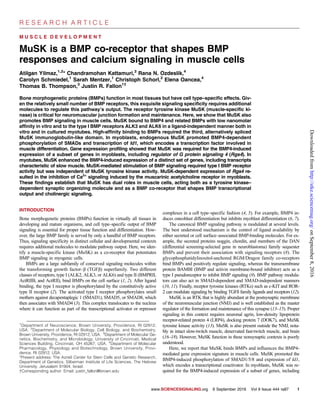
![the guanosine triphosphatase (GTP)–activating protein (GAP) Rgs4. This
MuSK-dependent, BMP4-responsive GAP regulated muscarinic acetylcho-
line receptor (mAChR)–mediated Ca2+
signaling in these cells. In myotubes,
MuSK stimulated the BMP4-induced expression of a large set of genes,
including the slow muscle–enriched genes myosin heavy chain 15 (Myh15)
and carbonic anhydrase 3 (Car3). MuSK bound to the type I BMP recep-
tors ALK3 and ALK6 and the type I activin receptor ALK4 in a ligand-
independent manner. Type I BMP receptor activity was necessary for the
regulation of MuSK-dependent transcripts, but MuSK tyrosine kinase ac-
tivity was dispensable. We propose that MuSK acts as a BMP co-receptor
to confer cell type–specific signaling in muscle. These findings also es-
tablish that MuSK has dual roles in muscle cells, acting as both a tyrosine
kinase–dependent synaptic organizing molecule and a BMP co-receptor
that shapes transcriptional output and Ca2+
signaling in myogenic cells.
RESULTS
MuSK binds to BMPs
Because both BMP4 and MuSK are important for muscle differentiation
(19), we tested whether MuSK and BMP interact in vitro. We preincu-
bated purified BMP4 with the His-tagged MuSK ectodomain (Fig. 1A)
or a control protein (His-tagged tobacco etch virus protease). After deple-
tion of the tagged MuSK or control protein with nickel beads, we assessed
BMP4 activity in the supernatants using a cell line harboring a luciferase
reporter gene under the control of the BMP-responsive elements of the Id1
promoter [C2C12BRA (20)]. Luciferase activity in the supernatants was
reduced about 50%, indicating that BMP4 had coprecipitated with MuSK
on the nickel beads (Fig. 1B). We observed no significant inhibition of
BMP4 activity with the control protein. Enzyme-linked immunosorbent
assay (ELISA) analysis of the bead-containing pellets confirmed the spe-
cific pull-down of BMP4 by MuSK (Fig. 1C). Thus, BMP4 and MuSK
bind in solution.
To determine the kinetics and binding affinity of the MuSK-BMP4
interaction, we used surface plasmon resonance (SPR). BMP4 was immo-
bilized as the ligand, and the MuSK ectodomain was used as the analyte.
A kinetic analysis using a heterogeneous ligand model revealed high-
affinity binding between these molecules, with a dissociation constant (Kd)
of 6.1 nM, similar in magnitude to the high-affinity interaction of BMP2
with its type I receptors (Fig. 1D and table S1) (21). We also used SPR
to determine whether the MuSK ectodomain binds to the closely related
family members BMP2 and BMP7 (22, 23). These proteins bound MuSK
with comparable affinity to BMP4 (5.6 and 11.8 nM for BMP2 and
BMP7, respectively) (fig. S1 and table S1). Thus, the MuSK ectodomain
binds with high affinity to a closely related set of the BMP family mem-
bers that includes BMP2, BMP4, and BMP7.
As a further test of the interaction between MuSK and BMP4, we treated
C2C12BRA cells either with BMP4 alone or with purified recombinant
MuSK ectodomain. Coincubation with 50 or 100 nM MuSK ectodomain
inhibited BMP4-induced luciferase activity by 50% (fig. S2). In contrast,
no inhibition was observed with the His-tagged control protein (200 nM;
fig. S2). Thus, the MuSK ectodomain inhibits the BMP4-induced activation
of the Id1 reporter in this cell line, possibly by sequestering BMP4.
The Ig3 domain of MuSK is required for high-affinity
BMP4 binding
We next determined which regions of the MuSK extracellular domain
were required for binding to BMP4. One candidate domain was suggested
by MuSK alternative splicing. A major splice isoform of MuSK lacks the
Ig3 domain in the extracellular portion of the molecule. The biological
importance of this DIg3 form of MuSK has been unclear because the nat-
urally occurring splice form with an intact cytoplasmic domain is sufficient
for forming postsynaptic specializations in muscle cells in culture and in
vivo (24). To test the potential role of this domain in MuSK-BMP4 binding,
we generated constructs encoding Fc-fusion ectodomain proteins either
containing or lacking the Ig3 domain (FL and DIg3, respectively; Fig.
1E) and tested their binding to soluble BMP4 over a range of concentra-
tions. BMP4 displayed saturable, high-affinity binding to FL-ecto-MuSK
with a half maximum of ~30 nM (Fig. 1F). In contrast, only low, nonsa-
turable BMP4 binding was observed with DIg3-ecto-MuSK.
We considered the possibility that the failure to observe specific bind-
ing of BMP4 to Fc-DIg3-ecto-MuSK might be due to artifactual misfolding
of the ectodomain. To control for such potential misfolding, we assessed
the binding of both proteins to biglycan, which associates with MuSK in a
manner that is independent of the Ig3 domains (25). Biglycan bound
equivalent amounts of both the FL-ecto-MuSK and DIg3-ecto-MuSK pro-
teins (fig. S3). Together, these data indicate that the alternatively spliced
Ig3 domain is necessary for MuSK binding to BMP4.
MuSK affects the expression of distinct sets of
BMP4-induced genes in myoblasts
BMPs are potent and selective regulators of gene transcription. We there-
fore determined whether MuSK modulates BMP-induced gene tran-
scription in myogenic cells. As a first test of this possibility, we treated
either wild-type or MuSK−/−
myoblasts with BMP4 and analyzed their
gene expression profiles using microarrays. We identified 120 transcripts
that increased in abundance in wild-type but not in MuSK−/−
cells after
BMP4 treatment [≥1.5-fold; false discovery rate (FDR)–corrected P < 0.05].
Thus, these BMP-regulated transcripts are MuSK-dependent (Fig. 2A and
table S2). In addition, 67 transcripts increased in abundance both in wild-
type and MuSK−/−
myoblasts and, therefore, were not qualitatively regulated
by MuSK (Fig. 2A and table S3). Notably, many of the genes induced in
both genotypes encoded proteins active in the canonical BMP pathway
including Id1, Id2, and Id3 and Smad6, Smad7, and Smad9. Finally, 42
transcripts increased abundance only in MuSK−/−
myoblasts (Fig. 2A and
table S4).
Because the arbitrary fold change and statistical significance cutoffs
might lead to false-positive hits in gene array studies, we validated
the responses revealed by the microarrays for a subset of transcripts. Using
quantitative reverse transcription polymerase chain reaction (qRT-PCR),
the relative amount of transcripts of these genes was measured and nor-
malized to 18S ribosomal RNA (rRNA). Fabp7, a previously unidentified
BMP4 target, was prominent among the 67 MuSK-independent tran-
scripts. qRT-PCR analysis showed that Fabp7 transcript abundance
increased more than 15-fold between untreated and treated wild-type myo-
blasts, as well as between untreated and treated MuSK−/−
myoblasts, after
an 8-hour BMP4 treatment (Fig. 2B). Next, we validated a group of
MuSK-dependent genes. BMP4 selectively increased the expression of
Ptgs2 and Ptger4 in wild-type as compared to MuSK−/−
myoblasts (Fig.
2, C and D). Ptger2 encodes cyclooxygenase-2 (COX2), the key enzyme
in the prostaglandin pathway, and Ptger4 encodes a prostaglandin re-
ceptor. BMP4 also induced an eightfold increase in the expression of
Rgs4, a regulator of heterotrimeric guanine nucleotide–binding protein
(G protein) signaling, in wild-type cells, but this transcript did not
change in abundance in MuSK−/−
cells (Fig. 2E). Finally, Rgs4 also
showed a strict MuSK dependence when cells were stimulated with
BMP4 for a shorter period (2 hours; fig. S4). Together, these results
confirm the microarray results and demonstrate that MuSK-dependent
transcripts show robust and selective responses to BMP stimulation in
myoblasts.
R E S E A R C H A R T I C L E
www.SCIENCESIGNALING.org 6 September 2016 Vol 9 Issue 444 ra87 2
onSeptember8,2016http://stke.sciencemag.org/Downloadedfrom](data:image/gif;base64,R0lGODlhAQABAIAAAAAAAP///yH5BAEAAAAALAAAAAABAAEAAAIBRAA7)