More Related Content
Similar to Seah_SURF (1) (20)
Seah_SURF (1)
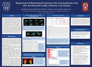
- 1. © 2016 Mayo Foundation for Medical Education and Research
Seah Buttar, Kharmen Bharucha, William A Faubion, Gwen Lomberk, Raul Urrutia
GIH Division, Department of Medicine, Biophysics, Biochemistry and Molecular Biology,
Mayo Clinic, Rochester, MN
Grzenda A, Ordog T, Urrutia R. Polycomb and the Emerging Epigenetics of
Pancreatic Cancer. Journal of gastrointestinal cancer.
2011;42(2):100-111. doi:10.1007/s12029-011-9262-4.
Jacobs S, Khorasanizadeh S. Structure of HP1 Chromodomain Bound to a
Lysine 9-Methylated Histone H3 Tail. Science. 2002:XXX(X):2080-
2083. doi: 10.1126/science.1069473
Kaustov L, Ouyang H, Amaya M, et al. Recognition and Specificity
Determinants of the Human Cbx Chromodomains. The Journal of
Biological Chemistry. 2011;286(1):521-529.
doi:10.1074/jbc.M110.191411.
Lomberk G, Wallrath L, Urrutia R. The Heterochromatin Protein 1
family.Genome Biology. 2006;7(7):228. doi:10.1186/gb-2006-7-7-
228.
Spassov VZ, Yan L. pH-selective mutagenesis of protein–protein
interfaces:In silico design of therapeutic antibodies with prolonged
half-life. Proteins. 2013;81(4):704-714. doi:10.1002/prot.24230.
Spassov, V.Z.; Yan, L. A fast and accurate computational approach to
protein ionization. Protein Science 2008, 17, 1955-1970.
Tajul-Arifin K, Teasdale R, Ravasi T, et al. Identification and Analysis of
Chromodomain-Containing Proteins Encoded in the Mouse
Transcriptome.Genome Research. 2003;13(6b):1416-1429.
doi:10.1101/gr.1015703.
Yap KL, Zhou M-M. Structure and Mechanisms of Lysine Methylation
Recognition by the Chromodomain in Gene
Transcription. Biochemistry. 2011;50(12):1966-1980.
doi:10.1021/bi101885m.
References
The highly similar chromodomain regions Heterochromatin Protein
1 (HP1) and Polycomb (PC) families play a critical role in
mediating chromatin remodeling. Biochemically, the chromodomain
of these proteins have the ability to bind to key methylated lysine
residues within the highly disordered histone tail and induce the
recruitment of protein complexes that bring nucleosomes together
into a heterochromatic organization. Modifications of proteins in
these regulatory pathways have been implicated in several forms of
cancer involved in events such as gene silencing, recombination,
DNA repair, chromosomal organization, cell proliferation, survival,
migration and senescence. Given their significance, the
chromodomain region of the HP1 and PC family proteins are of
utmost importance in the field of epigenomics. By enlisting
techniques of protein sequence analysis, molecular visualization,
docking, in silico mutagenesis and molecular dynamic simulation,
we can confidently discuss the biochemical and biophysical
properties of the highly similar chromodomain region between these
two protein families. We are optimistic that the collective use of this
knowledge may be applicable to better design mechanistic
experiments, understand naturally occurring mutations, as well as
build the trajectory toward the design of small drugs aimed at
modulating the function of these proteins for therapeutic purposes.
Abstract SEQUENCE ANALYSIS
Biophysical and Biochemical Properties of the Chromodomain of the
HP1 and Polycomb Family of Histone Code Readers
Background
Discovery: First discovered in Drosophila melanogaster, HP1 and PC
proteins were found to be regulators of higher order chromatin
structures, mediating position-effect variegation to create a mosaic
pattern of gene expression. Present day research shows us that HP1
and PC isoforms form a family of non-histone chromosomal proteins
including eight distinct members, CBX (chromobox) proteins 1 to 8.
Three proteins belong to the HP1 family, CBX1 (HP1b), CBX3
(HP1γ), and CBX5 (HP1a).The remaining proteins, encoded by the
Homo Sapien genome, belong to the PC family, CBX2, CBX4,
CBX6, CBX7, and CBX8.
Biochemical Basics: The evolutionarily conserved region of the
chromodomain is approximately 30-60 amino acids in length and
binds to lysine containing histone marks, MeK9H3 for HP1 and
MeK27H3 for Pc proteins, allowing the region to play an active role
in chromatin remodeling and gene silencing.
Histone Code Hypothesis: This hypothesis tries to explains the basic
coding capabilities of the histone-based epigenome by breaking down
the roles of writer, reader and erasers. Writer enzymes begin by
marking histones with chemical modifications, which are then
interpreted by reader domains as signals that are finally reversed by
eraser enzymes. The HP1 chromodomain family functions as readers
of both di- and tri-methylated lysine 9 in histone 3, which are written
by the histone methyltransferase enzymes G9a/GLP and
SUV39H1/SUV39H2, respectively and erased by Jumnji-containing
histone demethylases JMJD1A. Similarly, PC family of
chromodomains read the 3MeK27H3 mark deposited by writers
EZH1 and EZH2 histone methyltransferase enzymes and erased by
UTX/JMJD3 demethylases.
Relevance in Research: The epigenetic marks, methylation of K9
and K27 of H3, play a role in the signal formation of facultative and
constitutive heterochromatin, both critical in maintaining cellular
normality. Mutations, as well as increased and or decreased levels of
these marks, their writers, readers and erasers, directly cause or
accompany the course of common human disease. An understanding
of the biochemical and biophysical properties of these
chromodomains is critical in regulating normal cellular function and
restore altered homeostasis. Through computational biology and in
silico modeling, we have extended our knowledge of the HP1 and PC
chromodomains at the atomic resolution level in hopes to further the
reach of epigenomic research.
Phylogenetic Analysis
STRUCTURAL FEATURES OF CBX
CHROMODOMAINS
MOLECULAR DYNAMICS OF HISTONE MARK
READING
Sequence Identity
Multiple Sequence Alignment
Figure 4: The highly related CBX family can be divided into the HP1- and Pc-Type Group of
Chromodomains Based on Sequence Relationships: A. Phylogenetic tree made with Muscle3.8 using
the hierarchical clustering method UPGMA (Unweighted Pair Group Method with Arithmetic Mean)
shows the evolutionary relationship among these proteins. B. The Multiple Sequence Alignment of
the chromodomain of the CBX proteins was performed using MUSCLE 3.8, and formatted using
BoxShade. C. Table indicting the percentage of sequence identity among CBX family members
values. Protein sequences were obtained from NCBI.
Figure 1: Proteins cleaned and prepared in silico in Discovery Studio with corresponding PDB
reference: CBX1 – 3F2U, CBX2 – 3H91, CBX3 – 3TZD, CBX4 – 2K28, CBX5 – 2FDT, CBX6 –
3I90, CBX7 – 2L1B, CBX8 – 3I91. Chromodomains adopt a fold composed of three anti-parallel β-
sheet strands (β1, β2, β3) and a C-terminal alpha-helix. For all chromodomains, a random coil, four
residues in length, is sandwiched by beta sheet β1, nine residues in length, and beta sheet β2, seven
residues in length. Between β2 and β3 is a random coil eight residues in length.
Figure 2: Superimposition produced by Discovery Studio. A. The superimposition among HP1
Family (PDB codes 2F2U, 3FDT, 3QO2, 3TZD and 4QUF). B. The superimposition of Polycomb
Family (PDB codes 1PFB, 3H91, 2K28, 3I90, 2L1B and 3I91).C. The superimposition of members
of the HP1 Family and Polycomb family.
Figure 6: H3K27Me3 reading by CBX2. Heat
map representation showing that the importance
of the aromatic cage – Phe4, Trp25, Trp28 - in
binding to the trimethylated lysine of the H3 tail
during a 2 nsec MD simulation. The X axis
indicates the critical residues involved in
binding, highlighting the importance of the
aromatic cage. The Y axis indicates the different
conformations sampled during the simulation.
Figure 3: Root Mean
Squared Difference
(RMSD) calculated using I-
Tasser’s TM-Align
Program and measured in
Angstroms. Most similar
molecules, an RMSD of
0.54A, is CBX3 vs CBX5.
Most different molecules,
an RMSD of 1.28, is CBX3
vs Drosophilia PC.
Our comparative analyses of the structural properties of the
chromodomain regions of both the HP1 and PC protein families
allows us to validate their similarities, highlighting the residues that
are critical for defining their role as histone code readers. We are
optimistic that this information will assist investigators involved in
the development of small molecules aimed at inhibiting their
function to aid the design of novel therapies against diseases in
which these proteins are found to be altered, such as cancer.
Conclusions
Acknowledgements
This work was supported by funding from the National Institute of
Health grants CA178627 (GL) and DK52913 (RU), the Mayo Clinic
Center for Cell Signaling in Gastroenterology (P30DK084567) and
the Mayo Clinic SPORE in Pancreatic Cancer (P50 CA102701).
A special thank you to the Mayo Clinic Graduate School of
Medicine’s SURF program.
Figure 7: Root Mean Square Fluctuation
vs Residue Index of CBX2 depicts on
the x axis, the primary hits of the
aromatic residues showing that the
molecule stabilizes around that region.
Lines highlight the aromatic cage
residues.
MECHANISMS UNDERLYING THE ROLE OF
CBX CHROMODOMAIN AS HISTONE MARK
READER
Figure 5: Molecular graphics display the
binding of the HP1 and PC-type of
chromodomain to the tri-methylated lysine
containing histone mark (reading). A. Binding
of the CBX2 an aromatic cage within the
chromodomain– Tyr4, Trp25, Phe28 – to the
H3K27me3 mark. B. Binding of the aromatic
cage of the CBX5 chromodomain– Phe4,
Trp25, Trp28 – recognizing the H3K9me3
mark.
Editor's Notes
- Times New Roman: 12 pt, 10pt, 12pt