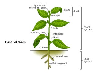
4. All Plant Cells are surrounded by an
extracellular matrix known as the Cell Wall
•a polysaccharide-rich matrix that surrounds all
plant cells
•plays multiple roles in plant growth,
development and defence responses
•there are two types of wall: primary &
secondary
5. Primary Wall
• first wall laid down
• surrounds growing cells
• surrounds meristematic cells
• cells in succulent tissues
• found at the junction of cells and at the outer edges of
secondary walls
• composed of ~ 90% carbohydrate and 10% protein
Secondary walls
• surround cells that differentiate to form specialized functions
(i.e. wood cells, xylem cells)
• have altered polysaccharide composition
• often are lignified
6. Polysaccharides are the main components of
the primary plant cell wall
Cross section of Nelumbo nucifera petiole showing primary cell wall
90% polysaccharide
10% protein
from Katherine Esau, Anatomy of Seed Plants, 1977
7. Cellulose
Hemicellulose
Pectin
Three classes of polysaccharides make up the primary wall
Walls from round and elongated carrot suspension cultured cells.
(Fast-freeze, deep-etch, rotary-shadowed replicas; McCann et al., 1993, J. Cell Science
106:1347)
8. Composition of primary cell walls of suspension-
cultured sycamore cells
Wall Component Mass % of Cell Wall
Pectic polysaccharides 34
Hemicellulose 24
Cellulose 23
Protein 19
McNeil et al., 1979, Fortschritte der Chemie organischer Naturstoffe, Volume 37, 191.
9. Type I primary walls
(all flower plants except the grass family)
Cellulose
Hemicellulose (xyloglucan)
Pectin (~22-35%) (homogalacturonan, HGA;
Rhamnogalacturonan I, RG-I; Rhamnogalacturonan II; RG-II)
Type II primary walls (the grass family, Poaceae):
Cellulose
Hemicellulose (glucuronoarabinoxylan)
Pectin (~10%) (HGA, RG-I, RG-II)
Primary walls can be divided into two types:
12. • World’s most abundant biopolymer
• Polymer of β1-,4-linked glucose
• Individual glucan chains associate via H-bonds to form
microfibrils that are largely crystalline.
• Cellulose I (the type of cellulose found in nature), glucan
chains are aligned parallel to each other
• Length of the glucan chains varies depending upon the
organism from DP ~2000 to up to DP ~15,000
• Size of microfibril also varies depending upon the organism
and can range from the elementary fibril (~ 36 glucan chains)
up to very large fibrils (> 200 chains) found in cellulosic algae
• As plant cells mature from 10 to 20 walls, cellulose can be
found as associates of macrofibrils or bundles
CELLULOSE
13.
14.
15.
16. •Cellulose gives tensile strength to the wall.
•In planta the cellulose microfibrils complex with
hemicellulosic polysaccharides such as xyloglucan.
•The pattern of cellulose deposition in the wall
determines the pattern of plant development.
•Generally, cellulose deposition is transverse to the
direction of cell elongation.
•X-ray diffraction studies indicate that Cellulose I exists
in a 2-fold ribbon-like helix 2(5.15) with 2 residues per
turn, a residue distance of 5.15 Å, and is stabilized by a
series of O3…05 H-bonds.
17.
18. Several organisms in addition to plants synthesize
cellulose.
These include several bacteria (e.g. Acetobacter
xylinum and Agrobacterium tumefaciens), the slime
mold (Dictyostelium discoideum) and the water mold
(Saprolegnia).
19. Genes for plant cellulose
synthase catalytic subunit were
identified in cotton based on
deduced amino acid sequence
homology to bacterial cellulose
synthase (cesA). CesA belongs
to multigene families in plants
(i.e. Arabidopsis may have at
least 17 members in the cesA
gene family).
Based on homology a “cesAlike
superfamily has been
identified. This family has four
conserved motifs: U1,U2, U3,
and U4 that are thought to be
involved in substrate binding
and/or catalysis.
20. Freeze fracture replicas of rosettes associated with cellulose microfibril biogenesis.
The rosettes after the fracture event exist in the leaflet of the plasma membrane bilayer
that is nearest the cytoplasm (the PF face). In the main micrograph, several rosettes are shown
(three surrounded by circles) in the plasma membrane of a differentiating tracheary element of
Zinnia elegans; differentiating tracheary elements deposit abundant cellulose into patterned
secondarywall thickenings. The inset shows one rosette at higher magnification and after high
resolution rotary shadowing at ultracold temperature with a minimum amount of
platinum/carbon. (Main micrograph, 222,000 x; inset, 504,545 x; both micrographs courtesy of
Mark J Grimson and Candace H Haigler, Department of Biological Sciences, Texas Tech University,
Lubbock, Texas.)
23. HEMICELLULOSE
•Class of structurally diverse polysaccharides that, in part, hydrogen bond to
cellulose
•Includes
Xyloglucan
Glucuronoarabinoxylan
Xylan
Mixed linkage glucans
“callose”
Galactomannans
• major hemicellulosic polysaccharide in most flowering plant primary walls
(except the grasses) is xyloglucan.
•Xyloglucan: a β-1,4-glucan substituted by α1,6-Xyl; some Xyl residues a β1,2-
linked Gal that is further substituted with an α-1,2-linked Fuc
•Galactomannan: food reserve polysaccharide in endosperm of legume seeds
& in endosperm walls and cell lumens; reserve carbohydrates used during
seed germination, protect the seed from desiccation, and are used as
thickeners and stabilizers in the food industry. Galactomannans are β1,4-linked
mannans substituted by α1,6-linked Gal.
24. All higher plants except the grass family have walls
of 30-35% pectin
The wall of the grass family contain ~10% pectin
25. PECTIN
Pectin is a family of complex carbohydrates found in all plant primary
walls that play structural and informational roles in plant cells.
Homogalacturonan (HG), the most abundant pectic polysaccharide, is a
homopolymer of α1,4-linked galacturonic acid that may be
methylesterified at C6 and acetylated or xylosylated at C3.
X-ray diffraction studies indicated that HG adopts a 3(4.45) right-handed
helix. Pectin forms gels in the presence of divalent cations (e.g. Ca++) or in
acidic conditions in the presence of high solute concentrations (e.g.
sucrose).
Pectin gels are important in the food, pharmaceutical, and cosmetic
industries.
Oligosaccharides (oligogalacturonides of DP 12-15), released from HGA by
endopolygalacturonases, induce plant defense responses and regulate
plant growth and development.
26. Pectin
family of polysaccharides that contains
α-4-linked galactosyluronic acid (GalA)
•Homogalacturonan (HG) (57-69%)
•Rhamnogalacturonan I (RG-I) (20-33%)
•Substituted galacturonans
Rhamnogalacturonan II (RG-II) (~10%)
Xylogalacturonan
Apiogalacturonan
27. • Cell Wall Structure / Assembly
• Cell-Cell Adhesion
• Cell Expansion
• Cell Wall Porosity
• Ion, growth factors, enzyme binding
• Biomechanics: regulation of water flow
• Reservoir of Biologically Active Oligosaccharides
• Pollen tube growth
• Seed hydration
• Leaf abscission
• Fruit development
Proposed functions of pectins in plants
28. Phenotype of known pectin structural mutants
•Dwarfed
•Brittle leaves
•Reduced numbers of shoots and flowers
•Reduced cell-cell adhesions