
Recommended
More Related Content
What's hot
What's hot (20)
Similar to larynx anatomy.pptx
Similar to larynx anatomy.pptx (20)
Recently uploaded
Recently uploaded (20)
larynx anatomy.pptx
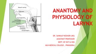
- 1. ANANTOMY AND PHYSIOLOGY OF LARYNX DR. SANKALP KESHARI (MS) ASSISTANT PROFESSOR DEPT. OF ENT & HNS MLN MEDICAL COLLEGE , PRAYAGRAJ
- 2. INTRODUCTION The larynx is the upper extended part of the lower respiratory tract. Made up of series of cartilages that are inter connected by ligaments and fibrous membranes Lies in the upper part of neck, anteriorly in the midline Extends from root of tongue to trachea Lies in front of 3rd to 6th cervical vertebra Before puberty size is almost same in males and females At puberty larynx grows rapidly in males and attend a bigger size than females
- 3. Thyroid cartilage is prominent in males- ADAM’S APPLE It moves vertically and in anteroposterior direction during swallowing and phonation. It can also be passively moved from side to side producing a characteristic grating sensation called laryngeal crepitus.
- 4. PHYSIOLOGY OF LARYNX 1. Protection of lower airways 2. Phonation 3. Respiration 4. Fixation of the chest.
- 6. LARYNGEAL CARTILAGES Unpaired: Thyroid, cricoid and epiglottis. Paired: Arytenoid, corniculate and cuneiform
- 7. Thyroid Cartilage It is the largest of all. Its two alae meet anteriorly forming an angle of 90° in males and 120° in females. Its function is to shield larynx from injury and provide an attachment to vocal cords Shied shaped, open posteriorly, angulated anteriorly
- 8. Cricoid Cartilage Signet ring shaped Stronger than thyroid cartilage. Lamina – 2 to 3 cm from above downwards, considerably broader than anterior arch.
- 9. Important from structural & functional point of view Base for entire larynx Support to arytenoid Attachment to intrinsic muscles Only part of cartilagenous framework that forms continuous 360 degree ring Once injured or strictured , difficult to resect while preserving laryngeal function
- 10. 3. Epiglottis. It is a leaf-like, yellow, elastic cartilage forming anterior wall of laryngeal inlet. A stalk-like process of epiglottis (petiole) attaches the epiglottis to the thyroid angle just above the attachment of vocal cords. Anterior surface of epiglottis is separated from thyrohyoid membrane and upper part of thyroid cartilage by a potential space filled with fat— the pre-epiglottic space.
- 11. 4. Arytenoid cartilages. They are paired. Each arytenoid cartilage is pyramidal in shape. It has a base which articulates with cricoid cartilage. 5. Corniculate cartilages (of Santorini) (Corn = horn). They are paired. Each articulates with the apex of arytenoid cartilage as if forming its horn. 6. Cuneiform cartilages (of Wrisberg). They are rod shaped. Each is situated in aryepiglottic fold in front of corniculate cartilage and provides passive supports to the fold.
- 12. Thyroid, cricoid and most of the arytenoid cartilages are hyalin cartilages epiglottis, corniculate, cuneiform and tip of arytenoid near the corniculate cartilage are elastic fibrocartilage.
- 14. Supraglottis Consists of ventricles, false cords, laryngeal surface of epiglottis, aryepiglottic folds and the mucosal expanse. Posterior tapering shape reduces area of mucosa in posterior region So majority of SG tumors are epiglottic
- 15. Glottis Consists of true cords, anterior commissure and posterior commissure Narrow triangular space between the true cords is called rima glottis Anterior 2/3 is membranous Posterior third consists of vocal processes of arytenoids Posterior 1/3 of cords and covering mucosa are called posterior commissure
- 16. Sub-glottis Begins about 5mm below free margins of VC Consists of a mobile upper and fixed lower part
- 17. Mucosa Mucosa of glottic and Supraglottic regions is stratified squamous epithelium. Mucosa of ventricles and sub-glottic regions is pseudo-stratified ciliated epithelium Supra and sub glottic regions particularly ventricles are rich in submucosal mucous or minor salivary glands while glottis is not.
- 18. LARYNGEAL JOINTS Cricothyroid Joint. It is a synovial joint. Each is formed by the inferior cornua of thyroid cartilage with a facet on the cricoid cartilage. Cricoid cartilage rotates at these joints on a transverse axis which passes transversely through these joints. Cricoarytenoid Joint. It is a synovial joint surrounded by capsular ligament. It is formed between the base of arytenoid and a facet on the upper border of cricoid lamina. Two types of movements occur: (i) rotatory (ii) gliding
- 20. LARYNGEAL MEMBRANES 1. Extrinsic membranes and ligaments (a) Thyrohyoid membrane (b) Cricotracheal membrane (c) Hyoepiglottic ligament
- 21. 2. Intrinsic membranes and ligaments (a) Cricovocal membrane. It is a triangular fibroelastic membrane. Its upper border is free and stretches between middle of thyroid angle to the vocal process of arytenoid and forms the vocal ligament Its lower border attaches to the arch of cricoid cartilage. thus, with its fellow on the opposite side, forms conus elasticus where subglottic foreign bodies sometimes get impacted.
- 23. b) Quadrangular membrane. It lies deep to mucosa of aryepiglottic folds and is not well-defined. (c) Cricothyroid ligament. The anterior part of cricothyroid membrane is thickened to form the ligament and its lateral part forms the cricovocal membrane. (d) Thyroepiglottic ligament. It attaches epiglottis to thyroid cartilage.
- 24. CAVITY OF THE LARYNX Inlet of Larynx. It is an oblique opening bounded anteriorly by free margin of epiglottis; on the sides, by aryepiglottic folds and posteriorly by interarytenoid fold Vestibule. It extends from laryngeal inlet to vestibular folds. anterior wall is formed by posterior surface of epiglottis; sides by the aryepiglottic folds and posterior wall by mucous membrane over the anterior surface of arytenoids.
- 26. Glottis (Rima Glottidis). It is the elongated space between vocal cords anteriorly, and vocal processes and base of arytenoids posteriorly glottis is about 24 mm in men and 16 mm in women. It is the narrowest part of laryngeal cavity. Anterior two-thirds of glottis is also called phonatory glottis as it is concerned with phonation posterior one-third called respiratory glottis.
- 27. Spaces of the Larynx 1. Pre-Epiglottic Space of Boyer. 2. Paraglottic Space. 3. Reinke’s Space
- 28. Pre-Epiglottic Space Bound sup by hyo- epiglottic ligament, ant by thyrohyoid memb. & thyroid cartilage and posteriorly by epiglottis Filled with fat and areolar tissue Continuous with para- glottic space Cx of laryngeal surface of epiglottis readily spread to PreEpiSpace
- 29. Paraglottic space. It is bounded by the thyroid cartilage laterally, conus elasticus inferomedially, the ventricle and quadrangular membrane medially, and mucosa of pyriform fossa posteriorly. It is continuous with pre-epiglottic space. Growths which invade this space can present in the neck through cricothyroid space
- 31. Reinke’s Space Mucosa over the vocal ligament loosely attached to ligaments Thus there is a submucosal space along most of the length of truer VC
- 32. PAEDIATRIC LARYNX 1. Infant’s larynx is positioned high in the neck level of glottis being opposite to C3 or C4 at rest and reaches C1 or C2 during swallowing. This high position allows the epiglottis to meet soft palate and make a nasopharyngeal channel for nasal breathing during suckling. 2. Laryngeal cartilages are soft and collapse easily. Epiglottis is omega shaped 3. Thyroid cartilage in an infant is flat. It also overlaps the cricoid cartilage and is in turn overlapped by the hyoid bone. 4. Infant’s larynx is small and conical. 5. Submucosal tissues of infant’s larynx are comparatively loose and easily undergo oedematous change with trauma or inflammation leading to obstruction
- 33. Infant’s larynx shows two spurts in growth: first 3 years of life, larynx grows in width and length The second spurt in growth occurs during adolescence when the thyroid angle develops. In childhood, vocal cord is 6 mm in females and 8 mm in males. It increases to 15–19 mm in adult female and 17–23 in adult male.
- 34. THANKYOU