👉 Chennai Sexy Aunty’s WhatsApp Number 👉📞 7427069034 👉📞 Just📲 Call Ruhi Colle...
Dislipdemia dan ldlr fungsi Rahmi Lisdeni Folder
1. Low-Density Lipoprotein Receptor—Its
Structure, Function, and Mutations
Joep C. Defesche, Ph.D.1
ABSTRACT
Uptake of cholesterol, mediated by the low-density lipoprotein (LDL)-receptor,
plays a crucial role in lipoprotein metabolism. The LDL-receptor is responsible for the
binding and subsequent cellular uptake of apolipoprotein B– and E–containing lipopro-
teins. To accomplish this, the receptor has to be transported from the site of synthesis, the
membranes of the rough endoplasmatic reticulum (ER), through the Golgi apparatus, to
its position on the surface of the cellular membrane. The translation of LDL-receptor
messenger RNA into the polypeptide chain for the receptor protein takes place on the
surface-bound ribosomes of the rough ER. Immature O-linked carbohydrate chains are
attached to this integral precursor membrane protein. The molecular weight of the receptor
at this stage is 120.000 d. The precursor-protein is transported from the rough ER to the
Golgi apparatus, where the O-linked sugar chains are elongated until their final size is
reached. The molecular weight has then increased to 160.000 d. The mature LDL-receptor
is subsequently guided to the ‘‘coated pits’’ on the cell surface. These specialized areas of the
cell membrane are rich in clathrin and interact with the LDL-receptor protein. Only here
can the LDL-receptor bind LDL-particles. Within 3 to 5 minutes of its formation, the
LDL-particle-receptor complex is internalized through endocytosis and is further meta-
bolized through the receptor-mediated endocytosis pathway. Mutations in the gene coding
for the LDL-receptor can interfere to a varying extent with all the different stages of the
posttranslational processing, binding, uptake, and subsequent dissociation of the LDL-
particle-LDL-receptor complex, but invariably the mutations lead to familial hypercho-
lesterolemia. Thus, mutations in the LDL-receptor gene give rise to a substantially varying
clinical expression of familial hypercholesterolemia.
KEYWORDS: Familial hypercholesterolemia, mutation class, genotype-phenotype
Educational Objectives: Upon completion of this article, the reader should be able to (1) summarize the basic structure of the low-
density lipoprotein receptor protein and its function, (2) appreciate the vast molecular heterogeneity in inherited hypercholesterolemia,
and (3) recognize the involvement of multiple genes in the aetiology of familial hypercholesterolemia.
Familial hypercholesterolemia (FH) was de-
scribed for the first time more than 125 years ago.
Initially, the disorder, with its visual characteristics
such as xanthomas and xanthelasmas, was thought to
be a disease of the skin.1
Only later did it became
apparent that FH was associated with a high incidence
of premature atherosclerosis, resulting in coronary, cere-
bral, or peripheral vascular disease.2
Clinical Manifestations, Laboratory Diagnosis, and Molecular Biology of Familial Hypercholesterolemia: Clinical Management and Prevention;
Editor in Chief, Jan Jacques Michiels, M.D., Ph.D.; Guest Editor, Joep C. Defesche, Ph.D. Seminars in Vascular Medicine, Volume 4, Number 1,
2004. Address for correspondence and reprint requests: Dr. J.C. Defesche, Department of Vascular Medicine, Academic Medical Center, Rm. G1-
112B, P.O. Box 22 660, NL–1100 DD Amsterdam, The Netherlands. E-mail: j.defesche@amc.uva.nl. 1
Department of Vascular Medicine,
Academic Medical Center at the University of Amsterdam, The Netherlands. Copyright # 2004 by Thieme Medical Publishers, Inc., 333 Seventh
Avenue, New York, NY 10001, USA. Tel: +1(212) 584-4662. 1528-9648,p;2004,04,01,005,011,ftx,en;svm00171x.
5
Downloadedby:UniversiteLaval.Copyrightedmaterial.
2. In the following decade, family studies by Wilkinson laid
the hereditary basis for FH, after which Khachadurian
demonstrated the autosomal dominant mode of inheri-
tance.3,4
The heterozygous and homozygous forms of
FH, with their vastly different clinical courses, were also
recognized at this time.
Yet another decade, later it became clear that an
increased level of plasma low-density lipoprotein
(LDL)-cholesterol was the hallmark of the disease.5,6
This observation lead to the discovery of a receptor for
LDL particles on the outer membrane of different cell
types.7
It was postulated, and later proven, that the
underlying molecular defect of FH consisted of muta-
tions in the gene that coded for the LDL-receptor
protein. Mutations in this gene result in failure to
produce LDL-receptor protein or in reduction of
LDL-receptor activity, with increased levels of LDL-
cholesterol in plasma appearing as a consequence.8
LDL-RECEPTOR STRUCTURE
The LDL-receptor protein is, in human, bovine, and
rodent species, an evolutionary highly conserved, inte-
gral membrane glycoprotein. After removal of the 21
amino acids–long signal peptide, the remaining protein
comprising 839 amino acids can be subdivided into five
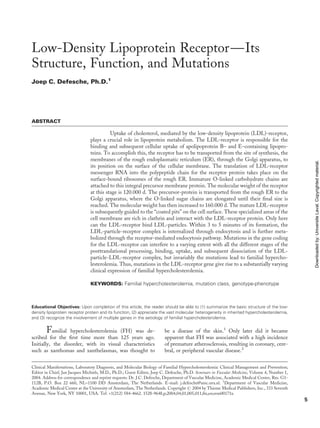
structurally different domains (Fig. 1): the ligand bind-
ing domain, the epidermal growth factor (EGF) pre-
cursor homology domain, the O-linked sugars domain,
the membrane spanning domain, and the cytoplasmatic
domain.
The first domain of the LDL-receptor is respon-
sible for interaction with the ligand protein apolipopro-
tein B100 (apoB) for the binding of LDL, or with
apolipoprotein E (apoE) for the binding of very low-
density lipoprotein (VLDL).9,10
Binding is mediated by
a stretch of 292 amino acids at the amino terminus of the
mature protein. The ligand binding domain consists of
seven identical modules of $40 aminoacids each, called
LDL-receptor modules. The seven modules are ar-
ranged in a head-to-tail arrangement, and because of
its relatively large amount of cysteine aminoacids, the
protein is stereometrically folded in such a way that the
negatively charged aminoacids are located on the outside
of the protein. The predicted tertiary structure shows
that in each segment, a central and inaccessible hydro-
phobic cysteine cluster, which forms three disulfide
bonds, and a calcium ion stabilize the negatively
charged hydrophillic aminoacids at the easily accessible
surface.11,12
The EGF precursor homology domain is located
next to the ligand binding domain and contains $400
amino acids. This domain consists of two EGF-repeats,
A and B, and a YWTD-6 bladed b-propeller flanked by
a third EGF-repeat, C. Each repeat is $40 amino acids
long and contains three disulfide bonds. The EGF
precursor homology domain is also responsible for the
binding of LDL and VLDL, and in addition, it controls
the ligand release at low pH in the lysosomes and the
recycling of the receptor to the cell surface.13
The O-linked polysaccharide domain is found
just outside the plasma membrane. It stretches over
Figure 1 Different domains in the low-density-lipopro-
tein-receptor protein are encoded by specific regions in
the low-density-lipoprotein-receptor gene.
6 SEMINARS IN VASCULAR MEDICINE/VOLUME 4, NUMBER 1 2004
Downloadedby:UniversiteLaval.Copyrightedmaterial.
3. 58 amino acids and contains several polysaccharide
chains. It is very likely that all hydroxylated branches
of the 18 serines and threonines are glycosylated. The
O-linked oligosaccharides—the N-acetyl galactosa-
mines—undergo a posttranslational modification in
which the galactosyl and sialyl groups are added to the
sidechains.
The glycosylated domain stretches out the three-
dimensional structure of the ligand protein to facilitate
its binding to the LDL-particle and protects the LDL-
receptor against proteolytic cleavage.14,15
The membrane-spanning domain consists of 25
hydrophobic amino acids that span the cellular mem-
brane and anchor the LDL-receptor protein. Evolution-
ary, this is the least conserved part of the LDL-receptor,
and in that sense, its having a function different from
anchoring is not very plausible. A deletion of the exon
coding for this domain in naturally occurring mutations
causes the LDL-receptor to dissociate from the cell
membrane into the surrounding medium.16,17
The cytoplasmatic domain or the carboxyterminal
part of the LDL-receptor molecule protrudes into the
cytoplasm of the cell. It contains 50–amino acid residues
with a carboxy-terminal alanine. This domain plays a
role in the direction of the LDL-receptor to the coated
pits. Naturally occurring mutations in this domain of the
LDL-receptor interfere with the clustering of receptor
molecules in the coated pits and with the subsequent
internalization.18
In vitro mutagenesis experiments have shown that
amino acid residues 791 to 812 are of crucial importance
for this process. Intracellular proteins, interacting with
the cytoplasmatic tail of the LDL-receptor, form part of
the so-called ‘‘fuzzy coat,’’ the protein lining of the
cytoplasmatic side of the coated pits. A substantial part
of the coated pits is formed by clathrin, an intriguing
protein that specifically interacts with the plasma mem-
brane and cytoskeleton. The composition of the cyto-
plasmatic part of transmembrane proteins determines to
a high extent the final localization of the proteins in the
cell. The LDL-receptor is transported to the cell’s
basolateral membrane and is then directed to the coated
pits. The information required for the routing through
the cell is stored in the amino acid sequence of the
cytoplasmatic part of the protein.19
LDL-RECEPTOR FUNCTION
LDL accounts for 75% of the cholesterol transport and
the majority of LDL, $70%, is cleared from the plasma
by LDL-receptors on the surface of liver cells. There-
fore, LDL-receptor-mediated uptake of cholesterol
plays a crucial role in lipoprotein metabolism. The
LDL-receptor is responsible for the binding and sub-
sequent cellular uptake of apoB and apoE containing
lipoproteins.
The translation of LDL-receptor messenger
RNA into the polypeptide chain for the receptor protein
takes place on the surface-bound ribosomes of the
endoplasmatic reticulum (ER). Immature O-linked car-
bohydrate chains are attached to this integral precursor
membrane protein.20
The molecular weight of the re-
ceptor at this stage is 120.000 d. The precursor-protein is
transported from the rough ER to the Golgi apparatus,
where the O-linked sugar chains are elongated until
their final size is reached. The molecular weight has then
increased to 160.000 d. The mature LDL-receptor is
subsequently guided to the ‘‘coated pits’’ on the cell
surface. These specialized areas of the cell membrane
are rich in clathrin and interact with the LDL-receptor
protein. Only here can the LDL-receptor bind LDL-
particles. Within 3 to 5 minutes of its formation, the
LDL-particle-receptor complex is internalized through
endocytosis and is further metabolized through the
receptor-mediated endocytosis pathway.21
LDL-RECEPTOR GENE
The LDL-receptor locus is located on the distal part of
the short arm of chromosome 19, on bands p13.1 to
p13.3.22
The locus stretches over 45,000 base pairs (bp)
or 45 kilobases and comprises 18 coding regions (exons)
and 17 intervening noncoding regions (introns) (Fig. 1).
The gene for the LDL-receptor is a so-called
housekeeping gene, which is, in almost all tissues, con-
tinuously translated into LDL-receptors. The transcrip-
tion is regulated by means of the negative feedback
mechanisms of certain sterols. The elements, regulating
the transcription, are located in the so-called promoter
region, upstream (50
) in the gene. In case of the LDL-
receptor gene, this region extends over 177 bp, from base
pair À58 to À234. Within the promoter region there are
two segments, each 7 bp long, in which the sequences
TATA and CT can be discerned.23
These sequences are
essential for the expression of most genes in eukaryotic
cells. Furthermore, there are three, not completely iden-
tical, elements of 16 bp each, which are also essential for
expression. These elements, regulated by sterols, are
composed of one conditional transcription enhancer,
the sterol regulatory element-1 (SRE-1), which is situ-
ated in the middle of the three repetitive elements. SRE-
1 enhances the transcription of the LDL-receptor gene
in the absence of sterols.24,25
The two remaining repetitive elements are bind-
ing sites for the general transcription enhancer factor
Sp1 and are, together with SRE-1, essential for tran-
scription at the maximal level. An increased cholesterol
level in cells is detected by a SRE-binding protein, which
detaches from SRE-1, thus inhibiting transcription of
the LDL-receptor gene.26
This means that end-product
repression is involved in the transcription of the LDL-
receptor gene. The end-products, sterols in this case,
LOW-DENSITY LIPOPROTEIN RECEPTOR—ITS STRUCTURE, FUNCTION, AND MUTATIONS/DEFESCHE 7
Downloadedby:UniversiteLaval.Copyrightedmaterial.
4. interfere with the action of positive transcription en-
hancers, which bind to the promoter region.
Exon 1 contains the information for the amino-
terminal part of the protein, mostly a signal-peptide.
The seven LDL-receptor modules in the ligand-binding
domain are coded for by exons 2 to 6. Exons 7 to 14 code
for the EGF precursor homology domain. These eight
exons are organized in a manner almost identical to the
exons in the gene coding for the EGF precursor pro-
tein.27
Exon 15 codes for the glycosylated domain only.
Exon 16 and part of exon 17 are the coding regions for
the transmembrane part of the receptor, whereas the
remaining segment of exon 17 and the beginning of exon
18 code for the cytoplasmatic part of the receptor. A
special feature of the LDL-receptor gene is the fact that
the last part of exon 18 is transcribed into a 2.5-kilobase
messenger RNA segment, which is eventually not trans-
lated into protein.
In general, the LDL-receptor gene is made up of
coding sequences that are shared not only with the EGF
precursor but also with coagulation proteins, comple-
ment factors, and many other proteins.28
Undoubtedly,
large parts of the LDL-receptor gene were created by
‘‘exon-shuffling’’ (the exchange of exons between differ-
ent genes of not directly related proteins), and therefore
the LDL-receptor gene is a member of several so-called
‘‘super gene’’ families. The underlying principle is that
the assembly of complex mosaic-proteins was possible
during evolution by means of the insertion of several
entire modules within certain primordial proteins that
already existed. Many of these modules have been identi-
fied: kringle-, growth-factor-, zinc-finger-, calcium-bind-
ing-, and LDL-receptor-modules. These modules could
only be inserted into other genes if the introns of the
receiving genes were ‘‘in phase.’’29
Being ‘‘in phase’’ means
that an intronstarts as well as ends in the same position ofa
codon, so that addition of an exon does not result in
alterationoftheaminoacidsequenceoftheoriginalprotein.
Ithasbeenshown thatthe exonsofthe first two domains of
the LDL-receptor gene are completely ‘‘in phase,’’ and the
same is true for certain coagulation proteins and comple-
ment factors.29
The special structure of the LDL-receptor
gene, evolved in this way, must also have introduced a
susceptibility for mutations.
LDL-RECEPTOR GENE MUTATIONS
By means of cell biological and immunological techni-
ques, it has been possible to divide the naturally occur-
ring mutations of the LDL-receptor initially into five,
and later into six, classes.30,31
Only in a later stage have
two of these functional classes again been subdivided.
The purification of the normal LDL-receptor protein
and the development of a monoclonal antibody have
made this subdivision possible.32
Immuno-precipitation
experiments rapidly showed that the trait of FH was not
caused by one, but by several, LDL-receptor mutations.
In Figure 2, point mutations in the LDL-receptor gene,
Figure 2 Point mutations in the low-density-lipoprotein-receptor gene characterized in Dutch patients with familial hypercholester-
olemia. Update December 2003: 255 point mutations in the low-density-lipoprotein-receptor gene and (not shown) 12 mutations in the
apoB gene.
8 SEMINARS IN VASCULAR MEDICINE/VOLUME 4, NUMBER 1 2004
Downloadedby:UniversiteLaval.Copyrightedmaterial.
5. characterized in Dutch patients with FH, are schemati-
cally presented.33
Class 1: Synthesis of Receptor or Precursor
Protein is Absent
The so-called null-allele is a prevalent class of mutations
and is generally associated with very high LDL-choles-
terol levels. The molecular basis of this type of mutation
shows a wide variety: point mutations introducing a stop
codon, mutations in the promoter region completely
blocking transcription, mutations giving rise to incorrect
excision of messenger RNA, and finally, large deletions
preventing the assembly of a normal receptor.
Class 2: Absent or Impaired Formation
of Receptor Protein
This class comprises mutations in which the normal
routing through the cell is not, or is only very slowly,
completed. Usually there is a complete blockade of
transport, and LDL-receptors are unable to leave the
ER. The Golgi apparatus is not reached, and the increase
of 40,000 d in molecular weight does not take place.
Truncated proteins, as a result of a premature stop
codons, and misfolded proteins, as a result of mutations
in cysteine-rich regions leading to free or unpaired
cysteine residues, are retained in the ER. However,
quality control by the ER is not perfect, given that
sometimes misfolded proteins do leave the ER but are
processed more slowly. Such mutations give rise to class
2B mutations, in contrast to the class 2A mutations that
cause complete retaining in the ER.
Class 3: Normal Synthesis of Receptor
Protein, Abnormal LDL-Binding
Receptors, characterized by this class of alleles, show the
normal rate of synthesis, exhibit normal conversion into
receptor protein, and are transported to the cell surface,
but binding to LDL is impaired. It is obvious that
mutations in the binding domain underlie this class of
receptors.
Class 4: Clustering in Coated Pits and
Internalization of the Receptor Complex
Does Not Take Place
The receptors in this class lack the property to cluster in
coated pits (class 4A). This phenomenon, which makes
interaction of receptors with the fuzzy coat impossible, is
caused by mutations in the carboxy-terminal part of the
receptor protein. These mutated receptors are synthe-
sized normally, and folding and transport is normal, but
clustering in coated pits is impossible, and sometimes the
receptors are even secreted after they have reached the
cell surface (class 4B).
Class 5: Receptors Are Not Recycled and
Are Rapidly Degraded
All mutations in this class are localized in the EGF-
precursor homologous domain of the LDL-receptor
protein. This domain seems to be involved in the acid-
dependent dissociation of the receptor-ligand complex
in endosomes, after which the receptor can be recycled.
When the entire EGF-precursor homologous domain is
deleted by site-directed mutagenesis, or when such a
deletion occurs naturally in a homozygous FH patient,
the receptor is trapped in the endosomes, and subse-
quently rapid degradation is observed.
Class 6: Receptors Fail to be Targeted
to the Basolateral Membrane
The class of mutations was recently discovered and is
caused by alterations in the cytoplasmatic tail of the
protein. Such receptors do not reach the liver cell’s
membrane and are probably rapidly degraded.31
These six mutation classes describe the effect of a
certain molecular defect in the DNA of the gene on the
function of the LDL-receptor protein. With regard to
molecular defects, one can discern base pair substitu-
tions, small and large deletions and insertions, splice site
mutations, and the generation of premature stop codons.
The actual DNA defect, however, cannot simply predict
the class in which the defect will result. A premature stop
codon or a splice site mutation will in many cases give
rise to a class 1 mutation, but it can also lead to a
truncated protein, resulting in a class 2 to 6 mutation,
depending on what part of the protein is lacking.
The effect of a molecular defect on protein func-
tion can be studied, of course, by performing immuno-
logical and cell biological studies. However, this is
complicated by the vast number of mutations in the
LDL-receptor gene currently characterized and by the
observation that in many cases, a mutation can belong to
more than one class.30
At present, more than 900
different mutations have been described.34–36
Because of the difficulty in predicting the effect of
a certain molecular defect, it is far simpler to classify
mutations into two groups: LDL-receptor deficient (in
fact, null-alleles that do not produce LDL-receptor
protein), and LDL-receptor defective mutations that
affect LDL-receptor activity.
MUTATIONS IN OTHER GENES
Apart from the vast array of mutations in the LDL-
receptor gene, mutations in other genes are also known
to cause inherited hypercholesterolemia, a condition that
LOW-DENSITY LIPOPROTEIN RECEPTOR—ITS STRUCTURE, FUNCTION, AND MUTATIONS/DEFESCHE 9
Downloadedby:UniversiteLaval.Copyrightedmaterial.
6. is clinically indistinguishable from FH.37
First of all,
structural rearrangements in the domain of apoB that
interacts with the LDL-receptor, caused by mutations in
exon 26 and 29 of the apoB-gene, interfere with the
binding of the LDL-particle with the LDL-receptor and
result in elevated LDL-cholesterol levels in plasma.38
Because the hypercholesterolemia is caused by defective
LDL-particles that cannot be bound by the LDL-
receptor, this disorder is referred to as familial defective
apolipoprotein B.
When a large group of patients with a definite
clinical diagnosis of FH, for example, children with
inherited hypercholesterolemia, is extensively investi-
gated down to the molecular level, up to 15% of the
cases fail to be explained by mutations in the LDL-
receptor and apoB genes.39
This demonstrates that still
other genes must be involved in the development of
inherited hypercholesterolemia. Indeed, recently a third
gene was shown to be involved: Mutations in the gene
coding for Neural Apoptosis Regulated Convertase-1
are associated with autosomal dominant hypercholester-
olemia.40
Although Neural Apoptosis Regulated Conver-
tase-1 is known to be involved in cholesterol home-
ostasis, its precise role and the pathogenicity of
mutations in this gene remain unclear.
GENOTYPE–PHENOTYPE RELATIONS
Numerous studies have been conducted to investigate
the relation between specific mutations or mutation
classes and the clinical expression of the disease in terms
of rate of LDL-cholesterol elevation, time of onset of
and severity of cardiovascular disease, presence of
the typical physical symptoms, and therapeutic re-
sponse.41–48
In these studies, however, patient popula-
tions were small and invariably selected through lipid
clinics. Thus, other risk factors for CVD were likely to
determine the excess mortality from FH, whereas the
type of mutation had seemingly little or no relevant
contribution. In a recent study, a large group of patients
with FH, free from selection for CVD, was investigated
and a relevant genotype–phenotype effect on lipids and
cardiovascular burden was established.49
However, the
importance of other, probably not only lipid-related, risk
factors for CVD in FH has to be taken into account.49–52
REFERENCES
1. Fagge CH. General xantheiasma or ritiligoldae. Transactions
of the Pathological Society, London 1837;24:242–250
2. Mu¨ller C. Xanthomata, hypercholesterolemia, angina pectoris.
Acta Med Scand 1938;89:75–84
3. Wilkinson CF, Hand EA, Fliegelman MT. Essential familial
hypercholesterolemia. Ann Intern Med 1948;29:671–676
4. Khachadurian AK. The inheritance of essential familial
hypercholesterolemia. Am J Med 1964;37:402–407
5. Goldstein JL, Brown MS. Familial hypercholesterolemia:
identification of a defect in the regulation of 3-hydroxy-3-
methylglutaryl Coenzyme A reductase activity with over-
production of cholesterol. Proc Natl Acad Sci USA 1973;70:
2804–2809
6. Brown MS, Goldstein JL. Expression of the familial
hypercholesterolemia gene in heterozygotes: mechanism for
a dominant disorder in man. Science 1974;185:61–63
7. Anderson RGW, Goldstein JL, Brown MS. Localization of
low density lipoprotein receptors on plasma membrane of
normal human fibroblasts and their absence in cells from a
familial hypercholesterolemia homozygote. Proc Natl Acad
Sci USA 1976;73:2434–2438
8. Brown MS, Goldstein JL. Receptor-mediated pathway for
cholesterol homeostasis. Science 1986;232:34–47
9. Su¨dhoff TC, Goldstein JL, Brown MS, Russell DW. The
LDL-receptor gene. A mosaic of exons shared with different
proteins. Science 1985;228:815–822
10. Esser V, Limbird LE, Brown MD, Goldstein JL, Russell
DW. Mutational analysis of the ligand binding domain of the
low-density lipoprotein receptor. J Biol Chem 1988;263:
13282–13290
11. Fass D, Blacklow S, Kim PS, Berger JM. Molecular basis of
familial hypercholesterolemia from structure of LDL-receptor
module. Nature 1997;388:691–693
12. North C, Blacklow SC. Structural independence of ligand-
binding modules five and six of the LDL-receptor. Biochem-
istry 1999;38:3926–3935
13. Davis CG, Goldstein JL, Su¨dhoff TC, Anderson RGW,
Russell DW, Brown MS. Growth factor homology region in
LDL receptor mediates acid-dependent dissociation and
receptor recycling. Nature 1987;326:760–764
14. Davis CG, Elhammer A, Russell DW, et al. Deletion of
clustered O-linked carbohydrates does not impair function
of low density lipoprotein receptor in transfected fibroblasts.
J Biol Chem 1986;261:2828–2038
15. Kozarsky K, Kingsley D, Krieger M. Use of a mutant cell line
to study the kinetics and function of the O-linked glycosyla-
tion of low-density lipoprotein receptors. Proc Natl Acad Sci
USA 1988;85:4335–4339
16. Lehrman MA, Schneider WJ, Su¨dhof T, Brown MS,
Goldstein JL, Russell DW. Mutations in LDL-receptor
Alu-Alu recombinations delete exons encoding transmem-
brane and cytoplasmic domains. Science 1985;227:140–146
17. Lehrman MA, Russell DW, Goldstein JL, Brown MS. Alu-
Alu recombination deletes splice acceptor sites and produces
secreted LDL receptor in a subject with FH. J Biol Chem
1987;262:3354–3361
18. Davis CG, Van Driel IR, Russell DW, Brown MS,
Goldstein JL. The LDL-receptor: identification of aminoa-
cids in cytoplasmic domain required for rapid endocytosis.
J Biol Chem 1987;262:4075–4079
19. Matter K, Yamamoto EM, Mellman I. Structural require-
ments and sequence motifs for polarized sorting and
endocytosis of LDL and Fc receptors in MDCK cells. J Cell
Biol 1994;126:991–1004
20. Anderson RGW, Brown MS, Goldstein JL. Biosynthesis of
the N- and O-linked oligosaccharides of the low-density
lipoprotein receptor. J Biol Chem 1983;258:15261–15273
21. Goldstein JL, Brown MS, Anderson RGW, Russell DW,
Schneider WJ. Receptor-mediated endocytosis: concepts
10 SEMINARS IN VASCULAR MEDICINE/VOLUME 4, NUMBER 1 2004
Downloadedby:UniversiteLaval.Copyrightedmaterial.
7. emerging from the LDL receptor system. Annu Rev Cell Biol
1985;1:1–39
22. Lindgren V, Luskey KL, Russell DW, Francke U. Human
genes involved in cholesterol metabolism: chromosomal
mapping of the loci for the low-density lipoprotein receptor
and 3-hydroxy-3-methylglutaryl-coenzyme A reductase with
cDNA probes. Proc Natl Acad Sci USA 1985;82:8567–
8571
23. Su¨dhoff TC, Van der Westhuyzen DR, Goldstein JL, Brown
MS, Russell DW. Three direct repeats and a TATA-like
sequence are required for regulated expression of the human
LDL-receptor gene. J Biol Chem 1987;262:10773–10779
24. Su¨dhoff TC, Russell DW, Brown MS, Goldstein JL. 42 bp
element from LDL receptor gene confers end-product
repression by sterols when inserted into viral TK promoter.
Cell 1987;48:1061–1069
25. Goldstein JL, Brown MS. Regulation of the mevalonate
pathway. Nature 1990;343:425–430
26. Rajavashisth TB, Taylor AK, Andalibi A, Svenson KL, Lusis
AL. Identification of a zinc finger protein that binds to the
sterol regulatory element. Science 1989;245:640–643
27. Russell DW, Schneider JW, Yamamoto T, Luskey KL,
Brown MS, Goldstein JL. Domain map of the LDL-receptor:
sequence homology with the epidermal growth factor
precursor. Cell 1984;37:577–585
28. Patthy L. Evolution of the proteases of blood coagulation and
fibrinolysis by assembly from modules. Cell 1985;41:657–663
29. Patthy L. Intron-dependent evolution; preferred types of
exons and introns. FEBS Lett 1987;214:1–7
30. Hobbs HH, Brown MS, Goldstein JL. Molecular genetics of
the LDL receptor gene in familial hypercholesterolemia.
Hum Mutat 1992;1:445–466
31. Koivisto UM, Hubbard AL, Mellman I. A novel cellular
phenotype for familial hypercholesterolemia due to a defect in
polarized targeting of LDL-receptor. Cell 2001;105:575–585
32. Schneider WJ, Beisiegel U, Goldstein JL, Brown MS.
Purification of the low density lipoprotein receptor, an acidic
glycoprotein of 164.000 molecular weight. J Biol Chem
1982;257:2664–2673
33. Fouchier SW, Defesche JC, Umans-Eckenhausen MAW,
Kastelein JJP. The molecular basis of familial hypercholester-
olemia in the Netherlands. Hum Genet 2001;109:602–615
34. LDL-Receptor Database. Available at: http://www.ucl.ac.uk/
fh. Accessed December 10, 2003
35. LDL-Receptor Database. Available at: http://www.
jojogenetics.nl
36. LDL-Receptor Database. Available at: http://www.umd.
necker.fr
37. Defesche JC, Pricker KL, Hayden MR, van der Ende BE,
Kastelein JJ. Familial defective apolipoprotein B-100 is
clinically indistinguishable from familial hypercholesterole-
mia. Arch Intern Med 1993;153:2349–2356
38. Boren J, Ekstrom U, Agren B, Nilsson-Ehle P, Innerarity TL.
The molecular mechanism for the genetic disorder familial
defective apolipoprotein B100. J Biol Chem 2001;276:9214–
9218
39. Wiegman A, Rodenburg J, De Jongh S, et al. Family history
and cardiovascular risk in familial hypercholesterolemia: data
in more than 1000 children. Circulation 2003;107:1473–
1478
40. Abifadel M, Varret M, Rabes JP, et al. Mutations in PCSK9
cause autosomal dominant hypercholesterolemia. Nat Genet
2003;34:154–156
41. Jeenah M, September W, Graadt van Roggen F, et al.
Influence of specific mutations at the LDL-receptor gene
locus on the response to simvastatin therapy in Afrikaner
patients with heterozygous familial hypercholesterolemia.
Atherosclerosis 1993;98:51–58
42. Koivisto PVI, Koivisto UM, Kovanen PT, Gylling H,
Miettinen TA, Kontula T. Deletion of exon 15 of the LDL
receptor gene is associated with a mild form of familial
hypercholesterolemia FHEspoo. Arterioscler Thromb 1993;13:
1680–1688
43. Gudnason V, Day INM, Humphries SE. Effect on plasma
lipid levels of different classes of mutations in the low-density
lipoprotein receptor gene in patients with familial hyperch-
olesterolemia. Arterioscler Thromb 1994;14:1717–1722
44. Sun XM, Patel DD, Bhatnagar D, et al. Characterization of a
splice-site mutation in the gene for the LDL-receptor
associated with an unpredictably severe clinical phenotype in
English patients with heterozygous FH. Arterioscler Thromb
Vasc Biol 1995;15:219–227
45. Vohl MC, Gaudet D, Moorjani S, et al. Comparison of the
effect of low-density lipoprotein receptor class mutations on
coronary heart disease among French-Canadian patients
heterozygous for familial hypercholesterolaemia. Eur J Clin
Invest 1997;27:366–373
46. Sijbrands EJG, Lombardi MP, Westendorp RGJ, et al.
Similar response to simvastatin in patients heterozygous for
familial hypercholesterolemia with mRNA negative and
mRNA positive mutations. Atherosclerosis 1998;136:247–
254
47. Graham CA, McClean E, Ward AJ, et al. Mutation screening
and genotype: phenotype correlation in familial hypercholes-
terolaemia. Atherosclerosis 1999;147:309–316
48. Gaudet D, Vohl MC, Couture P, et al. Contribution of
receptor negative versus receptor defective mutations in the
LDL-receptor gene to angiographically assessed coronary
artery disease among young (25–49 years) versus middle-aged
(50–64 years) men. Atherosclerosis 1999;143:153–161
49. Umans-Eckenhausen MAW, Sijbrands EJG, Kastelein JJP,
Defesche JC. Low-Density Lipoprotein-receptor gene muta-
tions and cardiovascular risk in a large genetic cascade
screening population. Circulation 2002;106:3031–3036
50. Jansen ACM, Van Wissen S, Defesche JC, Kastelein JJP.
Phenotypic variability in familial hypercholesterolemia: an
update. Curr Opin Lipidol 2002;13:165–171
51. Sijbrands EJG, Westendorp RGJ, Defesche JC, et al.
Mortality over two centuries in a large pedigree with familial
hypercholesterolaemia: family tree mortality study. BMJ
2001;322:1019–1022
52. de Sauvage Nolting PR, Defesche JC, Buirma RJ, Hutten BA,
Lansberg PJ, Kastelein JJ. Prevalence and significance of
cardiovascular risk factors in a large cohort of patients with
familial hypercholesterolemia. J Intern Med 2003;253:161–
168
LOW-DENSITY LIPOPROTEIN RECEPTOR—ITS STRUCTURE, FUNCTION, AND MUTATIONS/DEFESCHE 11
Downloadedby:UniversiteLaval.Copyrightedmaterial.