HEART ANATOMY AND PHYSIOLOGY.pptx
•Download as PPTX, PDF•
0 likes•22 views
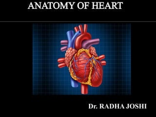
human heart anatomy and physiology
Report
Share
Report
Share

Recommended
Recommended
More Related Content
Similar to HEART ANATOMY AND PHYSIOLOGY.pptx
Similar to HEART ANATOMY AND PHYSIOLOGY.pptx (20)
Anatomy and physiology of the heart by Adeboye Oluwajuyitan

Anatomy and physiology of the heart by Adeboye Oluwajuyitan
Anatomy of heart dr nikunj shekhada (mbbs,ms gen surg ,dnb cts SR) 11 6-18

Anatomy of heart dr nikunj shekhada (mbbs,ms gen surg ,dnb cts SR) 11 6-18
Recently uploaded
PEMESANAN OBAT ASLI : +6287776558899
Cara Menggugurkan Kandungan usia 1 , 2 , bulan - obat penggugur janin - cara aborsi kandungan - obat penggugur kandungan 1 | 2 | 3 | 4 | 5 | 6 | 7 | 8 bulan - bagaimana cara menggugurkan kandungan - tips Cara aborsi kandungan - trik Cara menggugurkan janin - Cara aman bagi ibu menyusui menggugurkan kandungan - klinik apotek jual obat penggugur kandungan - jamu PENGGUGUR KANDUNGAN - WAJIB TAU CARA ABORSI JANIN - GUGURKAN KANDUNGAN AMAN TANPA KURET - CARA Menggugurkan Kandungan tanpa efek samping - rekomendasi dokter obat herbal penggugur kandungan - ABORSI JANIN - aborsi kandungan - jamu herbal Penggugur kandungan - cara Menggugurkan Kandungan yang cacat - tata cara Menggugurkan Kandungan - obat penggugur kandungan di apotik kimia Farma - obat telat datang bulan - obat penggugur kandungan tuntas - obat penggugur kandungan alami - klinik aborsi janin gugurkan kandungan - ©Cytotec ™misoprostol BPOM - OBAT PENGGUGUR KANDUNGAN ®CYTOTEC - aborsi janin dengan pil ©Cytotec - ®Cytotec misoprostol® BPOM 100% - penjual obat penggugur kandungan asli - klinik jual obat aborsi janin - obat penggugur kandungan di klinik k-24 || obat penggugur ™Cytotec di apotek umum || ®CYTOTEC ASLI || obat ©Cytotec yang asli 200mcg || obat penggugur ASLI || pil Cytotec© tablet || cara gugurin kandungan || jual ®Cytotec 200mcg || dokter gugurkan kandungan || cara menggugurkan kandungan dengan cepat selesai dalam 24 jam secara alami buah buahan || usia kandungan 1_2 3_4 5_6 7_8 bulan masih bisa di gugurkan || obat penggugur kandungan ®cytotec dan gastrul || cara gugurkan pembuahan janin secara alami dan cepat || gugurkan kandungan || gugurin janin || cara Menggugurkan janin di luar nikah || contoh aborsi janin yang benar || contoh obat penggugur kandungan asli || contoh cara Menggugurkan Kandungan yang benar || telat haid || obat telat haid || Cara Alami gugurkan kehamilan || obat telat menstruasi || cara Menggugurkan janin anak haram || cara aborsi menggugurkan janin yang tidak berkembang || gugurkan kandungan dengan obat ©Cytotec || obat penggugur kandungan ™Cytotec 100% original || HARGA obat penggugur kandungan || obat telat haid 1 bulan || obat telat menstruasi 1-2 3-4 5-6 7-8 BULAN || obat telat datang bulan || cara Menggugurkan janin 1 bulan || cara Menggugurkan Kandungan yang masih 2 bulan || cara Menggugurkan Kandungan yang masih hitungan Minggu || cara Menggugurkan Kandungan yang masih usia 3 bulan || cara Menggugurkan usia kandungan 4 bulan || cara Menggugurkan janin usia 5 bulan || cara Menggugurkan kehamilan 6 Bulan
________&&&_________&&&_____________&&&_________&&&&____________
Cara Menggugurkan Kandungan Usia Janin 1 | 7 | 8 Bulan Dengan Cepat Dalam Hitungan Jam Secara Alami, Kami Siap Meneriman Pesanan Ke Seluruh Indonesia, Melputi: Ambon, Banda Aceh, Bandung, Banjarbaru, Batam, Bau-Bau, Bengkulu, Binjai, Blitar, Bontang, Cilegon, Cirebon, Depok, Gorontalo, Jakarta, Jayapura, Kendari, Kota Mobagu, Kupang, LhokseumaweCara Menggugurkan Kandungan Dengan Cepat Selesai Dalam 24 Jam Secara Alami Bu...

Cara Menggugurkan Kandungan Dengan Cepat Selesai Dalam 24 Jam Secara Alami Bu...Cara Menggugurkan Kandungan 087776558899
TEST BANK For Porth's Essentials of Pathophysiology, 5th Edition by Tommie L Norris, Verified Chapters 1 - 52, Complete Newest VersionTEST BANK For Porth's Essentials of Pathophysiology, 5th Edition by Tommie L ...

TEST BANK For Porth's Essentials of Pathophysiology, 5th Edition by Tommie L ...rightmanforbloodline
Recently uploaded (20)
Obat Aborsi Ampuh Usia 1,2,3,4,5,6,7 Bulan 081901222272 Obat Penggugur Kandu...

Obat Aborsi Ampuh Usia 1,2,3,4,5,6,7 Bulan 081901222272 Obat Penggugur Kandu...
Difference Between Skeletal Smooth and Cardiac Muscles

Difference Between Skeletal Smooth and Cardiac Muscles
VIP ℂall Girls Kothanur {{ Bangalore }} 6378878445 WhatsApp: Me 24/7 Hours Se...

VIP ℂall Girls Kothanur {{ Bangalore }} 6378878445 WhatsApp: Me 24/7 Hours Se...
Cara Menggugurkan Kandungan Dengan Cepat Selesai Dalam 24 Jam Secara Alami Bu...

Cara Menggugurkan Kandungan Dengan Cepat Selesai Dalam 24 Jam Secara Alami Bu...
Circulatory Shock, types and stages, compensatory mechanisms

Circulatory Shock, types and stages, compensatory mechanisms
MOTION MANAGEMANT IN LUNG SBRT BY DR KANHU CHARAN PATRO

MOTION MANAGEMANT IN LUNG SBRT BY DR KANHU CHARAN PATRO
TEST BANK For Porth's Essentials of Pathophysiology, 5th Edition by Tommie L ...

TEST BANK For Porth's Essentials of Pathophysiology, 5th Edition by Tommie L ...
Creeping Stroke - Venous thrombosis presenting with pc-stroke.pptx

Creeping Stroke - Venous thrombosis presenting with pc-stroke.pptx
Part I - Anticipatory Grief: Experiencing grief before the loss has happened

Part I - Anticipatory Grief: Experiencing grief before the loss has happened
Dr. A Sumathi - LINEARITY CONCEPT OF SIGNIFICANCE.pdf

Dr. A Sumathi - LINEARITY CONCEPT OF SIGNIFICANCE.pdf
ANATOMY AND PHYSIOLOGY OF REPRODUCTIVE SYSTEM.pptx

ANATOMY AND PHYSIOLOGY OF REPRODUCTIVE SYSTEM.pptx
HEART ANATOMY AND PHYSIOLOGY.pptx
- 2. Lies within the pericardium in middle mediastinum. Behind the body of sternum and from 2nd to 5th inter-costal spaces. A third of it lies to the right of median plane and 2/3 to the left.
- 3. A hollow muscular organ, pyramidal in shape, Consists of four chambers( right and left atria, right and left ventricles). Cardiac Apex is formed by left ventricle and is directed downwards and forwards to the left. It lies at the level of the 5th left intercostal space. EXTERNAL CHARACTERISTIC
- 4. ►Approximately the size of your fist. ►Wt.= 250-300 grams ►Cardiac base is formed by the left atrium and to a small extent by the right atrium. It faces backward, upward and to the right.
- 5. Two surfaces Sternocostal surface Diaphragmatic surface Three borders Right border Left border Inferior border
- 6. FOUR GROOVES ■ Coronary sulcus which marks the the division between atria and ventricles ■ Interatrial groove separates the two atria ■ Interventricular groove- anterior and posterior, marks the division between ventricles
- 7. ●Pericardium- a double walled sac around the heart. Composed of: A superficial fibrous pericardium A deep two-layer serous pericardium Interatrial septum Located between right and left atria. Contains fossa ovalis Interventricular septum Located between right and left atria
- 8. Epicardium- visceral pericardium Myocardium- cardiac muscle layer forming the bulk of the heart Endocardium- endothelial layer of the inner myocardial surface
- 9. Atria- receiving chambers of the heart. ■Receive venous blood returning to heart. ■Separated by an interatrial septum. ■Blood enters right atria from superior and inferior vena cava and coronary sinus. ■Blood enters left atria from pulmonary veins.
- 10. VENTRICLE OF THE HEART Ventricles- are the discharging chambers of the heart. ■Separated by an interventricular septum Contains components of the conduction system. ■Right ventricle pumps blood into the pulmonary trunk. ■Left ventricle pumps blood into the aorta.
- 11. Two major types: ■Atrioventricular valves ■Semilunar valves Atrioventricular(AV) valves lie between the atria and the ventricles R-AV VALVE = Tricuspid valve L-AV VALVE= Bicuspid or mitral valve
- 12. ■Semilunar valves prevent backflow of blood into the ventricles. ■Aortic semilunar valve lies between the Ventricles and pulmonary trunk. ■Pulmonary semilunar valve lies between the right ventricle and pulmonary trunk. ■Heart sounds (“lub-dub”) due to valves Closing “Lub”- closing of atrioventricular valve “Dub”- closing of semilunar valves
- 13. The heart is supplied by sympathetic and parasympathetic fibers via the cardiac plexus situated below the arch of aorta. The beating of the heart is regulated by the Intrinsic conduction (Nodal) system. 1. Sinoatrial(SA)node[Pacemaker] located in the right atrium it generates the impulse. 2. Atrioventricular(AV)node is located at the junction of the atria and the ventricles 3. Atrioventricular(AV)bundle(bundle of his) is located in the interventricular septum.
- 14. The arterial supply of the heart is provided by the right and left coronary arteries Drains into the right atrium through coronary sinus. It is continuation of great cardiac vein
- 15. Clinical correlation of various anatomical structures 1.Congenital Atrial Septal Defects Failure of fusion of septum primum and septum secundum leads to a patent foramen ovale after birth. 2. Endocarditis Inflammation of endocardium is referred to endocarditis and that of myocardium as myocarditis. 3. Mitral stenosis Due to infection and subsequent fibrosis , the cusps become thick with reduced mobility often fuse with each other.
- 16. 4.Pericarditis Inflammation of pericardium 5.Pericardial effusion The pericardial cavity may be filled by fluid 6.Aortic aneurysms A dilatation of a segment of aorta is called as aneurysm 7.Myocardial infarction Complete blockage of a branch of a coronary artery leads to death of a part of myocardium supplied by that branch. 8.Atherosclerosis Building up of fats, cholesterol and other substances in and on artery walls.
- 17. The heartbeat originates in a specialized cardiac conduction system. The heart beats normally in an orderly sequence: Contraction of atria (atrial systole) is followed by contraction of the ventricles (ventricular systole) and during diastole all four chambers are relaxed.
- 18. Action potentials (electrical impulses) in the heart originate in specialized cardiac muscle cells, called autorhythmic cells. These cells are self-excitable, able to generate an action potential without external stimulation by nerve cells. The autorhythmic cells serve as a pacemaker to initiate the cardiac cycle and provide a conducting system to coordinate the contraction of muscle cells throughout the heart.
- 19. The autorhythmic cells are concentrated in the structures that make up the conduction system The sinoatrial node (SA node) The internodal atrial pathways The atrioventricular node (AV node) The bundle of His and its branches The purkinje system.
- 20. Sino atrial node It is a part of the wall of right atrium close to the opening of superior venacava. It generates impulses approximately at the rate of 72 times/min. Atrio ventricular node It is a part of the wall of right atrium close to the atrioventricular septum and near to the tricuspid valve. It generates impulses approximately at the rate of 60 times/min.
- 21. Bundle of His It is a thick band of muscle fibers starting from AV node. It generates impulses approximately at the rate of 40 times/min. Purkinje Fibers These fibers arise from the branches of bundle of His. This fibers pierce into the ventricular myocardium.