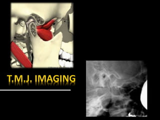
SlideShare a Scribd company logo
1 of 139
 Synovial joint
 Temporal part
 Mandibular part
 inferior aspect of squamous part of temporal bone
 Composed of
 Articular fossa and Articular eminence
 fibrous connective tissue located between the
condyle head and mandibular fossa
 Biconcave in shape
 10 mm anteroposterier
 20 mm mediolateral
 Superior
 Inferior
 Superior compartment
 3 times larger than the inferior compartment
 Anterior recess and posterior recess
 Inferior compartment
 Encloses the entire neck of the mandible
 Divided into anterior and posterior recesses.
 THICK ANTERIOR BAND
 Attached to superior head of lateral pterygoid muscle
 THIN CENTRAL BAND
 serve as articulating cushion between condyle and
articulating eminence
 THICKER POSTERIOR BAND
 Attached to retrodiskal tissue
 Bilaminar zone of vascularised and innervated loose
fibro elastic tissue
 Superior lamina inserts into posterior wall of mandibular fossa
 Inferior lamina attached to posterior surface of condyle
 Posterior attachment is covered with synovial membrane
 secretes synovial fluid
 lubricates the joint
 Evaluating Integrity Of Structures
 Determine The Extent Of Disease
 Monitor Disease Progression
 Monitor Effectiveness Of Treatment
 Exclude Other Causes For Symptoms
 Specific Clinical Problem
 Diagnostic Information Available
 Cost Of Examination
 Contraindications
 Availability Of Equipment
 Transcranial
 Transpharyngeal
 Transorbital
 Submentovertex
 Reverse Towne’s
 Orthopantomography
 Conventional Tomography
 Computer Tomography
 Cone Beam C.T.
 Magnetic Resonance Imaging
 Nuclear Medicine
 S.P.E.C.T.
 P.E.T.
 Ultrasonography
 Arthrography
 Arthroscopy
 Schüllers Technique
 Sagittal view of Lateral part of the condyle
and temporal components
 X-ray directed parallel to long axis of condyle
 Film is positioned against facial skin on side of
interest parallel to sagittal plane
 X ray tube head is projected from opposite side
 Central beam is projected downwards 25⁰ and
centered on T.M.J.
 Central beam is projected through cranium above
petrous ridge of temporal bone on film side
through T.M.J. in line with long axis of condyle
 Horizontal angle not more than 10⁰
 Vertical angle not more than 25⁰
Open mouth Closed mouth
 Deviation in horizontal axis
 Anteroposterior distortion
 Deviation in vertical axis
 shortening or elongation of condyle
 Superimposition
 Parma projection
 Mcqueen projection
 Infracranial projection
 Sagittal view of medial pole of condyle
 Open mouth technique to avoid superimposition
of condyle on temporal components
 Cassette is positioned against the side of patient head
 parallel to sagittal plane
 Tube head is placed on side of skull
 opposite the T.M.J. to be imaged
 Central beam is directed cranially 5-10⁰ and posteriorly 10⁰
 X ray pass through the sigmoid notch below the base of skull
through oropharynx
 to the film and oblique to long axis of condyle
patient is holding the film against the left T.M.J., the mouth is open
and the X-ray beam is aimed across the pharynx.
The side of the face with various anatomical structures zygomatic arch, condyle, sigmoid
notch and coronoid process drawn in to clarify the centering point of the X-ray beam
positioning from the front showing the film parallel to the sagittal plane and the
X-ray beam aimed across the pharynx
positioning from above showing the X-ray beam aimed slightly posteriorly
across the pharynx
 Provide gross visualization of condylar process
 Helps in diagnosing condylar fractures and gross
alteration in form
 If the angulation of condylar axis are low
 condylar profile will be better seen in this view
 If no mouth opening, superior portion of condyle
will be superimposed by Articular eminence
 No information about glenoid fossa
 Radiation dose is high when target to film
distance is short
 Temporal component is not well imaged
 Transmaxillary
 Zimmer projection
 Provides anterior view of T.M.J.
 X ray is directed perpendicular to long axis of condyle
 It have minimum superimposition
 Patient is seated in upright position
 Head is tipped downward for 10⁰
 Tube head is positioned infront of the patient
 Cassette is placed behind the patient
 X ray passes through the ipsilateral orbit through T.M.J.
of interest existing from skull behind mastoid process
 Patient is asked to open mouth widely to allow
profile visualization of entire lateromedial range
of articulating surface of condyle and Articular
eminence
 Simpler technique
 Less superimposition
 Erosive condylar changes are seen
 Open mouth position will show major portion of
condyle
 Glenoid fossa is not clear
 Superimposition in compromised mouth opening
 Less accurate
 Reproducibility is difficult
 skull base and condyle
 Angulation of long axis of condylar head
 Provides a view of T.M.J. in lateral plane
 evaluating
▪ Displaced Condyles
▪ Rotation Of Horizontal Plane Associated With Trauma Or Facial
Asymmetry
Tracing of angles between the long axis of each condyle and the midsagittal plane.
For tomography views the patient is rotated according to the measured angles to
produce an undistorted radiographic view of each T.M.J.
 To investigate the Articular surface of the
condyles and disease within the joint
 Fractures of the condylar heads and necks
 Patient is positioned facing the film
 Head tipped downward
 Forehead and nose touching the cassette
 X ray tube is aimed upward at 30⁰ from behind
 Used for screening purposes
 Both condyles can be visualized
 Gross osseous changes can be identified
 Disadvantage of superimposition
Ankylosis treated with total joint prosthesis
Panoramic view shows prosthesis (arrow), consisting of artificial fossa (fixed with six
titanium screws in temporal bone), and artificial condylar process (fixed with seven titanium
screws to mandibular ramus
 Multiple thin image slices, permitting visualization
of anatomic structures
 Exposed in sagittal plane
 Condylar long axis position is determined with
respect to mid sagittal plane using S.M.V.
projection
Frontal tomographic section of the mandibular condyle.
 Both x ray beam and film moves in opposite direction
 Moving x ray beam focus on a fixed region and project
it on moving film at same area
 Structures located out of focus is blurred
 Structures located within the focus is clear
1.Conventional tomography 1 year previously showed normal position of condyle in
fossa and normal bone; note in particular the eminence (arrow).
2.Tomography now shows condyle displaced anteriorly (probably due to joint
effusion) and erosion in articular eminence (arrow)
 Before C.T.,S.M.V. is taken to assess orientation of
condylar long axis
 Because images are obtained after individual
adjustments based on condylar axis at different
position and different mandibular postures
Dorsal displacement of the right, and ventral displacement of the left condyle suggest a
rotatory movement around the right condyle. After using an axial film to check the
inclination of the condylar axis with regard to the median sagittal plane (black) and to
the frontal plane (white), axially adjusted and precisely positioned tomography can
provide excellent results, and in some cases may even depict the Articular disc.
 Linear tomography
 Multidirectional hypocycloidal tomography
 Multidirectional computer-controlled spiral
tomography
Spiral tomography of the same patient
Left: A normally structured and positioned condyle.
Right: The ventral position and the advanced arthrosis of the condyle are
clearly visible in this case with perforated disc.
Two 6- mm thick coronal tomographs of the same left condylar head.
 Imaging method that combines multiple X rays
taken at different angles to create cross-sectional
images of the body.
 Each image is considered a ‘‘slice’’ and can be
reformatted to create a 3-Dimensional image
Psoriatic Arthropathy.
Oblique coronal (A) and oblique sagittal (C) CT images show punched-out erosion in
lateral part of condyle (arrow).
Similar CT sections of contralateral joint show normal bone
Coronal CT shows enlarged condyle with irregular outline and mineralization (arrow).
 The shape of the condyle and the condition of the
Articular surface
 The condition of the glenoid fossa and eminence
 The position and shape of the disc
 The integrity of the disc and its soft tissue
attachments
 The nature of any condylar head disease
Axial CT scan in bone windows. White arrows indicate bilateral condyle fractures with
anteromedial dislocation
Coronal CT scan in bone windows. White arrows indicate bilateral condyle fractures
with medial displacement
 Uses a cone shaped x ray beam
 Beam performs a single rotation around the
head of patient at an constant angle, producing
volumetric data set, which is laser reconstructed
into 3-dimensional pictures
Cone beam CT bone anatomy of normal temporomandibular joint upper right axial)
lower left oblique sagittal
lower right oblique coronal
Coronoid hyperplasia.
3D CT images at closed (A) and opened mouth (B) show enlarged coronoid process
(arrow) which does not allow normal motion of mandible
 Can depict tiny bony structures with fine details
 Sharper than conventional
 Whole head can be obtained
 Can be represented in 3D format-using
Multiplanar Reconstruction
 Radiation Exposure
 Expense
 Accessibility
 Size Of The Equipment
 Most accurate radiographic imaging modality
 disk position and associated soft tissue structures
 images are presented in T1- and T2-weighted sequences
 T1-weighted for visualization of osseous and disk tissues
 T2-weighted demonstrate inflammation and effusions
 Allows construction of image in sagittal and coronal
planes
 Images are acquired in open and closed mandibular
position using surface coils to improve image
resolution
upper left oblique sagittal MRI, upper right oblique coronal MRI,
lower left oblique sagittal section, lower right oblique coronal section,
 Analyze The Position Of The Articular Disk In Sagittal And Coronal Planes
 Dynamic Assessment Of Condylar Translation
 Disk Movement During Opening And Closing
 Disk Morphology
 Joint Effusions
 Synovitis
 Osseous Erosions
 Degenerative Joint Disease
 Internal Derangement
 Thickening Of Tendon Attachments,
 Rupture Of
 Retrodiscal Tissues,
 Osteoarthritic Changes Such As Condylar Flattening Or Osteophyte Formation
MRI shows completely destroyed disc, replaced by fibrous or vascular pannus and cortical
punched-out erosion (arrow) with sclerosis (shown by CT) in condyle.
B Autopsy specimen shows no disc structure but pannus, and erosion in condyle (arrow) from
patient with known long-standing rheumatoid arthritis
T1-weighted axial MRI. White arrow indicates cystic lesion of the left condyle with fluid levels.
T2-weighted axial MRI. White arrow indicates cystic lesion of the left condyle with fluid
levels. MS, maxillary sinus
T1-weighted sagittal MRI of T.M.J.. Solid white arrow indicates articular disk anteriorly
displaced. Broken white arrow indicates a joint effusion. C, condyle; LP, lateral pterygoid;
M, mastoid air cells.
T2-weighted sagittal view of T.M.J.. Solid white arrow indicates articular disk anteriorly
displaced. Broken white arrow indicates a joint effusion. C, condyle; LP, lateral pterygoid;
M, mastoid air cells.
T2-weighted sagittal MRI. White arrow indicates a joint effusion. C, condyle
 Motion images during opening and closing
can be obtained by having the patient open in
a series of stepped distance and using rapid
image acquisition(fast scan technique)
ADVANTAGES
 Precise information of
lesions
 Detects minute changes
 Non invasive method
DISADVANTAGES
 Expensive
 Skilled professionals
 Contraindications
 Metal prosthesis
 pacemakers
 Scintigraphy aids to discover early changes in
T.M.J. as a direct result of biochemical
alterations at cellular and sub cellular levels
 used as a physiologic adjunct to the anatomic
detail provided by other imaging modalities
 Uses radionuclide labeled tracers injected I.V.
 Emits gamma radiation
 Most commonly used are Technetium Diphosphonate
 Low Radiation
 Short Half Life
 A gamma scintillation camera ,which can
fluorescence on interaction with gamma rays will
detect the emitted radiation
 Fluorescence is then amplified by photomultiplier
to produce image
 Radionuclide Imaging Or Nuclear Scintigraphy
 Acquires multiple images by rotating gamma
scintillation camera 360⁰ around patient
 Slices is then stacked to give 3-D representation
 Improved resolution and better anatomic localization
 Uses Positron Emitting Isotopes
 Positron interact with adjacent electrons to produce 2
gamma rays travelling in opposite directions
 Multiple detectors are placed with P.E.T. scanners
 So several gamma emissions can be detected at nearly
the same time with the process of Annihilation
Coincidence Detection
 Assessing skeletal growth
 Condylar hyperplasia
 Synovitis
 Quantification of arthritis in R.A.
 determine joint stability before dental rehabilitation
 Diagnosing fibro osseous lesions, metastatic diseases
 Inability to reveal morphology of osseous components
 Inability to Reveal disk displacements
 Non specificity
Coronal view of SPECT bone scan.
White arrow indicates increased uptake in the lesion in the left condyle.
Axial view of SPECT bone scan.
White arrow indicates increased uptake in the lesion in the left condyle.
 Radiographic study where contrast material has
been injected into lower joint compartment or
lower and upper compartments to visualize soft
tissues such as Articular disc and joint capsule
Double-contrast dual space arthrotomography, i.e., both joint compartments injected
separately; normal disc position in half open mouth view as indicated by anterior band
(arrowhead) anterior to condyle (C) and posterior band (arrow) posterior to condyle; joint
spaces also filled with air and thus appear radiolucent. Articular eminence indicated
Single-contrast dual space arthrotomography; upper space filled through perforation (not
seen) in area of disc/posterior attachment, open mouth view; disc anteriorly displaced
without reduction as indicated by its posterior band (arrow) in front of condyle (C). Articular
eminence indicated (E)
Single-contrast lower space arthrotomography; open mouth view shows contrast
material in front of condyle (C) demonstrating anteriorly displaced disc (arrow)
without reduction. Articular eminence indicated (E).
Same joint, closed mouth view; contrast material in extended anterior recess
showing lower surface of posterior band (arrow) of anteriorly displaced disc in front
of condyle (C). Articular eminence indicated (E)
 Position And Morphology Of Discs
 Perforation Of Articular Discs
 Joint Adhesion
 Soft Tissue Integrity
 Iodine Hypersensitivity
 Acute Infections
 Position of mandibular fossa and head is confirmed
by palpation on a point of 10mm from the tragus
on the line between midpoint of tragus and lateral
angle of eyes
 Tracing puncture point
 Infiltrating anaesthesia
 Puncture point reached by fluoroscopy
 Lower compartment can be reached from side of face by
directing needle forward back of condyle through
external auditory meatus
 Upper compartment from side of face under zygomatic
arch when mouth is opened
 Injection of medium
 Imaging under fluoroscopy using x ray video system
 Monitoring
 Confirmation of puncture site
 Confirmation of injection of contrast medium
into upper and lower Articular cavities
 Observation of morphology and position of
disc perforation
 Inspection of joint surfaces by performing
minimally invasive surgical procedures
percutaneously using an Arthroscope
 Allows direct visualization of joint
 Diagnosis of adhesions and Synovitis are the main
applications for arthroscopy.
Upper joint compartment; normal joint as indicated by smooth surfaces of
both disc and eminence
Upper joint compartment; fibrous adhesion between disc and eminence (arrow
Upper joint compartment; synovial proliferation (arrowhead) in disc perforation
area,hyperemia (small arrowhead), and part of disc (arrow
Same joint; synovial proliferation (arrowhead) in disc perforation area,
and part of disc (arrow)
 Ultrasonography uses sound waves of high
frequency to produce images of body.
 Serves for diagnosis and comparison of
therapeutic results in treating internal joint
defects
 Scanning transducer of 7.5-12 MHz is used
 Depicts narrow space of joint
 Position of disc
 Fluid and ligament adhesion
 Transducer is placed on skin above joint parallel to long
axis of mandible
 Disc seen as thin homogenous hypoechoic areas
 Condylar borders and Articular eminence shows a
hyperechogenic lines
 it is possible to directly observe the joint disc
movement, when the mouth is opening and closing
 Recently, high frequency large diameter transducer is
introduced
 Able to penetrate easily through small aperture between
glenoid fossa and condyle
 High focus depth and narrow wave beam
 When evaluating T.M.J. disk position for internal
derangement, ultrasonography has shown some benefit,
especially when high-resolution, dynamic, real-time
ultrasonography is used
g
linear transducer positioned against the patents face in a horizontal direction overlying
the zygomatic arch and t.m.j. & 25° to mid sagittal plane
 Reduced Cost
 Accessibility
 Fast Results
 Decreased Examination Time
 Lack Of Radiation Exposure.
 MRI continues to be the gold standard for
imaging soft tissues
 CT is the ideal imaging choice for evaluating
hard tissues
 use of the new cone-beam CT. For more specific
T.M.J. pathology,
 As advancements in this area continue,
our understanding of this complex joint
and its pathology will follow, which will
lead to more defined imaging indications
and ultimately, to improved treatment
TMJ IMAGING.pptx
TMJ IMAGING.pptx
TMJ IMAGING.pptx

More Related Content

What's hot (20)

Intra oral radiographs
Intra oral radiographsIntra oral radiographs
Intra oral radiographs
 
SIALOGRAPHY.pptx
SIALOGRAPHY.pptxSIALOGRAPHY.pptx
SIALOGRAPHY.pptx
 
Orthopantomogram
OrthopantomogramOrthopantomogram
Orthopantomogram
 
TMJ Radiology & Applied Aspect
TMJ Radiology & Applied AspectTMJ Radiology & Applied Aspect
TMJ Radiology & Applied Aspect
 
Quality assurance in dental radiography
Quality assurance in dental radiographyQuality assurance in dental radiography
Quality assurance in dental radiography
 
Cbct
CbctCbct
Cbct
 
Nuclear imaging in dentistry
Nuclear imaging in dentistryNuclear imaging in dentistry
Nuclear imaging in dentistry
 
CBCT.pptx
CBCT.pptxCBCT.pptx
CBCT.pptx
 
Panoramic radiography
Panoramic radiographyPanoramic radiography
Panoramic radiography
 
Ideal radiography
Ideal radiographyIdeal radiography
Ideal radiography
 
radiology-bitewing-technique
 radiology-bitewing-technique radiology-bitewing-technique
radiology-bitewing-technique
 
radiology-paralleling-technique
radiology-paralleling-techniqueradiology-paralleling-technique
radiology-paralleling-technique
 
Panoramic Radiography
Panoramic RadiographyPanoramic Radiography
Panoramic Radiography
 
Cbct
CbctCbct
Cbct
 
Opg
OpgOpg
Opg
 
Extraoral radiography
Extraoral radiographyExtraoral radiography
Extraoral radiography
 
Extra Oral Radiographic Techniques I
Extra Oral Radiographic Techniques IExtra Oral Radiographic Techniques I
Extra Oral Radiographic Techniques I
 
Cephalometrics in orthodontics
Cephalometrics in orthodonticsCephalometrics in orthodontics
Cephalometrics in orthodontics
 
Panaromic radiography
Panaromic radiographyPanaromic radiography
Panaromic radiography
 
Cone beam ct
Cone beam ctCone beam ct
Cone beam ct
 

Similar to TMJ IMAGING.pptx

temporomandibularjointimaging-180225163412 2.pdf
temporomandibularjointimaging-180225163412 2.pdftemporomandibularjointimaging-180225163412 2.pdf
temporomandibularjointimaging-180225163412 2.pdfhashimedavath
 
1_updated_temporomandibularjointimaging-180225163412 2.pdf
1_updated_temporomandibularjointimaging-180225163412 2.pdf1_updated_temporomandibularjointimaging-180225163412 2.pdf
1_updated_temporomandibularjointimaging-180225163412 2.pdfhashimedavath
 
Temporomandibular joint imaging
Temporomandibular joint imagingTemporomandibular joint imaging
Temporomandibular joint imagingDr Jinki Singha
 
Presentation (1) omr.pptx
Presentation (1) omr.pptxPresentation (1) omr.pptx
Presentation (1) omr.pptxAswathySuresh37
 
Radiology of ear - DR. ROHIT BHARDWAJ.pptx
Radiology of ear - DR. ROHIT BHARDWAJ.pptxRadiology of ear - DR. ROHIT BHARDWAJ.pptx
Radiology of ear - DR. ROHIT BHARDWAJ.pptxRohit Bhardwaj
 
B.Sc RADIOLOGY QUESTION AND ANSWER BANK - POSITIONING
B.Sc RADIOLOGY QUESTION AND ANSWER BANK - POSITIONING B.Sc RADIOLOGY QUESTION AND ANSWER BANK - POSITIONING
B.Sc RADIOLOGY QUESTION AND ANSWER BANK - POSITIONING Ganesan Yogananthem
 
Radiographic Features of Temporomandibular Joint and Disorders
Radiographic Features of Temporomandibular Joint and DisordersRadiographic Features of Temporomandibular Joint and Disorders
Radiographic Features of Temporomandibular Joint and DisordersHadi Munib
 
Temporomandibular joint imaging 2 /certified fixed orthodontic courses by Ind...
Temporomandibular joint imaging 2 /certified fixed orthodontic courses by Ind...Temporomandibular joint imaging 2 /certified fixed orthodontic courses by Ind...
Temporomandibular joint imaging 2 /certified fixed orthodontic courses by Ind...Indian dental academy
 
SANTOSH jaiswal Radiology of Nose n PNS.pptxbackup.pptx
SANTOSH jaiswal Radiology of Nose n PNS.pptxbackup.pptxSANTOSH jaiswal Radiology of Nose n PNS.pptxbackup.pptx
SANTOSH jaiswal Radiology of Nose n PNS.pptxbackup.pptxSantosh Jaiswal
 
IMAGING OF TEMPORAL BONE
IMAGING OF TEMPORAL BONEIMAGING OF TEMPORAL BONE
IMAGING OF TEMPORAL BONESameer Peer
 
Presentation1.pptx, ultrasound examination of the shoulder joint.
Presentation1.pptx, ultrasound examination of the shoulder joint.Presentation1.pptx, ultrasound examination of the shoulder joint.
Presentation1.pptx, ultrasound examination of the shoulder joint.Abdellah Nazeer
 
Anatomy and Radiography of shoulder and arm
Anatomy and Radiography of shoulder and armAnatomy and Radiography of shoulder and arm
Anatomy and Radiography of shoulder and armPrasanta Nath
 
MRI procedure of pelvis and hip suman duwal
MRI procedure of pelvis and hip suman duwalMRI procedure of pelvis and hip suman duwal
MRI procedure of pelvis and hip suman duwalsuman duwal
 
MRI KNEE JOINT ANATOMY
MRI KNEE JOINT ANATOMYMRI KNEE JOINT ANATOMY
MRI KNEE JOINT ANATOMYNikhil Bansal
 

Similar to TMJ IMAGING.pptx (20)

Tmj joint imaging
Tmj joint imagingTmj joint imaging
Tmj joint imaging
 
temporomandibularjointimaging-180225163412 2.pdf
temporomandibularjointimaging-180225163412 2.pdftemporomandibularjointimaging-180225163412 2.pdf
temporomandibularjointimaging-180225163412 2.pdf
 
1_updated_temporomandibularjointimaging-180225163412 2.pdf
1_updated_temporomandibularjointimaging-180225163412 2.pdf1_updated_temporomandibularjointimaging-180225163412 2.pdf
1_updated_temporomandibularjointimaging-180225163412 2.pdf
 
Temporomandibular joint imaging
Temporomandibular joint imagingTemporomandibular joint imaging
Temporomandibular joint imaging
 
Presentation (1) omr.pptx
Presentation (1) omr.pptxPresentation (1) omr.pptx
Presentation (1) omr.pptx
 
Radiology of ear - DR. ROHIT BHARDWAJ.pptx
Radiology of ear - DR. ROHIT BHARDWAJ.pptxRadiology of ear - DR. ROHIT BHARDWAJ.pptx
Radiology of ear - DR. ROHIT BHARDWAJ.pptx
 
B.Sc RADIOLOGY QUESTION AND ANSWER BANK - POSITIONING
B.Sc RADIOLOGY QUESTION AND ANSWER BANK - POSITIONING B.Sc RADIOLOGY QUESTION AND ANSWER BANK - POSITIONING
B.Sc RADIOLOGY QUESTION AND ANSWER BANK - POSITIONING
 
Tmj imaging
Tmj imagingTmj imaging
Tmj imaging
 
Radiographic Features of Temporomandibular Joint and Disorders
Radiographic Features of Temporomandibular Joint and DisordersRadiographic Features of Temporomandibular Joint and Disorders
Radiographic Features of Temporomandibular Joint and Disorders
 
Imaging of TMJ
Imaging of  TMJImaging of  TMJ
Imaging of TMJ
 
Temporomandibular joint imaging 2 /certified fixed orthodontic courses by Ind...
Temporomandibular joint imaging 2 /certified fixed orthodontic courses by Ind...Temporomandibular joint imaging 2 /certified fixed orthodontic courses by Ind...
Temporomandibular joint imaging 2 /certified fixed orthodontic courses by Ind...
 
Elbow joint
Elbow joint Elbow joint
Elbow joint
 
SANTOSH jaiswal Radiology of Nose n PNS.pptxbackup.pptx
SANTOSH jaiswal Radiology of Nose n PNS.pptxbackup.pptxSANTOSH jaiswal Radiology of Nose n PNS.pptxbackup.pptx
SANTOSH jaiswal Radiology of Nose n PNS.pptxbackup.pptx
 
IMAGING OF TEMPORAL BONE
IMAGING OF TEMPORAL BONEIMAGING OF TEMPORAL BONE
IMAGING OF TEMPORAL BONE
 
Swimmer’s view.pptx
Swimmer’s view.pptxSwimmer’s view.pptx
Swimmer’s view.pptx
 
Presentation1.pptx, ultrasound examination of the shoulder joint.
Presentation1.pptx, ultrasound examination of the shoulder joint.Presentation1.pptx, ultrasound examination of the shoulder joint.
Presentation1.pptx, ultrasound examination of the shoulder joint.
 
Anatomy and Radiography of shoulder and arm
Anatomy and Radiography of shoulder and armAnatomy and Radiography of shoulder and arm
Anatomy and Radiography of shoulder and arm
 
Dr Harshil radio spine
Dr Harshil radio spineDr Harshil radio spine
Dr Harshil radio spine
 
MRI procedure of pelvis and hip suman duwal
MRI procedure of pelvis and hip suman duwalMRI procedure of pelvis and hip suman duwal
MRI procedure of pelvis and hip suman duwal
 
MRI KNEE JOINT ANATOMY
MRI KNEE JOINT ANATOMYMRI KNEE JOINT ANATOMY
MRI KNEE JOINT ANATOMY
 

More from Royal Dental College Library

DENTAL EVIDENCES IN CRIME SCENES AND THE PROCEDURES .pptx......pptx
DENTAL EVIDENCES IN CRIME SCENES AND THE PROCEDURES .pptx......pptxDENTAL EVIDENCES IN CRIME SCENES AND THE PROCEDURES .pptx......pptx
DENTAL EVIDENCES IN CRIME SCENES AND THE PROCEDURES .pptx......pptxRoyal Dental College Library
 

More from Royal Dental College Library (20)

Thyroid Gland Dr Sheeba.pptx
Thyroid Gland Dr Sheeba.pptxThyroid Gland Dr Sheeba.pptx
Thyroid Gland Dr Sheeba.pptx
 
EYE PRESENTATION Dr Sheeba.ppt
EYE PRESENTATION   Dr Sheeba.pptEYE PRESENTATION   Dr Sheeba.ppt
EYE PRESENTATION Dr Sheeba.ppt
 
transport of resp gases Reeshma.ppt
transport of resp gases Reeshma.ppttransport of resp gases Reeshma.ppt
transport of resp gases Reeshma.ppt
 
spermatogenesis Reeshma.PPT
spermatogenesis Reeshma.PPTspermatogenesis Reeshma.PPT
spermatogenesis Reeshma.PPT
 
alveolar bone.pptx
alveolar bone.pptxalveolar bone.pptx
alveolar bone.pptx
 
DENTIN.pptx
DENTIN.pptxDENTIN.pptx
DENTIN.pptx
 
Specialized Mucosa.pptx
Specialized Mucosa.pptxSpecialized Mucosa.pptx
Specialized Mucosa.pptx
 
Pulp.pptx
Pulp.pptxPulp.pptx
Pulp.pptx
 
sequle of pulpitis.pptx
sequle of pulpitis.pptxsequle of pulpitis.pptx
sequle of pulpitis.pptx
 
DENTAL EVIDENCES IN CRIME SCENES AND THE PROCEDURES .pptx......pptx
DENTAL EVIDENCES IN CRIME SCENES AND THE PROCEDURES .pptx......pptxDENTAL EVIDENCES IN CRIME SCENES AND THE PROCEDURES .pptx......pptx
DENTAL EVIDENCES IN CRIME SCENES AND THE PROCEDURES .pptx......pptx
 
Amelogenesis Slideshare.pptx
Amelogenesis Slideshare.pptxAmelogenesis Slideshare.pptx
Amelogenesis Slideshare.pptx
 
P D L Slideshare.ppt
P D L Slideshare.pptP D L Slideshare.ppt
P D L Slideshare.ppt
 
odontogenic tumor 2022.pptx
odontogenic tumor 2022.pptxodontogenic tumor 2022.pptx
odontogenic tumor 2022.pptx
 
Dental Caries.pptx
Dental Caries.pptxDental Caries.pptx
Dental Caries.pptx
 
Ethics in Research.ppt
Ethics in Research.pptEthics in Research.ppt
Ethics in Research.ppt
 
Dental Ethics.ppt
Dental Ethics.pptDental Ethics.ppt
Dental Ethics.ppt
 
SSC.pptx
SSC.pptxSSC.pptx
SSC.pptx
 
ORAL HABITS.ppt
ORAL HABITS.pptORAL HABITS.ppt
ORAL HABITS.ppt
 
space management.ppt
space management.pptspace management.ppt
space management.ppt
 
Medically Compromised.pptx
Medically Compromised.pptxMedically Compromised.pptx
Medically Compromised.pptx
 

Recently uploaded

VIP Call Girls Pune Vani 9907093804 Short 1500 Night 6000 Best call girls Ser...
VIP Call Girls Pune Vani 9907093804 Short 1500 Night 6000 Best call girls Ser...VIP Call Girls Pune Vani 9907093804 Short 1500 Night 6000 Best call girls Ser...
VIP Call Girls Pune Vani 9907093804 Short 1500 Night 6000 Best call girls Ser...Miss joya
 
(Rocky) Jaipur Call Girl - 9521753030 Escorts Service 50% Off with Cash ON De...
(Rocky) Jaipur Call Girl - 9521753030 Escorts Service 50% Off with Cash ON De...(Rocky) Jaipur Call Girl - 9521753030 Escorts Service 50% Off with Cash ON De...
(Rocky) Jaipur Call Girl - 9521753030 Escorts Service 50% Off with Cash ON De...indiancallgirl4rent
 
Aspirin presentation slides by Dr. Rewas Ali
Aspirin presentation slides by Dr. Rewas AliAspirin presentation slides by Dr. Rewas Ali
Aspirin presentation slides by Dr. Rewas AliRewAs ALI
 
Bangalore Call Girl Whatsapp Number 100% Complete Your Sexual Needs
Bangalore Call Girl Whatsapp Number 100% Complete Your Sexual NeedsBangalore Call Girl Whatsapp Number 100% Complete Your Sexual Needs
Bangalore Call Girl Whatsapp Number 100% Complete Your Sexual NeedsGfnyt
 
High Profile Call Girls Coimbatore Saanvi☎️ 8250192130 Independent Escort Se...
High Profile Call Girls Coimbatore Saanvi☎️  8250192130 Independent Escort Se...High Profile Call Girls Coimbatore Saanvi☎️  8250192130 Independent Escort Se...
High Profile Call Girls Coimbatore Saanvi☎️ 8250192130 Independent Escort Se...narwatsonia7
 
♛VVIP Hyderabad Call Girls Chintalkunta🖕7001035870🖕Riya Kappor Top Call Girl ...
♛VVIP Hyderabad Call Girls Chintalkunta🖕7001035870🖕Riya Kappor Top Call Girl ...♛VVIP Hyderabad Call Girls Chintalkunta🖕7001035870🖕Riya Kappor Top Call Girl ...
♛VVIP Hyderabad Call Girls Chintalkunta🖕7001035870🖕Riya Kappor Top Call Girl ...astropune
 
Call Girls Service Navi Mumbai Samaira 8617697112 Independent Escort Service ...
Call Girls Service Navi Mumbai Samaira 8617697112 Independent Escort Service ...Call Girls Service Navi Mumbai Samaira 8617697112 Independent Escort Service ...
Call Girls Service Navi Mumbai Samaira 8617697112 Independent Escort Service ...Call girls in Ahmedabad High profile
 
Call Girls Horamavu WhatsApp Number 7001035870 Meeting With Bangalore Escorts
Call Girls Horamavu WhatsApp Number 7001035870 Meeting With Bangalore EscortsCall Girls Horamavu WhatsApp Number 7001035870 Meeting With Bangalore Escorts
Call Girls Horamavu WhatsApp Number 7001035870 Meeting With Bangalore Escortsvidya singh
 
VIP Russian Call Girls in Varanasi Samaira 8250192130 Independent Escort Serv...
VIP Russian Call Girls in Varanasi Samaira 8250192130 Independent Escort Serv...VIP Russian Call Girls in Varanasi Samaira 8250192130 Independent Escort Serv...
VIP Russian Call Girls in Varanasi Samaira 8250192130 Independent Escort Serv...Neha Kaur
 
Call Girls Darjeeling Just Call 9907093804 Top Class Call Girl Service Available
Call Girls Darjeeling Just Call 9907093804 Top Class Call Girl Service AvailableCall Girls Darjeeling Just Call 9907093804 Top Class Call Girl Service Available
Call Girls Darjeeling Just Call 9907093804 Top Class Call Girl Service AvailableDipal Arora
 
VIP Mumbai Call Girls Hiranandani Gardens Just Call 9920874524 with A/C Room ...
VIP Mumbai Call Girls Hiranandani Gardens Just Call 9920874524 with A/C Room ...VIP Mumbai Call Girls Hiranandani Gardens Just Call 9920874524 with A/C Room ...
VIP Mumbai Call Girls Hiranandani Gardens Just Call 9920874524 with A/C Room ...Garima Khatri
 
VIP Service Call Girls Sindhi Colony 📳 7877925207 For 18+ VIP Call Girl At Th...
VIP Service Call Girls Sindhi Colony 📳 7877925207 For 18+ VIP Call Girl At Th...VIP Service Call Girls Sindhi Colony 📳 7877925207 For 18+ VIP Call Girl At Th...
VIP Service Call Girls Sindhi Colony 📳 7877925207 For 18+ VIP Call Girl At Th...jageshsingh5554
 
VIP Call Girls Pune Vrinda 9907093804 Short 1500 Night 6000 Best call girls S...
VIP Call Girls Pune Vrinda 9907093804 Short 1500 Night 6000 Best call girls S...VIP Call Girls Pune Vrinda 9907093804 Short 1500 Night 6000 Best call girls S...
VIP Call Girls Pune Vrinda 9907093804 Short 1500 Night 6000 Best call girls S...Miss joya
 
Bangalore Call Girls Marathahalli 📞 9907093804 High Profile Service 100% Safe
Bangalore Call Girls Marathahalli 📞 9907093804 High Profile Service 100% SafeBangalore Call Girls Marathahalli 📞 9907093804 High Profile Service 100% Safe
Bangalore Call Girls Marathahalli 📞 9907093804 High Profile Service 100% Safenarwatsonia7
 
Low Rate Call Girls Patna Anika 8250192130 Independent Escort Service Patna
Low Rate Call Girls Patna Anika 8250192130 Independent Escort Service PatnaLow Rate Call Girls Patna Anika 8250192130 Independent Escort Service Patna
Low Rate Call Girls Patna Anika 8250192130 Independent Escort Service Patnamakika9823
 
Call Girl Number in Vashi Mumbai📲 9833363713 💞 Full Night Enjoy
Call Girl Number in Vashi Mumbai📲 9833363713 💞 Full Night EnjoyCall Girl Number in Vashi Mumbai📲 9833363713 💞 Full Night Enjoy
Call Girl Number in Vashi Mumbai📲 9833363713 💞 Full Night Enjoybabeytanya
 
College Call Girls Pune Mira 9907093804 Short 1500 Night 6000 Best call girls...
College Call Girls Pune Mira 9907093804 Short 1500 Night 6000 Best call girls...College Call Girls Pune Mira 9907093804 Short 1500 Night 6000 Best call girls...
College Call Girls Pune Mira 9907093804 Short 1500 Night 6000 Best call girls...Miss joya
 
Kesar Bagh Call Girl Price 9548273370 , Lucknow Call Girls Service
Kesar Bagh Call Girl Price 9548273370 , Lucknow Call Girls ServiceKesar Bagh Call Girl Price 9548273370 , Lucknow Call Girls Service
Kesar Bagh Call Girl Price 9548273370 , Lucknow Call Girls Servicemakika9823
 
Call Girls Cuttack Just Call 9907093804 Top Class Call Girl Service Available
Call Girls Cuttack Just Call 9907093804 Top Class Call Girl Service AvailableCall Girls Cuttack Just Call 9907093804 Top Class Call Girl Service Available
Call Girls Cuttack Just Call 9907093804 Top Class Call Girl Service AvailableDipal Arora
 

Recently uploaded (20)

VIP Call Girls Pune Vani 9907093804 Short 1500 Night 6000 Best call girls Ser...
VIP Call Girls Pune Vani 9907093804 Short 1500 Night 6000 Best call girls Ser...VIP Call Girls Pune Vani 9907093804 Short 1500 Night 6000 Best call girls Ser...
VIP Call Girls Pune Vani 9907093804 Short 1500 Night 6000 Best call girls Ser...
 
(Rocky) Jaipur Call Girl - 9521753030 Escorts Service 50% Off with Cash ON De...
(Rocky) Jaipur Call Girl - 9521753030 Escorts Service 50% Off with Cash ON De...(Rocky) Jaipur Call Girl - 9521753030 Escorts Service 50% Off with Cash ON De...
(Rocky) Jaipur Call Girl - 9521753030 Escorts Service 50% Off with Cash ON De...
 
Aspirin presentation slides by Dr. Rewas Ali
Aspirin presentation slides by Dr. Rewas AliAspirin presentation slides by Dr. Rewas Ali
Aspirin presentation slides by Dr. Rewas Ali
 
Bangalore Call Girl Whatsapp Number 100% Complete Your Sexual Needs
Bangalore Call Girl Whatsapp Number 100% Complete Your Sexual NeedsBangalore Call Girl Whatsapp Number 100% Complete Your Sexual Needs
Bangalore Call Girl Whatsapp Number 100% Complete Your Sexual Needs
 
High Profile Call Girls Coimbatore Saanvi☎️ 8250192130 Independent Escort Se...
High Profile Call Girls Coimbatore Saanvi☎️  8250192130 Independent Escort Se...High Profile Call Girls Coimbatore Saanvi☎️  8250192130 Independent Escort Se...
High Profile Call Girls Coimbatore Saanvi☎️ 8250192130 Independent Escort Se...
 
♛VVIP Hyderabad Call Girls Chintalkunta🖕7001035870🖕Riya Kappor Top Call Girl ...
♛VVIP Hyderabad Call Girls Chintalkunta🖕7001035870🖕Riya Kappor Top Call Girl ...♛VVIP Hyderabad Call Girls Chintalkunta🖕7001035870🖕Riya Kappor Top Call Girl ...
♛VVIP Hyderabad Call Girls Chintalkunta🖕7001035870🖕Riya Kappor Top Call Girl ...
 
Call Girls Service Navi Mumbai Samaira 8617697112 Independent Escort Service ...
Call Girls Service Navi Mumbai Samaira 8617697112 Independent Escort Service ...Call Girls Service Navi Mumbai Samaira 8617697112 Independent Escort Service ...
Call Girls Service Navi Mumbai Samaira 8617697112 Independent Escort Service ...
 
Call Girls Horamavu WhatsApp Number 7001035870 Meeting With Bangalore Escorts
Call Girls Horamavu WhatsApp Number 7001035870 Meeting With Bangalore EscortsCall Girls Horamavu WhatsApp Number 7001035870 Meeting With Bangalore Escorts
Call Girls Horamavu WhatsApp Number 7001035870 Meeting With Bangalore Escorts
 
VIP Russian Call Girls in Varanasi Samaira 8250192130 Independent Escort Serv...
VIP Russian Call Girls in Varanasi Samaira 8250192130 Independent Escort Serv...VIP Russian Call Girls in Varanasi Samaira 8250192130 Independent Escort Serv...
VIP Russian Call Girls in Varanasi Samaira 8250192130 Independent Escort Serv...
 
Russian Call Girls in Delhi Tanvi ➡️ 9711199012 💋📞 Independent Escort Service...
Russian Call Girls in Delhi Tanvi ➡️ 9711199012 💋📞 Independent Escort Service...Russian Call Girls in Delhi Tanvi ➡️ 9711199012 💋📞 Independent Escort Service...
Russian Call Girls in Delhi Tanvi ➡️ 9711199012 💋📞 Independent Escort Service...
 
Call Girls Darjeeling Just Call 9907093804 Top Class Call Girl Service Available
Call Girls Darjeeling Just Call 9907093804 Top Class Call Girl Service AvailableCall Girls Darjeeling Just Call 9907093804 Top Class Call Girl Service Available
Call Girls Darjeeling Just Call 9907093804 Top Class Call Girl Service Available
 
VIP Mumbai Call Girls Hiranandani Gardens Just Call 9920874524 with A/C Room ...
VIP Mumbai Call Girls Hiranandani Gardens Just Call 9920874524 with A/C Room ...VIP Mumbai Call Girls Hiranandani Gardens Just Call 9920874524 with A/C Room ...
VIP Mumbai Call Girls Hiranandani Gardens Just Call 9920874524 with A/C Room ...
 
VIP Service Call Girls Sindhi Colony 📳 7877925207 For 18+ VIP Call Girl At Th...
VIP Service Call Girls Sindhi Colony 📳 7877925207 For 18+ VIP Call Girl At Th...VIP Service Call Girls Sindhi Colony 📳 7877925207 For 18+ VIP Call Girl At Th...
VIP Service Call Girls Sindhi Colony 📳 7877925207 For 18+ VIP Call Girl At Th...
 
VIP Call Girls Pune Vrinda 9907093804 Short 1500 Night 6000 Best call girls S...
VIP Call Girls Pune Vrinda 9907093804 Short 1500 Night 6000 Best call girls S...VIP Call Girls Pune Vrinda 9907093804 Short 1500 Night 6000 Best call girls S...
VIP Call Girls Pune Vrinda 9907093804 Short 1500 Night 6000 Best call girls S...
 
Bangalore Call Girls Marathahalli 📞 9907093804 High Profile Service 100% Safe
Bangalore Call Girls Marathahalli 📞 9907093804 High Profile Service 100% SafeBangalore Call Girls Marathahalli 📞 9907093804 High Profile Service 100% Safe
Bangalore Call Girls Marathahalli 📞 9907093804 High Profile Service 100% Safe
 
Low Rate Call Girls Patna Anika 8250192130 Independent Escort Service Patna
Low Rate Call Girls Patna Anika 8250192130 Independent Escort Service PatnaLow Rate Call Girls Patna Anika 8250192130 Independent Escort Service Patna
Low Rate Call Girls Patna Anika 8250192130 Independent Escort Service Patna
 
Call Girl Number in Vashi Mumbai📲 9833363713 💞 Full Night Enjoy
Call Girl Number in Vashi Mumbai📲 9833363713 💞 Full Night EnjoyCall Girl Number in Vashi Mumbai📲 9833363713 💞 Full Night Enjoy
Call Girl Number in Vashi Mumbai📲 9833363713 💞 Full Night Enjoy
 
College Call Girls Pune Mira 9907093804 Short 1500 Night 6000 Best call girls...
College Call Girls Pune Mira 9907093804 Short 1500 Night 6000 Best call girls...College Call Girls Pune Mira 9907093804 Short 1500 Night 6000 Best call girls...
College Call Girls Pune Mira 9907093804 Short 1500 Night 6000 Best call girls...
 
Kesar Bagh Call Girl Price 9548273370 , Lucknow Call Girls Service
Kesar Bagh Call Girl Price 9548273370 , Lucknow Call Girls ServiceKesar Bagh Call Girl Price 9548273370 , Lucknow Call Girls Service
Kesar Bagh Call Girl Price 9548273370 , Lucknow Call Girls Service
 
Call Girls Cuttack Just Call 9907093804 Top Class Call Girl Service Available
Call Girls Cuttack Just Call 9907093804 Top Class Call Girl Service AvailableCall Girls Cuttack Just Call 9907093804 Top Class Call Girl Service Available
Call Girls Cuttack Just Call 9907093804 Top Class Call Girl Service Available
 

TMJ IMAGING.pptx

  • 1.
  • 2.  Synovial joint  Temporal part  Mandibular part
  • 3.
  • 4.
  • 5.
  • 6.
  • 7.  inferior aspect of squamous part of temporal bone  Composed of  Articular fossa and Articular eminence
  • 8.
  • 9.
  • 10.
  • 11.  fibrous connective tissue located between the condyle head and mandibular fossa  Biconcave in shape  10 mm anteroposterier  20 mm mediolateral
  • 12.
  • 13.  Superior  Inferior  Superior compartment  3 times larger than the inferior compartment  Anterior recess and posterior recess  Inferior compartment  Encloses the entire neck of the mandible  Divided into anterior and posterior recesses.
  • 14.
  • 15.  THICK ANTERIOR BAND  Attached to superior head of lateral pterygoid muscle  THIN CENTRAL BAND  serve as articulating cushion between condyle and articulating eminence  THICKER POSTERIOR BAND  Attached to retrodiskal tissue
  • 16.
  • 17.  Bilaminar zone of vascularised and innervated loose fibro elastic tissue  Superior lamina inserts into posterior wall of mandibular fossa  Inferior lamina attached to posterior surface of condyle  Posterior attachment is covered with synovial membrane  secretes synovial fluid  lubricates the joint
  • 18.
  • 19.
  • 20.  Evaluating Integrity Of Structures  Determine The Extent Of Disease  Monitor Disease Progression  Monitor Effectiveness Of Treatment  Exclude Other Causes For Symptoms
  • 21.  Specific Clinical Problem  Diagnostic Information Available  Cost Of Examination  Contraindications  Availability Of Equipment
  • 22.  Transcranial  Transpharyngeal  Transorbital  Submentovertex  Reverse Towne’s  Orthopantomography  Conventional Tomography
  • 23.  Computer Tomography  Cone Beam C.T.  Magnetic Resonance Imaging  Nuclear Medicine  S.P.E.C.T.  P.E.T.
  • 25.  Schüllers Technique  Sagittal view of Lateral part of the condyle and temporal components  X-ray directed parallel to long axis of condyle
  • 26.  Film is positioned against facial skin on side of interest parallel to sagittal plane  X ray tube head is projected from opposite side  Central beam is projected downwards 25⁰ and centered on T.M.J.
  • 27.
  • 28.  Central beam is projected through cranium above petrous ridge of temporal bone on film side through T.M.J. in line with long axis of condyle  Horizontal angle not more than 10⁰  Vertical angle not more than 25⁰
  • 29.
  • 31.
  • 32.  Deviation in horizontal axis  Anteroposterior distortion  Deviation in vertical axis  shortening or elongation of condyle  Superimposition
  • 33.  Parma projection  Mcqueen projection  Infracranial projection  Sagittal view of medial pole of condyle  Open mouth technique to avoid superimposition of condyle on temporal components
  • 34.  Cassette is positioned against the side of patient head  parallel to sagittal plane  Tube head is placed on side of skull  opposite the T.M.J. to be imaged  Central beam is directed cranially 5-10⁰ and posteriorly 10⁰  X ray pass through the sigmoid notch below the base of skull through oropharynx  to the film and oblique to long axis of condyle
  • 35. patient is holding the film against the left T.M.J., the mouth is open and the X-ray beam is aimed across the pharynx.
  • 36. The side of the face with various anatomical structures zygomatic arch, condyle, sigmoid notch and coronoid process drawn in to clarify the centering point of the X-ray beam
  • 37. positioning from the front showing the film parallel to the sagittal plane and the X-ray beam aimed across the pharynx
  • 38. positioning from above showing the X-ray beam aimed slightly posteriorly across the pharynx
  • 39.
  • 40.  Provide gross visualization of condylar process  Helps in diagnosing condylar fractures and gross alteration in form  If the angulation of condylar axis are low  condylar profile will be better seen in this view
  • 41.  If no mouth opening, superior portion of condyle will be superimposed by Articular eminence  No information about glenoid fossa  Radiation dose is high when target to film distance is short  Temporal component is not well imaged
  • 42.  Transmaxillary  Zimmer projection  Provides anterior view of T.M.J.  X ray is directed perpendicular to long axis of condyle  It have minimum superimposition
  • 43.  Patient is seated in upright position  Head is tipped downward for 10⁰  Tube head is positioned infront of the patient  Cassette is placed behind the patient  X ray passes through the ipsilateral orbit through T.M.J. of interest existing from skull behind mastoid process
  • 44.  Patient is asked to open mouth widely to allow profile visualization of entire lateromedial range of articulating surface of condyle and Articular eminence
  • 45.
  • 46.
  • 47.  Simpler technique  Less superimposition  Erosive condylar changes are seen  Open mouth position will show major portion of condyle
  • 48.  Glenoid fossa is not clear  Superimposition in compromised mouth opening  Less accurate  Reproducibility is difficult
  • 49.  skull base and condyle  Angulation of long axis of condylar head  Provides a view of T.M.J. in lateral plane  evaluating ▪ Displaced Condyles ▪ Rotation Of Horizontal Plane Associated With Trauma Or Facial Asymmetry
  • 50.
  • 51. Tracing of angles between the long axis of each condyle and the midsagittal plane. For tomography views the patient is rotated according to the measured angles to produce an undistorted radiographic view of each T.M.J.
  • 52.  To investigate the Articular surface of the condyles and disease within the joint  Fractures of the condylar heads and necks
  • 53.  Patient is positioned facing the film  Head tipped downward  Forehead and nose touching the cassette  X ray tube is aimed upward at 30⁰ from behind
  • 54.
  • 55.
  • 56.
  • 57.  Used for screening purposes  Both condyles can be visualized  Gross osseous changes can be identified  Disadvantage of superimposition
  • 58.
  • 59. Ankylosis treated with total joint prosthesis Panoramic view shows prosthesis (arrow), consisting of artificial fossa (fixed with six titanium screws in temporal bone), and artificial condylar process (fixed with seven titanium screws to mandibular ramus
  • 60.
  • 61.
  • 62.  Multiple thin image slices, permitting visualization of anatomic structures  Exposed in sagittal plane  Condylar long axis position is determined with respect to mid sagittal plane using S.M.V. projection
  • 63.
  • 64. Frontal tomographic section of the mandibular condyle.
  • 65.  Both x ray beam and film moves in opposite direction  Moving x ray beam focus on a fixed region and project it on moving film at same area  Structures located out of focus is blurred  Structures located within the focus is clear
  • 66. 1.Conventional tomography 1 year previously showed normal position of condyle in fossa and normal bone; note in particular the eminence (arrow). 2.Tomography now shows condyle displaced anteriorly (probably due to joint effusion) and erosion in articular eminence (arrow)
  • 67.  Before C.T.,S.M.V. is taken to assess orientation of condylar long axis  Because images are obtained after individual adjustments based on condylar axis at different position and different mandibular postures
  • 68. Dorsal displacement of the right, and ventral displacement of the left condyle suggest a rotatory movement around the right condyle. After using an axial film to check the inclination of the condylar axis with regard to the median sagittal plane (black) and to the frontal plane (white), axially adjusted and precisely positioned tomography can provide excellent results, and in some cases may even depict the Articular disc.
  • 69.  Linear tomography  Multidirectional hypocycloidal tomography  Multidirectional computer-controlled spiral tomography
  • 70. Spiral tomography of the same patient Left: A normally structured and positioned condyle. Right: The ventral position and the advanced arthrosis of the condyle are clearly visible in this case with perforated disc.
  • 71. Two 6- mm thick coronal tomographs of the same left condylar head.
  • 72.  Imaging method that combines multiple X rays taken at different angles to create cross-sectional images of the body.  Each image is considered a ‘‘slice’’ and can be reformatted to create a 3-Dimensional image
  • 73. Psoriatic Arthropathy. Oblique coronal (A) and oblique sagittal (C) CT images show punched-out erosion in lateral part of condyle (arrow). Similar CT sections of contralateral joint show normal bone
  • 74.
  • 75. Coronal CT shows enlarged condyle with irregular outline and mineralization (arrow).
  • 76.
  • 77.  The shape of the condyle and the condition of the Articular surface  The condition of the glenoid fossa and eminence  The position and shape of the disc  The integrity of the disc and its soft tissue attachments  The nature of any condylar head disease
  • 78. Axial CT scan in bone windows. White arrows indicate bilateral condyle fractures with anteromedial dislocation
  • 79. Coronal CT scan in bone windows. White arrows indicate bilateral condyle fractures with medial displacement
  • 80.  Uses a cone shaped x ray beam  Beam performs a single rotation around the head of patient at an constant angle, producing volumetric data set, which is laser reconstructed into 3-dimensional pictures
  • 81. Cone beam CT bone anatomy of normal temporomandibular joint upper right axial)
  • 82. lower left oblique sagittal
  • 84.
  • 85.
  • 86. Coronoid hyperplasia. 3D CT images at closed (A) and opened mouth (B) show enlarged coronoid process (arrow) which does not allow normal motion of mandible
  • 87.  Can depict tiny bony structures with fine details  Sharper than conventional  Whole head can be obtained  Can be represented in 3D format-using Multiplanar Reconstruction
  • 88.  Radiation Exposure  Expense  Accessibility  Size Of The Equipment
  • 89.  Most accurate radiographic imaging modality  disk position and associated soft tissue structures  images are presented in T1- and T2-weighted sequences  T1-weighted for visualization of osseous and disk tissues  T2-weighted demonstrate inflammation and effusions
  • 90.  Allows construction of image in sagittal and coronal planes  Images are acquired in open and closed mandibular position using surface coils to improve image resolution
  • 91.
  • 92. upper left oblique sagittal MRI, upper right oblique coronal MRI,
  • 93. lower left oblique sagittal section, lower right oblique coronal section,
  • 94.  Analyze The Position Of The Articular Disk In Sagittal And Coronal Planes  Dynamic Assessment Of Condylar Translation  Disk Movement During Opening And Closing  Disk Morphology  Joint Effusions  Synovitis  Osseous Erosions  Degenerative Joint Disease  Internal Derangement  Thickening Of Tendon Attachments,  Rupture Of  Retrodiscal Tissues,  Osteoarthritic Changes Such As Condylar Flattening Or Osteophyte Formation
  • 95. MRI shows completely destroyed disc, replaced by fibrous or vascular pannus and cortical punched-out erosion (arrow) with sclerosis (shown by CT) in condyle. B Autopsy specimen shows no disc structure but pannus, and erosion in condyle (arrow) from patient with known long-standing rheumatoid arthritis
  • 96. T1-weighted axial MRI. White arrow indicates cystic lesion of the left condyle with fluid levels.
  • 97. T2-weighted axial MRI. White arrow indicates cystic lesion of the left condyle with fluid levels. MS, maxillary sinus
  • 98. T1-weighted sagittal MRI of T.M.J.. Solid white arrow indicates articular disk anteriorly displaced. Broken white arrow indicates a joint effusion. C, condyle; LP, lateral pterygoid; M, mastoid air cells.
  • 99. T2-weighted sagittal view of T.M.J.. Solid white arrow indicates articular disk anteriorly displaced. Broken white arrow indicates a joint effusion. C, condyle; LP, lateral pterygoid; M, mastoid air cells.
  • 100. T2-weighted sagittal MRI. White arrow indicates a joint effusion. C, condyle
  • 101.  Motion images during opening and closing can be obtained by having the patient open in a series of stepped distance and using rapid image acquisition(fast scan technique)
  • 102. ADVANTAGES  Precise information of lesions  Detects minute changes  Non invasive method DISADVANTAGES  Expensive  Skilled professionals  Contraindications  Metal prosthesis  pacemakers
  • 103.  Scintigraphy aids to discover early changes in T.M.J. as a direct result of biochemical alterations at cellular and sub cellular levels  used as a physiologic adjunct to the anatomic detail provided by other imaging modalities
  • 104.  Uses radionuclide labeled tracers injected I.V.  Emits gamma radiation  Most commonly used are Technetium Diphosphonate  Low Radiation  Short Half Life
  • 105.  A gamma scintillation camera ,which can fluorescence on interaction with gamma rays will detect the emitted radiation  Fluorescence is then amplified by photomultiplier to produce image  Radionuclide Imaging Or Nuclear Scintigraphy
  • 106.  Acquires multiple images by rotating gamma scintillation camera 360⁰ around patient  Slices is then stacked to give 3-D representation  Improved resolution and better anatomic localization
  • 107.  Uses Positron Emitting Isotopes  Positron interact with adjacent electrons to produce 2 gamma rays travelling in opposite directions  Multiple detectors are placed with P.E.T. scanners  So several gamma emissions can be detected at nearly the same time with the process of Annihilation Coincidence Detection
  • 108.  Assessing skeletal growth  Condylar hyperplasia  Synovitis  Quantification of arthritis in R.A.  determine joint stability before dental rehabilitation  Diagnosing fibro osseous lesions, metastatic diseases
  • 109.  Inability to reveal morphology of osseous components  Inability to Reveal disk displacements  Non specificity
  • 110. Coronal view of SPECT bone scan. White arrow indicates increased uptake in the lesion in the left condyle.
  • 111. Axial view of SPECT bone scan. White arrow indicates increased uptake in the lesion in the left condyle.
  • 112.  Radiographic study where contrast material has been injected into lower joint compartment or lower and upper compartments to visualize soft tissues such as Articular disc and joint capsule
  • 113.
  • 114. Double-contrast dual space arthrotomography, i.e., both joint compartments injected separately; normal disc position in half open mouth view as indicated by anterior band (arrowhead) anterior to condyle (C) and posterior band (arrow) posterior to condyle; joint spaces also filled with air and thus appear radiolucent. Articular eminence indicated
  • 115. Single-contrast dual space arthrotomography; upper space filled through perforation (not seen) in area of disc/posterior attachment, open mouth view; disc anteriorly displaced without reduction as indicated by its posterior band (arrow) in front of condyle (C). Articular eminence indicated (E)
  • 116. Single-contrast lower space arthrotomography; open mouth view shows contrast material in front of condyle (C) demonstrating anteriorly displaced disc (arrow) without reduction. Articular eminence indicated (E).
  • 117. Same joint, closed mouth view; contrast material in extended anterior recess showing lower surface of posterior band (arrow) of anteriorly displaced disc in front of condyle (C). Articular eminence indicated (E)
  • 118.  Position And Morphology Of Discs  Perforation Of Articular Discs  Joint Adhesion  Soft Tissue Integrity
  • 119.  Iodine Hypersensitivity  Acute Infections
  • 120.  Position of mandibular fossa and head is confirmed by palpation on a point of 10mm from the tragus on the line between midpoint of tragus and lateral angle of eyes  Tracing puncture point  Infiltrating anaesthesia  Puncture point reached by fluoroscopy
  • 121.  Lower compartment can be reached from side of face by directing needle forward back of condyle through external auditory meatus  Upper compartment from side of face under zygomatic arch when mouth is opened  Injection of medium  Imaging under fluoroscopy using x ray video system
  • 122.  Monitoring  Confirmation of puncture site  Confirmation of injection of contrast medium into upper and lower Articular cavities  Observation of morphology and position of disc perforation
  • 123.  Inspection of joint surfaces by performing minimally invasive surgical procedures percutaneously using an Arthroscope  Allows direct visualization of joint  Diagnosis of adhesions and Synovitis are the main applications for arthroscopy.
  • 124. Upper joint compartment; normal joint as indicated by smooth surfaces of both disc and eminence
  • 125. Upper joint compartment; fibrous adhesion between disc and eminence (arrow
  • 126. Upper joint compartment; synovial proliferation (arrowhead) in disc perforation area,hyperemia (small arrowhead), and part of disc (arrow
  • 127. Same joint; synovial proliferation (arrowhead) in disc perforation area, and part of disc (arrow)
  • 128.  Ultrasonography uses sound waves of high frequency to produce images of body.  Serves for diagnosis and comparison of therapeutic results in treating internal joint defects  Scanning transducer of 7.5-12 MHz is used
  • 129.  Depicts narrow space of joint  Position of disc  Fluid and ligament adhesion
  • 130.  Transducer is placed on skin above joint parallel to long axis of mandible  Disc seen as thin homogenous hypoechoic areas  Condylar borders and Articular eminence shows a hyperechogenic lines  it is possible to directly observe the joint disc movement, when the mouth is opening and closing
  • 131.  Recently, high frequency large diameter transducer is introduced  Able to penetrate easily through small aperture between glenoid fossa and condyle  High focus depth and narrow wave beam  When evaluating T.M.J. disk position for internal derangement, ultrasonography has shown some benefit, especially when high-resolution, dynamic, real-time ultrasonography is used
  • 132. g linear transducer positioned against the patents face in a horizontal direction overlying the zygomatic arch and t.m.j. & 25° to mid sagittal plane
  • 133.
  • 134.  Reduced Cost  Accessibility  Fast Results  Decreased Examination Time  Lack Of Radiation Exposure.
  • 135.  MRI continues to be the gold standard for imaging soft tissues  CT is the ideal imaging choice for evaluating hard tissues  use of the new cone-beam CT. For more specific T.M.J. pathology,
  • 136.  As advancements in this area continue, our understanding of this complex joint and its pathology will follow, which will lead to more defined imaging indications and ultimately, to improved treatment