Hydrocephalus
•
4 likes•656 views

This document discusses hydrocephalus, which is characterized by dilated cerebral ventricles and increased CSF pressure. It can be congenital or acquired. CSF is produced by the choroid plexus and normally absorbed by arachnoid villi. In hydrocephalus, there is an accumulation of CSF in the ventricles causing their dilation and compression of brain tissue. It can be classified as communicating or obstructive. Clinical features include increased head circumference, fontanel bulging, irritability, and vomiting in infants. Diagnosis involves imaging like ultrasound, CT scan, or MRI. Treatment involves medical management with drugs to reduce CSF production or pressure, or surgical placement of a shunt to drain CSF from the ventricles

Recommended
More Related Content
What's hot
What's hot (20)
Similar to Hydrocephalus
Similar to Hydrocephalus (20)
More from RAVI RAI DANGI
More from RAVI RAI DANGI (20)
Recently uploaded
Recently uploaded (20)
Hydrocephalus
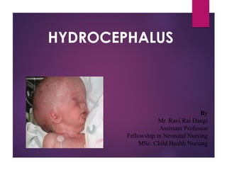
- 1. HYDROCEPHALUS By Mr. Ravi Rai Dangi Assistant Professor Fellowship in Neonatal Nursing MSc. Child Health Nursing
- 2. HYDROCEPHALUS Hydrocephalus is characterized by dilatation of the cerebral ventricles and increased CSF pressure . Incidence :- 2 out of 1000
- 4. CSF FLOW CSF is predominantly formed by the choroid plexus of the lateral, third and fourth ventricles by an active transport process across the endothelium of capillaries in the villous process of the choroid plexus. The arachnoids villus is the primary site of CSF absorption.
- 5. PATHOPHYSIOLOGY Etiological factors Accumulation of CSF in the ventricular system Dilatation of ventricles Compression of the brain tissue against the Surrounding bony cranium
- 6. PATHOPHYSIOLOGY Before the cranial sutures are fused in young infants , the skull may be able to expand to accommodate gradual increase in volume. With fusion of sutures the skull can no longer enlarges and ove signs of increasing ICP indicates the presence of hydrocephalus
- 7. CLASSIFICATION Non obstructive, or communicating Hydrocephalus occurs when the CSF flows out of the chambers of the brain (ventricles) and into the spinal canal, but it is not reabsorbed normally by the tissue surrounding the brain and spinal cord. Sometimes this type of hydrocephalus corrects itself.
- 8. CLASSIFICATION Obstructive, or non communicating, Hydrocephalus occurs when the CSF does not flow properly between or out of the brain ventricles because of an obstruction, such as from a malformation or narrowing.
- 9. CLINICAL FEATURES Early onset Vomiting Drowsiness Irritable Increase head circumference
- 10. CLINICAL FEATURES Frontal bossing Anterior fontanels is widely open Sunset sign
- 11. CLINICAL FEATURES Late Onset- Pappiledema (Optic disc swelling that is caused by increased intracranial pressure due to any cause) Head ache Vomiting Mc Ewen’s sign (Percussion on the skull at a particular spot (near the junction of the frontal, temporal and parietal bones) yields an unusually resonant sound in the presence of hydrocephalus or a brain abscess.)
- 12. DIAGNOSTIC FEATURES Ultrasonography CT scan MRI
- 13. TREATMENT GOALS Decreasing intracranial pressure Maximizing the potential for neurological development.
- 14. TREATMENT Medical therapy 1. In communicating Acetazolamide (50-100mg/kg/day )( To decrease the production of CSF ) Furosemide Mannitol is given to decrease the intracranial pressure.
- 15. SURGICAL MANAGEMENT Shunt surgery 1. Ventriculoperitoneal shunt In this shunt is placed between the enlarged ventricle that drains out csf in to the peritoneal cavity
- 17. Nursing management Preoperative care 1. Assess the signs of increased intracranial pressure. 2. Maimane of nutrition 3. Risk for injury 4. Parenteral anxiety
- 18. Nursing management Post operative care 1. Assessment of vital signs 2. Assess for increased ICP. 3. Treat for infection 4. Parent education
- 19. COMPLICATION Subdural hematoma Infection Malfunctioning of the tube Constipation