SlideShare a Scribd company logo
1 of 98
CHAIRMAN: DR. V.M. PATTANSHETTI
CO-CHAIRMAN: DR. PRAVEEN KAMATAGI
PRESENTER: DR. PRASHANT SHARMA
RECTAL PROLAPSE
HISTORICAL PERSPECTIVE
• Evidence of rectal prolapse (rectal procidentia) exists from ancient times.
• The first described mortality from rectal prolapse was in the 4th century.
• The initiating cause of prolapse is elusive. The path to rectal prolapse generally occurs
slowly over a period of years.
• Suggested contributing factors include standing, constipation, colic, coughing, sneezing,
weak anal sphincters and pelvic floor musculature, an abnormal cul-de-sac, and distal
rectal intussusception.
• The proposed treatments have changed significantly over time as well.
• Treatments such as cauterizing the rectal mucosa, binding patient’s legs together, and
applying trusses have been replaced with variations of resection and rectopexy today.
INTRODUCTION
• Rectal prolapse is an uncommon, disabling condition that has long fascinated surgeons.
• Few clinical disorders have generated such a large number of surgical procedures, with
varying degrees of surgical outcomes, as has rectal prolapse.
• Confusing terminology is a major problem in the study of rectal prolapse and the terms
that must be distinguished are:
1. Mucosal Prolapse
2. Internal Intussusception (Occult Prolapse)
3. Complete Rectal Prolapse (Procidentia)
• MUCOSAL PROLAPSE is caused by a looseness or breaking down of the connective
tissue between the submucosa of the rectum and anal canal and the underlying muscle.
•This usually starts in the anal canal and, in its earliest form, is represented by prolapsing
hemorrhoids.
•With progression, more anal canal mucosa and distal rectal mucosa protrude, leading to the
characteristic picture of linear mucosal furrows and absence of the perianal sulcus.
•Mucosal prolapse doesn’t progress to complete prolapse and is best considered part of the
spectrum of hemorrhoidal disease.
•INTERNAL INTUSSUSCEPTION of the rectum is a distinct clinical entity that may
represent the precursor of a complete rectal prolapse. It can only be diagnosed reliably by
obtaining a defecating proctogram.
Patients with internal intussusception shouldn’t undergo operation as the basic underlying
abnormalities in pelvic floor function cannot be addressed by surgery
•COMPLETE RECTAL PROLAPSE is defined as protrusion of the full thickness of the
rectal wall through the anal orifice.
•It is the entity to which the various surgical procedures are directed.
ANATOMY OF ANAL CANAL
ANAL CANAL STRUCTURE, ANUS, AND ANAL VERGE
•The anus or anal orifice is an anteroposterior cutaneous slit, that along with the anal canal
remains virtually closed at rest, as a result of tonic circumferential contraction of the
sphincters and the presence of anal cushions.
• The edge of the anal orifice, the anal verge or margin (Ano-cutaneous line of Hilton), marks
the lowermost edge of the anal canal and is sometimes the level of reference for
measurements taken during sigmoidoscopy.
• Others favor the dentate line as a landmark because it is more precise. The difference
between the anal verge and the dentate line is usually 1–2 cm.
• The epithelium distal to the anal verge acquires hair follicles, glands, including apocrine
glands, and other features of normal skin, and is the source of perianal hidradenitis
suppurativa, (inflammation of the apocrine glands).
ANATOMIC VS. SURGICALANAL CANAL
Two definitions are found describing the anal canal::
• The “anatomic” or “embryologic” anal canal is only 2.5 cm long, extending from the
anal verge to the dentate line, the level that corresponds to the proctodeal membrane.
• The “surgical” or “functional” anal canal is longer, extending for approximately 4.0 cm
(in men) from the anal verge to the anorectal ring (levator ani).
• The anorectal ring is at the level of the distal end of the ampullary part of the rectum and
forms the anorectal angle, and the beginning of a region of higher intraluminal pressure.
Therefore, this definition correlates with digital, manometric, and sonographic
examinations.
ANATOMIC RELATIONS OF THE ANAL CANAL
• Posteriorly, the anal canal is related to the coccyx and Anteriorly to the perineal body and
the lowest part of the posterior vaginal wall in the female, and to the urethra in the male.
• The ischium and the ischiorectal fossa are situated on either side.
• The fossa ischiorectal contains fat and the inferior rectal vessels and nerves, which cross it to
enter the wall of the anal canal.
MUSCLES OF THE ANAL CANAL
 The muscular component of the mechanism of continence can Be stratified into three
functional groups:
1. Lateral compression from the pubococcygeus,
2. Circumferential closure from the Internal and external anal sphincter, and
3. Angulation from the Puborectalis
INTERNALANAL SPHINCTER
• The internal anal sphincter represents the distal 2.5- to 4.0-cm condensation of the
circular muscle layer of the rectum.
• As a consequence of both intrinsic myogenic and extrinsic autonomic neurogenic
properties, the internal anal sphincter is a smooth muscle in a state of continuous
maximal contraction, and represents a natural barrier to the involuntary loss of stool and
gas.
• • The lower rounded edge of the internal anal sphincter can be felt on physical
examination, about 1.2 cm distal to the dentate line.
• The groove between the internal and external anal sphincter, the intersphincteric sulcus,
can be visualized or easily palpated.
• Endosonographically, the internal anal sphincter is a 2 to 3 mm thick circular band and
shows a uniform hypoechogenicity.
EXTERNALANAL SPHINCTER
• The external anal sphincter is the elliptical cylinder of striated muscle that envelops the
entire length of the inner tube of smooth muscle, but it ends slightly more distal than the
internal anal sphincter.
• The deepest part of the external anal sphincter is intimately related to the pubo- rectalis
muscle, which can actually be considered a component of both the levator ani and the
external anal sphincter muscle complexes.
• Endosonographically, the puborectalis and the external anal sphincter, despite their mixed
linear echogenicity, are both predominantly hyperechogenic, with a mean thickness of 6
mm (range, 5–8 mm).
• In the male, the upper half of the external anal sphincter is enveloped anteriorly by the
conjoined longitudinal muscle, whereas the lower half is crossed by it.
• In the female, the entire external anal sphincter is encapsulated by a mixture of fibers
derived from both longitudinal and internal anal sphincter muscles.
 The automatic continence mechanism is formed by the resting tone, maintained by the
internal anal sphincter, magnified by voluntary, reflex, and resting external anal
sphincter contractile activities.
• In response to conditions of threatened incontinence, such as increased intra-abdominal
pressure and rectal distension, the external anal sphincter and puborectalis reflexively and
voluntarily contract further to prevent fecal leakage.
 The external anal sphincter and the pelvic floor muscles, unlike other skeletal muscles,
which are usually inactive at rest, maintain unconscious resting electrical tone through
a reflex arc at the cauda equina level.
CONJOINED LONGITUDINAL MUSCLE
• Whereas the inner circular layer of the rectum gives rise to the internal anal sphincter, the
outer longitudinal layer, at the level of the anorectal ring, mixes with fibers of the levator
ani muscle to form the conjoined longitudinal muscle.
• This muscle descends between the internal and external anal sphincter, and ultimately
some of its fibers, referred to as the corrugator cutis ani muscle, traverse the lowermost
part of the external anal sphincter to insert into the perianal skin.
• Possible functions of the conjoined longitudinal muscle include:
1. Attaching the Anorectum to the pelvis; and
2. Acting as a Skeleton that supports and binds the internal and external sphincter complex
together.
EPITHELIUM OF THE ANAL CANAL
• The lining of the anal canal consists of an upper mucosal (endoderm) and a lower
cutaneous (ectoderm) segment.
• The dentate (pectinate) line is the “saw-toothed” junction between these two distinct
origins of venous and lymphatic drainage, nerve supply, and epithelial lining.
• Above this level, the intestine is innervated by the sympathetic and parasympathetic
systems, with venous, arterial, and lymphatic drainage to and from the hypogastric
vessels.
• Distal to the dentate line, the anal canal is innervated by the somatic nervous system, with
blood supply and drainage from the inferior hemorrhoidal system. These differences are
important when the classification and treatment of hemorrhoids are considered.
• The pectinate or dentate line corresponds to a line of anal valves that represent remnants
of the proctodeal membrane. Above each valve, there is a little pocket known as an Anal
Sinus or Crypt. These crypts are connected to a variable number of glands, in average 6
(range, 3–12).
• More than one gland may open into the same crypt, whereas half the crypts have no
communication.
• The anal gland ducts, in an outward and downward route, enter the submucosa; two-
thirds enter the internal anal sphincter, and half of them terminate in the intersphincteric
plane.
• Obstruction of these ducts, presumably by accumulation of foreign material in the crypts,
may lead to perianal abscesses and fistulas.
• Cephalad to the dentate line, 8–14 longitudinal folds, known as the rectal columns
(Columns of Morgagni), have their bases connected in pairs to each valve at the dentate
line.
• At the lower end of the columns are the anal papillae. The mucosa in the area of the
columns consists of several layers of cuboidal cells and has a deep purple color because
of the underlying internal hemorrhoidal plexus.
 This 0.5- to 1.0 cm strip of mucosa above the dentate line is known as the Anal Transition
or Cloacogenic Zone.
• Cephalad to this area, the epithelium changes to a single layer of columnar cells and
macroscopically acquires the characteristic pink color of the rectal mucosa.
 The cutaneous part of the anal canal consists of modified squamous epithelium that is
thin, smooth, pale, stretched, and devoid of hair and glands.
RECTUM
• Both proximal and distal limits of the rectum are controversial: the rectosigmoid junction
is considered to be at the level of the third sacral vertebra by anatomists but at the sacral
promontory by surgeons, and likewise, the distal limit is regarded to be the dentate line by
anatomists and the muscular anorectal ring by surgeons.
• The rectum measures 12–15 cm in length and has three lateral curves: the upper and
lower are convex to the right and the middle is convex to the left. These curves
correspond intraluminally to the folds or Valves of Houston.
 Although the rectal valves do not contain all muscle wall layers from a clinical point of
view, they are a good location for performing a rectal biopsies, because they are readily
accessible with minimal risk for perforation.
• The two left-sided folds are usually noted at 7–8 cm and at 12–13 cm, respectively, and the
one on the right is generally at 9–11 cm. The middle valve (Kohlrausch’s plica) is the most
consistent in presence and location and corresponds to the level of the anterior peritoneal
reflection.
• The rectum is characterized by its wide, easily distensible lumen, and the absence of
taeniae, epiploic appendices, haustrations, or a well defined mesentery.
• The word “mesorectum” has gained widespread popularity among surgeons to address
the perirectal areolar tissue, which is thicker posteriorly, containing terminal branches of
the inferior mesenteric artery and enclosed by the fascia propria.
 The “mesorectum” may be a metastatic site for a rectal cancer and is removed during
surgery for rectal cancer without neurologic sequelae, because no functionally
significant nerves pass through it.
• The upper third of the rectum is anteriorly and laterally invested by peritoneum; the
middle third is covered by peritoneum on its anterior aspect only.
• Finally, the lower third of the rectum is entirely extraperitoneal, because the anterior
peritoneal reflection occurs at 9.0–7.0 cm from the anal verge in men and at 7.5–5.0 cm
from the anal verge in women.
 Valve of Houston: Rectum
 (Spiral) Valve of Heister: Cystic Duct
 Valve of Hasner: Nasolacrimal Duct
ANATOMIC RELATIONS OF THE RECTUM
• The rectum occupies the sacral concavity and ends 2–3 cm anteroinferiorly from the tip of
the coccyx. At this point, it angulates backward sharply to pass through the levators and
becomes the anal canal.
• Anteriorly, in women, the rectum is closely related to the uterine cervix and posterior
vaginal wall; in men, it lies behind the bladder, vas deferens, seminal vesicles, and prostate.
Posterior to the rectum lie the median sacral vessels and the roots of the sacral nerve
plexus.
FASCIAL RELATIONSHIPS OF THE RECTUM
• The parietal endo-pelvic fascia lines the walls and floor of the pelvis and continues on the
internal organs as a visceral pelvic fascia.
• The lateral ligaments or stalks of the rectum are distal condensations of the pelvic fascia
that form a roughly triangular structure with a base on the lateral pelvic wall and an apex
attached to the lateral aspect of the rectum.
• The lateral stalks are comprised essentially of connective tissue and nerves, and the
middle rectal artery does not traverse them. Branches, however, course through in
approximately 25% of cases. Consequently, division of the lateral stalks during rectal
mobilization is associated with a 25% risk for bleeding.
 One theoretical concern in ligation of the stalks is leaving behind lateral mesorectal
tissue, which may limit adequate lateral or mesorectal margins during cancer surgery.
• The presacral fascia is a thickened part of the parietal endopelvic fascia that covers the
concavity of the sacrum and coccyx, nerves, the middle sacral artery, and presacral veins.
Operative dissection deep to the presacral fascia may cause troublesome bleeding from the
underlying presacral veins.
 Presacral hemorrhage occurs as frequently as 4.6–7.0% of resections for rectal
neoplasms, and despite its venous nature, can be life threatening. This is a consequence
of two factors:
 The difficulty in securing control because of retraction of the vascular stump into the
sacral foramen, and
 The high hydrostatic pressure of the presacral venous system.
• The rectosacral fascia is an anteroinferiorly directed thick fascial reflection from the
presacral fascia at the S-4 level to the fascia propria of the rectum just above the anorectal
ring.
 The rectosacral fascia, classically known as the Fascia of Waldeyer, is an
important landmark during posterior rectal dissection.
 The Visceral Pelvic Fascia of Denonvilliers is a tough fascial investment that separates
the extraperitoneal rectum anteriorly from the prostate and seminal vesicles or vagina.
ARTERIAL SUPPLY OF THE RECTUM AND ANAL CANAL
• The superior hemorrhoidal/rectal artery is the continuation of the inferior mesenteric
artery, once it crosses the left iliac vessels.
•The artery descends in the sigmoid mesocolon to the level of S-3 and then to the posterior
aspect of the rectum.
•In 80% of cases, it bifurcates into right, usually wider, and left terminal branches; multiple
branches are present in 17%. These divisions, once within the submucosa of the rectum, run
straight downward to supply the lower rectum and the anal canal.
• The superior and inferior hemorrhoidal/rectal arteries represent the major blood supply to
the anorectum. In addition, it is also supplied by the internal iliac arteries.
 The contribution of the middle hemorrhoidal artery varies with the size of the superior
hemorrhoidal artery; this may explain its controversial anatomy. Some authors report
absence of the middle hemorrhoidal artery in 40–88% whereas others identify it in 94–
100% of specimens.
 The middle hemorrhoidal artery is more prone to be injured during low anterior
resection, when anterolateral dissection of the rectum is performed close to the pelvic
floor and the prostate and seminal vesicles or upper part of the vagina are being
separated.
• The anorectum has a profuse intramural anastomotic network, which probably accounts
for the fact that division of both superior and middle hemorrhoidal arteries does not
result in necrosis of the rectum.
• The paired inferior hemorrhoidal arteries are branches of the internal pudendal artery,
which in turn is a branch of the internal iliac artery.
VENOUS DRAINAGE AND LYMPHATIC DRAINAGE OF THE RECTUM
AND ANAL CANAL
• The anorectum also drains, via middle and inferior hemorrhoidal/rectal veins, to the
internal iliac vein and then to the inferior vena cava.
 The external hemorrhoidal plexus, situated subcutaneously around the anal canal below
the dentate line, constitutes when dilated the external hemorrhoids.
• The internal hemorrhoidal plexus is situated submucosally, around the upper anal canal
and above the dentate line. The internal hemorrhoids originate from this plexus.
• Lymph from the upper two-thirds of the rectum drains exclusively upward to the inferior
mesenteric nodes and then to the paraaortic nodes.
• Lymphatic drainage from the lower third of the rectum occurs not only cephalad, along the
superior hemorrhoidal and inferior mesentery arteries, but also laterally, along the middle
hemorrhoidal vessels to the internal iliac nodes.
• In the anal canal, the dentate line is the landmark for two different systems of lymphatic
drainage: above, to the inferior mesenteric and internal iliac nodes, and below, along the
inferior rectal lymphatics to the superficial inguinal nodes, or less frequently along the
inferior hemorrhoidal artery.
 In the female, drainage at 5 cm above the anal verge in the lymphatic may also spread to
the posterior vaginal wall, uterus, cervix, broad ligament, fallopian tubes, ovaries, and
cul-de-sac, and at 10 cm above the anal verge, spread seems to occur only to the broad
ligament and cul-de-sac.
INNERVATION OF THE RECTUM AND ANAL CANAL
• Innervation of the Rectum – The sympathetic supply of the rectum and the left colon
arises from L-1, L-2, and L-3.
– Two main hypogastric nerves, on either side of the rectum, carry sympathetic innervation
from the hypogastric plexus to the pelvic plexus.
– The parasympathetic fibers to the rectum and anal canal emerge through the sacral foramen
and are called the nervi erigentes (S-2, S-3, and S-4).
– The periprostatic plexus, a subdivision of the pelvic plexus situated on Denonvilliers’
fascia, supplies the prostate, seminal vesicles, corpora cavernosa, vas deferens,
urethra, ejaculatory ducts, and bulbourethral glands.
– Sexual function is regulated by cerebrospinal, sympathetic, and parasympathetic
components. Erection of the penis is mediated by both parasympathetic (arteriolar
vasodilatation) and sympathetic inflow (inhibition of vasoconstriction).
– All pelvic nerves lie in the plane between the peritoneum and the endopelvic fascia and are
in danger of injury during rectal dissection.
 Permanent bladder paresis occurs in 7–59% of patients after abdomino-perineal resection
of the rectum; the incidence of impotence is reported to range from 15 to 45%, and that
of ejaculatory dysfunction from 32 to 42%.
 The overall incidence of sexual dysfunction after proctectomy has been reported to reach
100% when wide dissection is performed for malignant disease.
 Dissections performed for benign conditions are undertaken closer to the bowel wall,
thus reducing the possibility of nerve injury.
– Trauma to the autonomic nerves may occur at several points. During high ligation of the
inferior mesenteric artery, close to the aorta, the sympathetic preaortic nerves may be
injured.
– Division of both superior hypogastric plexus and hypogastric nerves may occur also during
dissection at the level of the sacral promontory or in the presacral region.
 In such circumstances, sympathetic denervation with intact nervi-erigentes results in
retrograde ejaculation and bladder dysfunction.
– The nervi erigentes are located in the posterolateral aspect of the pelvis, and at the point of
fusion with the sympathetic nerves are closely related to the middle hemorrhoidal artery.
 Injury to these nerves will completely abolish erectile function.
– The pelvic plexus may be damaged either by excessive traction on the rectum, particularly
laterally, or during division of the lateral stalks when this is performed close
to the lateral pelvic wall.
– Finally, dissection near the seminal vesicles and prostate may damage the periprostatic
plexus, leading to a mixed parasympathetic and sympathetic injury. This can result in
erectile impotence as well as a flaccid, neurogenic bladder.
– Sexual complications after rectal surgery are readily evident in men but are probably
underdiagnosed in women.
• ANAL CANAL
– The internal anal sphincter is supplied by sympathetic (L-5) and parasympathetic nerves
(S-2, S-3, and S-4) following the same route as the nerves to the rectum.
– The external anal sphincter is innervated on each side by the inferior rectal branch of the
pudendal nerve (S-2 and S-3) and by the perineal branch of S-4.
 Despite the fact that the puborectalis and external anal sphincter have somewhat different
innervations, these muscles seem to act as an indivisible unit.
– After unilateral transection of a pudendal nerve, external anal sphincter function is still
preserved because of the crossover of the fibers at the spinal cord level.
 Anal sensation is carried in the inferior rectal branch of the pudendal nerve and is thought
to have a role in maintenance of anal continence.
PELVIC FLOOR MUSCULATURE
 The muscles within the pelvis can be divided into three categories
(1) the anal sphincter complex;
(2) pelvic floor muscles; and
(3) muscles that line the sidewalls of the osseous pelvis.
LEVATOR ANI
• The levator ani muscle, or pelvic diaphragm, is the major component of the pelvic floor. It
is a pair of broad, symmetric sheets composed of three striated muscles: ileococcygeus,
pubococcygeus, and puborectalis.
• The pelvic floor is “incomplete” in the midline where the lower rectum, urethra, and either
the dorsal vein of the penis in men, or the vagina in women, pass through it. This defect is
called the levator hiatus and consists of an elliptic space situated between the two
pubococcygeus muscles.
• The puborectalis muscle is a strong, U-shaped loop of striated muscle that slings the
anorectal junction to the posterior aspect of the pubis.
THE ANORECTAL RING AND THE ANORECTALANGLE
• Two anatomic structures of the junction of the rectum and anal canal are related to the
puborectalis muscle: the anorectal ring and the anorectal angle.
The anorectal ring, a term coined by Milligan and Morgan, is a strong muscular ring that
represents the upper end of the sphincter, more precisely the puborectalis, and the upper
border of the internal anal sphincter, around the anorectal junction.
 It is of clinical relevance, because division of this structure during surgery for abscesses
or fistula inevitably results in fecal incontinence.
 The anteriorly directed pull of the puborectalis contributes to the angulation between the
rectum and anal canal, the anorectal angle.
• The Anorectal Angle is thought to be the result of the anatomic configuration of the U-
shaped sling of puborectalis muscle around the anorectal junction.
 Whereas the anal sphincters are responsible for closure of the anal canal to retain gas and
liquid stool, the puborectalis muscle and the anorectal angle are designed to maintain
gross fecal continence.
ANORECTAL
PHYSIOLOGY
 The normal function of the anorectum represents a complex interaction between
anatomical, neurological, sensory, myogenic and hormonal factors.
 Failure or weakness of any one part or combination of parts of these factors may lead to
symptoms of many common diseases seen in colorectal practice.
 The anorectal physiology laboratories are designed to provide informations about the
function of the neurological, sensory and anotomical components of anorectal function.
MECHANISM OF DEFECATION
• The development of a mass peristaltic wave by the left colon results in the delivery of
stool to the lower sigmoid colon and rectum. Once the stool content arrived in the rectum
several physiological events take place.
1. There is reflex relaxation of the internal sphincter and contraction of the distal external
sphincter. This is called Rectoanal Inhibitory Reflex (RAIR).
2. As the result of the reflex relaxation of the internal sphincter the content of the rectum
comes in contact with the highly sensitive lower end of the anal canal and the nature of
the content either solid, liquid or gas is determined accurately.
3. This increase in the volume of the rectum leads to an increase in the rectal pressure that
is maintained for 1-2 minutes before the intra-rectal pressure returns to normal by
process called accommodation which allows the relaxation of the rectum to
accommodate the increased rectal volume.
4. As the rectal volume increases there is a gradual rise in the rectal pressure and at a
particular pressure the urge to defecate occurs.
 If the urge occurs at a socially acceptable time to proceed with defecation or if the
accommodation has reaches its upper limit the process of defecation occurs and the
individual will normally assume a squatting or sitting position.
 By Valsalva maneuver the intra-abdominal and intra-rectal pressures are raised. The
anorectal angle is straightened the entire sphincter mechanism relaxes and the pelvic floor
forms funnel shape.
 Both straining and recto-sigmoid contraction aids in emptying of the rectum.
 If this process is accompanied by further mass peristalsis the entire left colon may be
evacuated. If not the evacuation may occur in a piecemeal fashion.
 Once the rectum is empty the external sphincter and puborectalis transiently contract (the
closing reflex).
 Following defecation, sphincter pressure, intra-abdominal pressure, return to their normal
resting pressure and the anorectal angle returns to its position.
 Constipation manifested as outlet obstruction may occur when any portion of this process is
interrupted.
 Such a situation may occur in the following conditions:
1. Failure of puborectalis to relax (Paradoxical puborectalis, Anismus, levator syndrome).
2. Abnormal high resting pressure (Nut-cracker anus).
3. Excessive accommodation (Mega rectum).
4. External compression of the rectum (Enterocele).
5. Incomplete evacuation (Larger rectocele).
6. Intussusception (Overt or Covert prolapse).
7. Excessive pelvic descent (descending Perineal syndrome). Whether the perineal descent is
the result of chronic straining due to outlet obstruction or a primary cause of the
constipation is debatable.
8. Constipation can also be a manifestation of dysfunctional colon mobility.
ALTERED ANORECTAL PHYSIOLOGY
• Altered anorectal physiology may lead to two important conditions, constipation and
incontinence.
I. In the case of constipation quality of feces, hard or soft, type (excessive straining or
failure of recognize a call to stool) and frequency may help direct to the initial
evaluation.
II. In the case of incontinence frequency and type of incontinence either to gas, liquid or
solid is important for the evaluation.
Factors that contribute to anal continence are:
1. The anal canal high pressure zone
2. Anal canal length
3. Anorectal angle
4. Rectal compliance
5. Anorectal sensation
6. Anal cushions
7. Stool consistency and frequency.
 The anal canal high pressure zone is due to internal anal sphincter, external anal
sphincter and the puborectalis muscle.
 The internal anal sphincter is responsibles for 85 percent of the resting pressure. The
external sphincter and puborectal muscle are responsible for squeeze pressure of the anal
canal. The anal canal pressure can be elevated voluntarily for about one minute only. The
same action of these muscles is responsible for the anal canal length.
 The anorectal angle is not constant and it varies according to the strain and increase in
intra-abdominal pressure.
 When the puborectalis muscle relaxes the angle is widened and defecation occurs. The
rectal compliance plays a roll in maintenance of continence by the accommodation
reflex.
 In disease like ulcerative colitis and irradiation proctitis the rectum becomes less
compliant and the ability to stretch the rectal wall may be decreased. As a result, the
rectal pressure remains high and when further increase in pressure occurs, the continence
becomes dependent upon external anal sphincter and puborectalis muscle which can last
for one minute only.
• The consistency of the stool is an important factor for continence. Solid feces can be
controlled and regulated even by weak sphincters and fluid stools can not be controlled
even by a normal sphincter.
• The sensory components of the mechanism of continence are critical in allowing a
satisfactory reaction time to read to a bolus of stool or gas entering the rectum.
 Sensation is associated with the sampling reflex. This reflex results in the transient
relaxation of the internal sphincter with contraction of distal external sphincter, allowing
for discrimination of the contents in the upper anal canal and rectum as either gas or liquid
or solid.
BASIC ANORECTAL PHYSIOLOGY—LABORATORY TESTS
In clinical practice, the basic tests which are useful include:
1. Anorectal manometry (ARM)
2. Anorectal electromyography (EMG)
3. Dynamic proctography (DPG)
4. Colon transit time (CTT)
5. Trans anal ultrasound (TAUS)
6. Estimation of neurological function (ENF).
 It is important to select carefully those tests that will provide useful clinical information
for an individual patient.
A. ANORECTAL MANOMETRY
• Anorectal manometry is an objective method for assessing anal muscular tone, rectal
compliance, and anorectal sensation and of verifying the integrity of the rectoanal
inhibitory reflex (RAIR).
• It is a useful investigation in patients with fecal incontinence to identify the presence of
sensory or muscular defects as well as to find out the functional weakness of internal and
external anal sphincter.
• It may be helpful in evaluation of Chronic Constipation and Hirschsprung's disease.
• It helps to determine whether sphincter weakness or dysfunction is likely to be the cause
of symptoms. Other tests such as electromyography, ultrasound or other radiological
study are needed to determine the underlying pathology.
• The most basic components of anorectal manometry are pressure transuding catheter and
a recorder.
• Different types of transuding catheters are available such as water perfused catheter,
micro transducer and balloon catheters.
• Regardless which catheter are used, manometric examination measure a resting pressure
(RP) a squeeze pressure (SP) and the rectoanal inhibitory reflex (RAIR) and the anal
canal length.
• Resting pressure and squeeze pressure are obtained by measuring the pressure at
multiple points in the ano-rectum. This can be done by pulling the catheter out at 0.5 cm
intervals and recording pressure at each point.
• The maximum squeeze pressure is twice the resting pressure and it indicates the function
of external sphincter.
• The anal canal length is determined by measuring from the point at which the pressure
suddenly rises higher than the basal rectal tone to that where the pressure suddenly drops
off at or near the anal verge.
• The anal canal length is generally shorter in incontinence patients. The anal canal length is
usually increased after post anal repair which predict the return of continence.
 The rectoanal inhibitory reflex (RAIR) is usually initiated by instillation of air or water
into the balloon in the rectum. This causes a reflex contraction of the external anal
sphincter with synchronous relaxation of the internal anal sphincter.
This reflex is absent in Hirschsprung's disease and some cases of scleroderma.
 Chronically relaxed internal sphincter (low resting pressure) and failure to further relax
may be associated with fecal impaction and overflow incontinence.
• To measure the rectal sensation and compliance an intra-rectal balloon is connected to a
pressure monitor is slowly filled with warm saline and the individual is asked to note
when distention is first feel (minimum perceived or threshold volume) and when the
further distention can not be tolerable (maximum tolerable volume).
By recording the intrarectal pressure at various volumes a compliance curve can he
obtained. The maximum tolerable volume is more useful in biofeedback treatment for anal
incontinence.
B. AMBULATORY ANORECTAL MANOMETRY
• In an effort to observe the anorectal continence mechanism in a more suitable
environment ambulatory anorectal manometry has been developed.
• Usually a micro transducer catheter used from which constant recordings are made.
Surface electromyography can also be coupled with it.
C. BALLOON RETENTION AND EXPULSION TEST
• The ability of the rectum to retain or expel an intra rectal balloon can be evaluated. The
intra rectal balloon is filled with water at 60 cc per minute till it is expelled or maximum
tolerable volume is reached.
• Water filled balloon stimulate like the solid or semisolid stool rather than free water. Some
research people use air filled rectal balloon at the end of the standard anorectal manometry
study.
• This test helps to discriminate which patient truly have anismus and will benefit from bio-
feedback therapy.
D. RECTAL TONE MEASUREMENT
• Recently a technique is described for measuring rectal tone (as opposed to compliance)
• Changes in the tone are measured by an electronic barostat that measures change in the
volume in an intrarectal balloon maintained at a constant pressure.
 Profound changes have been recorded in rectal tone that where not detected
manometrically.
E. SALINE INFUSION TEST
• This is an investigation done for the evaluation of fluid incontinent individuals.
• Isotonic saline 1500 cc at body temperature is infused per anum at 60 cc per minute and
the time of first leak and the total volume of leak are noted.
 The time of first leak is significally lower in incontinent patients.
ELECTROPHYSIOLOGICAL TESTS
• Anal electromyography (EMG) is the recording of the electrical activity from the muscle
fibers of the external sphincter and puborectalis complex during rest, during maximum
squeeze, during defecations and in response to various reflexes.
• It has been used for sphincter mapping in patients with faecal incontinence and also is of
use in assessing the function of the pelvic floor in individuals with constipation.
• Anorectal EMG consists of two basic evaluations, assess the conduction time and the
assessment of pelvic floor activity.
 To assess the conduction time in the Pudendal nerve from Alcock's canal to the end organ
in the external sphincter and this test is called as Pudendal Nerve Terminal Motor Latency
(PNTML).
• Normal conduction occurs in 2.0 + 0.2 milli seconds. Prolonged times suggest injury to the
large fast conducting fibres.
• In patients with neuropathtic incontinence PNTML is measured by using a specially
designed glove with a stimulating electrode on the finger tip and a recording electrode at
the base of the finger.
• The stimulating electrode is used intra rectally to stimulate the pudendal nerve as it exits
the Alcock's canal and the recording electrode documents the response in the external anal
sphincter.
• Prolonged PNTML is found in several disorders associated with difficult vaginal delivery
or chronic straining at stool and fecal incontinence. Severe constipation, prolapse rectum,
uterine prolapse and solitary rectal ulcer syndrome can also prolong PNTML.
 EMG can also be used for the activity of the external sphincter by inserting electrode in
all four quadrants to detect areas of electrically silent, which indicate a lack of muscle at
that spot.
• Single fiber EMC shows density of muscle fibers innervated by individual axon in the
muscle suspected to he damaged by a neurogenic process.
 A raised fiber density indicates reinnervation of the muscle.
DYNAMIC PROCTOGRAPHY (DPG) (DEFECOGRAPHY)
• Dynamic Proctography (DPG) is used to evaluate function of the pelvic floor at the time
of defecation.
• DPG has many variations in name as well as the procedures. Videoproctography,
Defecography, Defecating Scintigraphy, all accomplish the same essential assessment of
the ano-rectal angle, ano-rectal function, pelvic floor descent and the length of the anal
canal.
 Patients with colonic inertia often undergo defecography in order to exclude pelvic floor
dysfunction.
• DPG is performed by thick mixture of barium contrast and methyl cellulose in the rectum.
Additional liquid contrast is placed in the vagina generally the patient sits on a radiolucent
toilet seat when the test is done under videofluroscopic monitoring.
• This allows the evaluation to occur during three phases – Rest, Evacuation and Recovery.
• The technique can be enhanced with the use of 'Triple Contrast'. This technique combines
the use of the standard DPG with the contrast in the small bowel.
• The 3rd element of the triple contrast is contrast in the urinary bladder so that its
interaction with the pelvic floor at the time of straining can also be evaluated. This tripple
contrast DPG gives the most thorough evaluation of pelvic floor at the time of defecation.
• DPG can demonstrate paradoxical contraction of the puborectalis, internal
intussusception, enterocele, rectocele, pelvic floor hernia and incomplete evacuation.
TRANSANAL ULTRASONOGRAM (TAUS)
• This test is useful in the assessment of fecal incontinence.
 This provides the most accurate and complete evaluation for detecting the anal sphincter
defect.
• Ultrasonography is a safe test, without risk of radiation exposure. Therefore, it is ideal
for children and pregnant women. It is also a more cost-effective and safer technique.
• The problem of differentiating local recurrence from that of postoperative changes can
be minimize by performing a base line endorectal ultrasonography 3 months after
surgery by which time most acute inflammatory reaction has decreased.
• Generally a probe with 7-10 mhz, 360 degree rotating transducer is used.
• In TUS the internal sphincter is visualized as a hypoechogenicity band around the anal
canal and the external sphincter as a band of mixed echogenicity.
 Defects in either band can be readily identified and measured.
PRESENTATION AND WORK-UP
• Patients with rectal prolapse will often report a history of constipation and even
incontinence. Women are 6 times more likely than men to suffer from prolapse.
• The primary presenting complaint is often of a protuberance or bulge from the anus,
which is often mistaken for hemorrhoidal disease. It may spontaneously reduce or
remain prolapsed, causing considerable distress.
• Other complaints may be a feeling of incomplete evacuation of stool, bleeding, mucus
discharge, urinary incontinence, and pain.
• The prolapse may be apparent on initial exam; other times it will need to be elicited by
asking the patient to perform a Valsalva. At times, it is even necessary to examine the
patient after having them strain while squatting on the commode.
• Once the prolapse is produced, it is important to differentiate true full-thickness rectal
prolapse with concentric mucosal rings from simple hemorrhoidal disease, which is
identified by radial folds of hemorrhoidal tissue.
• Colonoscopy is required in all patients considered candidates for operative repair of
rectal prolapse to rule out a neoplastic lead point.
• On endoscopic examination, the rectal mucosa in a patient with chronic recurrent
prolapse will show signs of circumferential erythema.
• Other diagnostic tests include anal manometry, endorectal ultrasound, and defecography.
 Defecography is valuable in occult, intermittent prolapse to document marked mobility
of the rectosigmoid junction away from the normal point of fixation at the sacral
promontory.
ACUTE TREATMENT
• The immediate management of obvious prolapse is reduction. Before any attempts at
reducing the prolapse, it is of utmost importance to fully assess the viability of the
rectum.
• Ischemia or frank necrosis may be present in the neglected or demented patient. If
significant full-thickness ischemia is present, reduction should be avoided and operative
resection will likely be required.
• The majority of patients with viable prolapse will respond to continuous, steady
pressure in the office setting or emergency room. At times, intravenous sedation or even
general anesthesia in the operating room may be necessary for full reduction.
• Applying table salt or sugar to the mucosa can reduce the swelling of the incarcerated
rectum and aid in reduction.
• Another technique described for the stubborn prolapse involves encompassing the
rectum with an elastic compression wrap.
• Hyaluronidase injection into the prolapsed rectum has also been described to aid in
reduction.
TRANSABDOMINAL OPTIONS
• Abdominal approaches to definitive repair of prolapse consistently show lower recurrence
rates compared with perineal approaches.
• The transabdominal approach is the preferred repair in patients who can tolerate a
transabdominal operation.
• Ideally, the operation should be limited in morbidity and mortality, have a low recurrence
rate, and address the patient’s associated constipation or incontinence, if present.
• Care must also be given to evaluating the patient for the presence of concurrent anterior
compartment prolapse to assess the feasibility of repair of these prolapsing organs as well
in a combined operation.
• There are two primary technical components in the abdominal approach to rectal
prolapse: rectopexy and resection.
• The goal of rectopexy is to mobilize the rectum out of the pelvis to straighten the low
rectum in the curve of the sacrum and secure it proximally at the sacral promontory so
that prolapse cannot recur.
• In 1963, Ripstein described the posterior rectopexy: a posterior rectal dissection to the
level of the coccyx and a sling of fascia lata around the rectum anchored to the proximal
sacrum.
• Modifications using biologic and synthetic meshes have come and gone over time.
• Rectopexy can even be performed without the use of mesh by securing peritoneal wings
of the rectum to the sacral promontory with suture or tacks. This technique, however,
results in slightly higher recurrence rates than rectopexy with mesh.
• The extent of rectal mobilization required during rectopexy has also been an issue of
debate.
• Circumferential mobilization may decrease the rate of prolapse recurrence; however, a
known drawback of rectopexy is constipation, and division of the anterolateral ligaments
(parasympathetic innervations) during dissection.
• The most recent addition to the abdominal operation for rectal prolapse is the Ventral
Rectopexy that involves mobilization of the anterior portion of the rectum without a
posterior or lateral dissection.
• Mesh shaped in the form of a “hockey stick” is then sutured from the anterior rectal wall
to the sacral promontory and covered with peritoneum.
 This technique reduces the need for extensive posterior dissection. Some authors report
lower constipation rates, while maintaining recurrence rates similar to posterior
Rectopexy.
• The use of laparoscopy in the repair of rectal prolapse has been studied extensively. The
integrity of the repair has not been compromised with the laparoscopic approach, as
recurrence rates are similar.
 The short-term postoperative benefits of laparoscopic surgery are realized in patients
with rectal prolapse, including
 Decreased postoperative pain,
 Shorter hospital length of stay, and
 Quicker return of bowel function.
 These recovery benefits of laparoscopy may also allow more high-risk patients to be
approached transabdominally, who may otherwise have only been considered for the
less durable perineal approaches.
PERINEAL APPROACHES
• While abdominal approaches for rectal prolapse have the lowest recurrence rates with
better overall functional outcomes, the majority of patients with prolapse are elderly.
• Many of these patients have significant comorbidities that limit their ability to tolerate
an abdominal approach.
• Additionally, though a much smaller cohort, young patients, especially males, may
consider a perineal approach to limit the added risk of sexual dysfunction.
• Up to 60% of patients suffering from rectal prolapse will undergo a perineal approach.
• When rectal prolapse recurs after an abdominal approach, a second abdominal approach
can be technically difficult. In these circumstances, a perineal approach may be a viable
option.
• However, a perineal resection-rectopexy can spell doom if the blood supply of the
rectosigmoid was previously divided, and the perineal resection leaves an ischemic
segment of rectum in place.
PERINEAL PROCTOSIGMOIDECTOMY
(ALTEMEIER PROCEDURE)
• Perineal proctosigmoidectomy for rectal prolapse was originally introduced by Mikulicz
in 1899 and popularized by Altemeier et al.
• The procedure can be performed in lithotomy or prone jack-knife position. Anesthetic
options include regional or general anesthesia.
• The operation is begun by applying gentle retraction to the rectum so that the redundant
rectum is externalized, recreating the prolapse.
• The anal canal is held open with a Lone Star retractor (Lone Star Company, Frisco,
Texas).
• The dentate line is identified. An injection of dilute epinephrine solution is administered
circumferentially 1 to 2 cm proximal to the dentate line followed by a full-thickness,
circumferential rectal incision with electrocautery.
• The avascular intersphincteric plane is developed up to the pelvis, and the distal rectum
is pulled from the pelvis.
• Using either suture ligation or an energy device, the vascular supply of the exposed
bowel is then ligated as the dissection is continued proximally.
• The redundant rectum and sigmoid colon are extracted to the level of the left colon, and the
bowel is divided under mild stretch at the point of planned coloanal anastomosis.
• The more bowel removed, the better. A posterior rectopexy with a postanal levatorplasty
has been shown to improve anal continence.
• The rectopexy is performed by using a nonabsorbable suture to fix the posterior surface of
the nonprolapsed bowel to the pre-sacrococcygeal fascia. The normal anorectal angle is
reconstructed with a posterior levator-plasty to restore the anorectal angle.
• A hand-sewn colo-anal anastomosis is performed between left colon and dentate line.A
stapled anastomosis can be performed using two purse strings. However, some authors
report a higher probability of recurrence with a stapled anastomosis.
• Ultimately, patients selected for a perineal procto-sigmoidectomy should expect an
improved continence compared with times of prolapse and decreased constipation with
overall improved quality of life.
• However, a disadvantage of the perineal proctosigmoidectomy is the increased
recurrence rates of 12% to 24% compared with the abdominal approach. This can be
reduced by removing all of the redundant distal colon up to the left colon.
ANORECTAL MUCOSECTOMY WITH MUSCULAR PLICATION
(DELORME PROCEDURE)
• In 1900, Delorme described the anorectal mucosectomy with muscular plication for the
treatment of rectal prolapse.
 A significant advantage of the Delorme procedure is that no full-thickness bowel resection
with anastomosis is performed. Similar to the Altemeier procedure, the bowel is
completely prolapsed and dilute epinephrine solution is injected in a submucosal plane.
• Using electrocautery, a circumferential submucosal incision is then made 1 cm proximal
to the dentate line. Being careful not to violate the muscular layer of the rectal wall, a
submucosal dissection is performed and extended proximally.
• A mucosectomy twice the length of the prolapse is performed.
• After the mucosal sleeve is excised, longitudinal plicating sutures are placed in rows
along the length of the rectal wall musculature 1 cm apart.
• The sutures are not tied until all the sutures have been placed. As the longitudinal sutures
are tied around the circumference of the rectum, the cut edge of the mucosa is brought to
the level of the anal canal.
• Finally, the proximal and distal mucosal edges are re-approximated using absorbable
sutures at the dentate line.
• The muscular plication and mucosal anastomosis can be alternatively performed using a
circular stapler.
• In patients with a patulous anus on physical examination and in efforts to improve
continence, a posterior levatorplasty can also be performed.
• The Delorme procedure for rectal prolapse offers patients a safe procedure, with reported
low morbidity of 9.5% but reported recurrence rates of 12% to 31%.
 Significant preexisting long-term constipation has been associated with higher
recurrence rates, while pelvic floor repair with levator-plasty has been associated with
reduced recurrence rates.
 Patients may complain of the urge to defecate because of the reefed muscle in the anal
canal.
Anorectal mucosectomy with muscular plication (Delorme procedure).
THIERSCH PROCEDURE
• Anal encirclement for the treatment of rectal prolapse was introduced in 1891 by
Thiersch.
 In the original description, Thiersch used a silver wire to encircle the anus in an
effort to provide a mechanical replacement of the relaxed sphincter.
 The procedure does not correct the prolapse but simply mechanically supports the
anal sphincter function by narrowing the anal canal and preventing external rectal
prolapse.
• The procedure can be performed quite quickly. With the patient in prone jackknife
or lithotomy position, in reference to the center of the anus, two lateral perianal
skin incisions are made 180 degrees apart.
• The incisions are connected subcutaneously by tunneling through the ischiorectal
fossa and perineal body. Next, a piece of polypropylene mesh or silicone roll 1.5
cm in height is tunneled circumferentially around the entire anus.
 “The presence of a foreign body would incite an inflammatory reaction
and create an adhesive circumferential band to create support of the
perineum.”
• The mesh is tightened to a point that allows only the passage of one finger into the
anal canal, and the mesh is sutured to itself at this level of tension.
• Reported complications include recurrence rates of up to 75%, anal stenosis, fecal
impaction, and even erosion of the mesh or Silastic product into the anorectum.
 Because of the high recurrence rates and reported complications, indications for the
procedure are very narrow but include patients with mild to moderate rectal
prolapse and significant life-threatening comorbidities, with anticipated short life
expectancy, that prevent them from either abdominal or resective perineal
approaches.
COMPARISON BETWEEN THE DIFFERENT MODALITIES
• Historically, elderly patients with significant comorbidities were only candidates for
perineal approaches.
• The perineal approaches traditionally rendered less durable long-term control of
prolapse compared to abdominal approaches.
 Comparing the Altemeier procedure to Delorme procedure, a randomized controlled
study showed there were fewer recurrences after the Altemeier procedure (24% vs.
31%), but functional outcomes were similar.
 With abdominal rectopexy, there is no significant difference in terms of recurrence
between the methods used for fixation.
 In investigating the degree of rectal mobilization necessary, circumferential
mobilization rather than preservation of the lateral ligaments has been associated
with decreased recurrent prolapse. However, circumferential mobilization is also
associated with increased postoperative constipation.
• In comparison of resection rectopexy to rectopexy alone, lower rates of both
constipation and recurrence were encountered with resection rectopexy.
• Unfortunately, resection is associated with a finite anastomotic leak rate. Although it
has been a long-held belief that perineal approaches are associated with a
significantly higher recurrence rate than abdominal approaches, a 2015 Cochrane
review of 15 randomized controlled trials and over 1000 patients failed to display
superiority of any approach in terms of recurrence rates or quality of life.
 Therefore the surgeon’s experience and judgment are paramount to the choice of
operative approach based on patient characteristics, the clinical situation, and
expertise.
MANAGEMENT OF RECURRENCE
• It is important to understand patient and technical factors that are associated with
recurrence patterns.
• During perineal proctosigmoidectomy, stapled anastomosis, shorter specimen
length, and severe preexisting constipation were all associated with an increased
risk of recurrence.
• A postanal repair and levatorplasty were associated with lower recurrence rates.
• During abdominal rectopexy, variables identified as related to recurrence included
incontinence, constipation, the extent of rectal mobilization, and whether sigmoid
resection was performed in the setting of constipation.
• As mentioned previously, circumferential rectal mobilization was associated with
decreased recurrence.
• Overall, a high recurrence rate after either approach is likely due to technical
failures such as inadequate fixation or incomplete mobilization.
• Poor decision making and failure to recognize preexisting conditions such as colonic
dysmotility in a patient undergoing rectopexy without resection will ultimately
render suboptimal results.
• Repair of recurrent rectal prolapse should be undertaken only after consideration of
several complexities that guide choice of approach.
• Traditionally, if a perineal resection was initially performed, repeat perineal resection
is often the safest route to avoid compromising the remaining proximal blood supply.
• Similarly, a recurrence after abdominal resection rectopexy should not be
approached with a perineal resection, as there is great risk for rectal ischemia after
ligating both the proximal and then distal rectal blood supply. However, a perineal
proctosigmoidectomy is a very reasonable option if abdominal rectopexy without
resection was performed as the index procedure.
• A second abdominal approach is often a technically difficult procedure in the pelvis
that can increase the potential for added patient morbidity.
• Similarly, it is reasonable to consider abdominal rectopexy without resection after a
recurrence from a perineal resection.
 Therefore, in the case of recurrent prolapse, the index procedure often dictates the
reoperative surgical approach. Mesh suspension may be considered if tissues were
inadequate for pexy at the initial operation.
INTERNAL INTUSSUSCEPTION
(INTERNAL RECTAL PROLAPSE)
• Internal rectal intussusception is described as internal telescoping of the rectum
that does not result in protrusion from the anal canal. When the rectum protrudes
from the anal canal, rectal prolapse occurs.
• The Oxford grading system has been established to grade the severity of rectal
prolapse based on evacuation proctography.
• While internal intussusception has been identified in asymptomatic individuals and
has historically been treated as a normal variant, evidence now suggests that
patients with internal intussusception can suffer from a spectrum of disorders
ranging from obstructed defecation syndrome to fecal incontinence. As such,
detailed physical examination and often extensive diagnostic imaging work-up are
required.
HISTORY
• Women with internal rectal intussusception commonly suffer from a range of
symptoms.
• Historically, symptoms of internal intussusception included only symptoms of
obstructive defecation; however, evidence now suggests that fecal incontinence also
frequently plays a significant role in symptomatology.
• In fact, fecal incontinence can be the predominant complaint in patients with high-
grade internal intussusception. In females with internal intussusception, fecal
incontinence was reported in 56%, while complaints of incomplete evacuation
(45%), straining at defecation (34%), and digital assistance during defecation (34%)
were also commonly reported.
• The majority of patients had complaints of urge incontinence, while passive
incontinence and incontinence to flatus were reported less often. During the initial
clinical encounter of a patient with pelvic defecatory dysfunction, it is necessary to
quantify the severity of symptoms.
• The subsequent success of therapy can be gauged by comparison of function before
and after treatment using grading scales, which include Wexner constipation and
incontinence score, fecal incontinence severity index score, and quality-of-life.
PATHOPHYSIOLOGY
• Initial evidence pointed toward changes in the rectoanal inhibitory reflex (RAIR)
caused by the intussusceptum as the source of incontinence.
• Reducing the intussusceptum with rectopexy, the RAIR would return to normal, and
continence would be restored.
• Upon evaluation with anal manometry, initial results suggested a significant
reduction in the average maximum resting pressure (MRP) as the grade of rectal
intussusception increased. This predicted the role of incontinence was mainly
through a reduction in internal anal sphincter tone induced by the inhibitory reflex
as the rectum stretched the anal canal.
• Subsequent follow-up data, however, showed that the MRP between patients with
and without intussusception did not differ, suggesting that the reduction of MRP is
not permanent.
• Furthermore, urge incontinence associated with high-grade internal intussusception
was significantly associated with a decrease in rectal sensory volume thresholds.
One explanation for this observation involves the intussusceptum inciting a chronic
irritation on the rectal wall, rendering a reduction in wall compliance with
subsequent urge incontinence.
• . Unlike incontinence, constipation severity does not increase with increasing Oxford
grades of internal intussusception.
• Further research is necessary to understand the relationship between internal rectal
intussusception, the development of defecatory disorders, and the need for surgical
repair.
 The majority of mild to moderate intussusception seen on defecography is
asymptomatic, and symptoms can be alleviated with the administration of dietary
fiber supplements.
PHYSICAL EXAM
 Performing a digital rectal examination (DRE) is a necessary initial screening tool in
the work-up of patients presenting with defecatory dysfunction.
• Not only does the surgeon gain insight on anal sphincter pressures, but DRE also
shows high positive predictive values in detecting patients with other forms of
outlet obstruction such as paradoxical puborectalis contraction (PPC) or increased
perineal descent (IPD).
• Forward motion of the puborectalis shelf during attempts by the patient to push the
examining finger out indicates paradoxical contraction or nonrelaxation (PPC). By
having a high index of suspicion for PPC and IPD, physicians can appropriately guide
therapy.
• A sign of intussusception during proctoscopy and anoscopy includes invagination of
the circumference of the rectal wall. Low-grade intussusception remains in the
rectal vault while high-grade intussusception prolapses into the anal canal without
external prolapse.
• Valsalva maneuver can be helpful during examination to assist in diagnosis.
PHYSIOLOGIC TESTING AND
DIAGNOSTIC IMAGING
• Advanced physiologic and imaging procedures in the evaluation of patients suffering
from defecatory dysfunction are often required.
• Anal ultrasound can evaluate for sphincter injury or sphincter defects responsible
for fecal incontinence. When fecal incontinence is not explained by history of anal
obstetric trauma, fistulotomy, abnormal anal manometry, or ultrasound, further
work-up is necessary.
• Most centers do not routinely perform evacuation defecography in patients with
fecal incontinence; however, defecography is now often used in the work-up for
intussusception and quite valuable in this role.
• Physicians must remember the most common symptom in patients suffering
from internal intussusception is fecal incontinence.
• Using defecography, physicians have established a grading system for internal
intussusception.
• The Oxford grading scale was created to assist physicians in quantifying the severity
of intussusception.
• The system grades intussusception according to the lowest extent reached by the
apex of the prolapse, in relation to the anal canal or a rectocele.
 A rectocele is present in up to 89% of patients with internal intussusception.
• Evidence supports the higher the Oxford grade of prolapse, the more significant the
symptoms of urge incontinence.
• The presence of intussuscepted rectum in the upper anal canal results in mucus
drainage per anum.
TREATMENT
• Intervention and treatment of internal rectal intussusception is afforded in two
realms: Biofeedback Therapy And Surgery.
• Conservative measures of fiber supplementation, laxatives, and stool softeners
should be trialed prior to any operative approach to management.
• As with other forms of obstructed defecation and pelvic floor dysfunction, the
cornerstone of therapy in internal intussusception is biofeedback and pelvic floor
physiotherapy, from which up to 78% of patients benefit.
• Biofeedback is administered by a physical therapist trained specifically in the
technique, with a goal of reliable, reproducible relaxation of the puborectalis
muscle.
• A high degree of efficacy can be achieved with as few as five 1-hour sessions
performed in an outpatient setting.
• Surgery as a primary modality of treatment of internal intussusception has been
met with incoming and outgoing tides of enthusiasm. This was especially the case
with the introduction of the Stapled Trans-Anal Rectal Resection (STARR)
procedure.
• At its initial introduction, the procedure was reported to result in excellent outcomes
with respect to improvement in constipation while yielding no untoward effects on
rectal anatomy or physiology.
• Subsequent reports revealed reason for concern in light of severe complications
such as rectovaginal fistula, fecal incontinence, and rectal stricture.
 While still a viable choice for internal intussusception, STARR is best suited for
highly selected patients who have failed medical management in conjunction with
biofeedback.
• A similar wave of enthusiasm surrounds the performance of laparoscopic ventral
mesh rectopexy for internal intussusception.
• Originally described by D’Hoore et al., the procedure is now used in both full-
thickness rectal prolapse and internal intussusception.
• The rectum is mobilized in the ventral distribution without dividing the lateral rectal
attachments or performing a presacral dissection.
• A ventral mesh is insinuated into the rectovaginal septum after dissection of the
rectovaginal plane. The mesh is affixed to the rectum, vaginal apex, and then to the
sacral promontory.
• Care is taken to implant the mesh below a reapproximated peritoneum so as to avoid
contact with bowel other than the rectum.
• The reported benefits over a standard rectopexy or resection rectopexy are a reduced
rate of induced constipation, avoidance of anastomotic complication, and avoidance
of presacral bleeding.
• The laparoscopic ventral rectocolpopexy affords a favorable continence profile in
correcting the prolapse while subjecting the patient to less risk than a rectopexy
inclusive of posterior rectal mobilization.
CONCLUSION
 Abdominal approaches for the surgical management of rectal prolapse have
increasingly been used.
 However, recent evidence supports that perineal approaches may offer patients
acceptable outcomes not clearly inferior to abdominal approaches.
 A major consideration for the approach taken during the index operation, or in the
setting of recurrence, should then focus on the surgeon’s comfort level and
expertise combined with the patient’s surgical history and comorbid conditions.
 Additionally, while biofeedback and fiber therapy remain the cornerstone of
treatment in patients with internal rectal intussusception, isolated internal rectal
intussusception can now be considered for surgical therapy.
THANK YOU

More Related Content

What's hot

Laparoscopic Ventral Hernia Repair Ppt. DR DILIP S.RAJPAL
Laparoscopic Ventral Hernia Repair Ppt. DR DILIP S.RAJPALLaparoscopic Ventral Hernia Repair Ppt. DR DILIP S.RAJPAL
Laparoscopic Ventral Hernia Repair Ppt. DR DILIP S.RAJPALdiliprajpal
 
Rectal prolapse surgical approaches
Rectal prolapse  surgical approachesRectal prolapse  surgical approaches
Rectal prolapse surgical approachesDr. Kiran Pandey
 
Component separation for ventral hernias prof. ahm shamsul alam
Component separation for ventral hernias prof. ahm shamsul alamComponent separation for ventral hernias prof. ahm shamsul alam
Component separation for ventral hernias prof. ahm shamsul alamnoel alam
 
Management of duodenal trauma
Management of duodenal traumaManagement of duodenal trauma
Management of duodenal traumaUday Sankar Reddy
 
Safe laparoscopic cholecystectomy finale
Safe laparoscopic cholecystectomy finaleSafe laparoscopic cholecystectomy finale
Safe laparoscopic cholecystectomy finaleDrRahul Singh
 
Radical cystectomy
Radical cystectomyRadical cystectomy
Radical cystectomyBright Singh
 
Giant Duodenal perforation Management.pptx
Giant Duodenal perforation Management.pptxGiant Duodenal perforation Management.pptx
Giant Duodenal perforation Management.pptxFayyeeraaAbeetuu
 
EXTENDED TOTALLY EXTRA-PERITONEAL REPAIR {eTEP}
EXTENDED TOTALLY EXTRA-PERITONEAL REPAIR {eTEP}EXTENDED TOTALLY EXTRA-PERITONEAL REPAIR {eTEP}
EXTENDED TOTALLY EXTRA-PERITONEAL REPAIR {eTEP}Dr Jasbeer Singh
 
Component seperation technique for the repair of very large ventral hernias
Component seperation technique for the repair of very large ventral hernias Component seperation technique for the repair of very large ventral hernias
Component seperation technique for the repair of very large ventral hernias nikhilameerchetty
 
Laparoscopic anatomy of inguinal canal
Laparoscopic anatomy of inguinal canalLaparoscopic anatomy of inguinal canal
Laparoscopic anatomy of inguinal canalEaswar Moorthy
 
The Surgery for Rectal Cancer
The Surgery for Rectal CancerThe Surgery for Rectal Cancer
The Surgery for Rectal Cancerensteve
 
Management of perforated giant duodenal ulcer and patch failure.pptx
Management of perforated giant duodenal ulcer and patch failure.pptxManagement of perforated giant duodenal ulcer and patch failure.pptx
Management of perforated giant duodenal ulcer and patch failure.pptxDr Mengistu Kassa
 
Surgical Management of Chronic Pancreatitis
Surgical Management of Chronic PancreatitisSurgical Management of Chronic Pancreatitis
Surgical Management of Chronic PancreatitisHappykumar Kagathara
 
Liver resection indications & methods
Liver resection   indications & methodsLiver resection   indications & methods
Liver resection indications & methodsDr Harsh Shah
 
Rectal Prolapse-1 (2).pptx
Rectal Prolapse-1 (2).pptxRectal Prolapse-1 (2).pptx
Rectal Prolapse-1 (2).pptxHosayNoorzai
 

What's hot (20)

Laparoscopic Ventral Hernia Repair Ppt. DR DILIP S.RAJPAL
Laparoscopic Ventral Hernia Repair Ppt. DR DILIP S.RAJPALLaparoscopic Ventral Hernia Repair Ppt. DR DILIP S.RAJPAL
Laparoscopic Ventral Hernia Repair Ppt. DR DILIP S.RAJPAL
 
Rectal prolapse surgical approaches
Rectal prolapse  surgical approachesRectal prolapse  surgical approaches
Rectal prolapse surgical approaches
 
Component separation for ventral hernias prof. ahm shamsul alam
Component separation for ventral hernias prof. ahm shamsul alamComponent separation for ventral hernias prof. ahm shamsul alam
Component separation for ventral hernias prof. ahm shamsul alam
 
Rectal prolapse
Rectal prolapseRectal prolapse
Rectal prolapse
 
Management of duodenal trauma
Management of duodenal traumaManagement of duodenal trauma
Management of duodenal trauma
 
Types of mesh & complications
Types of mesh & complicationsTypes of mesh & complications
Types of mesh & complications
 
Safe laparoscopic cholecystectomy finale
Safe laparoscopic cholecystectomy finaleSafe laparoscopic cholecystectomy finale
Safe laparoscopic cholecystectomy finale
 
Laparoscopic ipom plus
Laparoscopic ipom plusLaparoscopic ipom plus
Laparoscopic ipom plus
 
Radical cystectomy
Radical cystectomyRadical cystectomy
Radical cystectomy
 
Giant Duodenal perforation Management.pptx
Giant Duodenal perforation Management.pptxGiant Duodenal perforation Management.pptx
Giant Duodenal perforation Management.pptx
 
Pancreatic Trauma
Pancreatic TraumaPancreatic Trauma
Pancreatic Trauma
 
EXTENDED TOTALLY EXTRA-PERITONEAL REPAIR {eTEP}
EXTENDED TOTALLY EXTRA-PERITONEAL REPAIR {eTEP}EXTENDED TOTALLY EXTRA-PERITONEAL REPAIR {eTEP}
EXTENDED TOTALLY EXTRA-PERITONEAL REPAIR {eTEP}
 
Component seperation technique for the repair of very large ventral hernias
Component seperation technique for the repair of very large ventral hernias Component seperation technique for the repair of very large ventral hernias
Component seperation technique for the repair of very large ventral hernias
 
Laparoscopic anatomy of inguinal canal
Laparoscopic anatomy of inguinal canalLaparoscopic anatomy of inguinal canal
Laparoscopic anatomy of inguinal canal
 
The Surgery for Rectal Cancer
The Surgery for Rectal CancerThe Surgery for Rectal Cancer
The Surgery for Rectal Cancer
 
Management of perforated giant duodenal ulcer and patch failure.pptx
Management of perforated giant duodenal ulcer and patch failure.pptxManagement of perforated giant duodenal ulcer and patch failure.pptx
Management of perforated giant duodenal ulcer and patch failure.pptx
 
Surgical Management of Chronic Pancreatitis
Surgical Management of Chronic PancreatitisSurgical Management of Chronic Pancreatitis
Surgical Management of Chronic Pancreatitis
 
Ventral hernia
Ventral herniaVentral hernia
Ventral hernia
 
Liver resection indications & methods
Liver resection   indications & methodsLiver resection   indications & methods
Liver resection indications & methods
 
Rectal Prolapse-1 (2).pptx
Rectal Prolapse-1 (2).pptxRectal Prolapse-1 (2).pptx
Rectal Prolapse-1 (2).pptx
 

Similar to Rectal Prolapse

Similar to Rectal Prolapse (20)

Surgical anatomy anal canal
Surgical anatomy  anal canalSurgical anatomy  anal canal
Surgical anatomy anal canal
 
Fistula in ano
Fistula in anoFistula in ano
Fistula in ano
 
Anorectal diseases
Anorectal diseases Anorectal diseases
Anorectal diseases
 
4337896.ppt
4337896.ppt4337896.ppt
4337896.ppt
 
Hindgut
HindgutHindgut
Hindgut
 
Hindgut
HindgutHindgut
Hindgut
 
Inguinal hernia and its management
Inguinal hernia and its managementInguinal hernia and its management
Inguinal hernia and its management
 
Fistulainanosiap 170820115528
Fistulainanosiap 170820115528Fistulainanosiap 170820115528
Fistulainanosiap 170820115528
 
Fistula in ANO
Fistula in ANOFistula in ANO
Fistula in ANO
 
Rectum And Anus
Rectum And AnusRectum And Anus
Rectum And Anus
 
Rectum And Anus
Rectum And  AnusRectum And  Anus
Rectum And Anus
 
hernia.pptx
hernia.pptxhernia.pptx
hernia.pptx
 
Large intestine/Ulcerative colitis/colorectal carcinoma/polyp/FAP/HNPCC
Large intestine/Ulcerative colitis/colorectal carcinoma/polyp/FAP/HNPCCLarge intestine/Ulcerative colitis/colorectal carcinoma/polyp/FAP/HNPCC
Large intestine/Ulcerative colitis/colorectal carcinoma/polyp/FAP/HNPCC
 
Hindgut
HindgutHindgut
Hindgut
 
Inguinal canal
Inguinal canalInguinal canal
Inguinal canal
 
Anorectal malformation
Anorectal malformationAnorectal malformation
Anorectal malformation
 
anal_canal_surgical_anatomy_pilonidal_sinus.ppt
anal_canal_surgical_anatomy_pilonidal_sinus.pptanal_canal_surgical_anatomy_pilonidal_sinus.ppt
anal_canal_surgical_anatomy_pilonidal_sinus.ppt
 
The Pharynx
The PharynxThe Pharynx
The Pharynx
 
esophagus.pptx
esophagus.pptxesophagus.pptx
esophagus.pptx
 
GROSS ANATOMY OF THE INGUINAL CANAL.ppt
GROSS ANATOMY OF THE INGUINAL CANAL.pptGROSS ANATOMY OF THE INGUINAL CANAL.ppt
GROSS ANATOMY OF THE INGUINAL CANAL.ppt
 

Recently uploaded

Hemostasis Physiology and Clinical correlations by Dr Faiza.pdf
Hemostasis Physiology and Clinical correlations by Dr Faiza.pdfHemostasis Physiology and Clinical correlations by Dr Faiza.pdf
Hemostasis Physiology and Clinical correlations by Dr Faiza.pdfMedicoseAcademics
 
VIP Call Girls Mumbai Arpita 9910780858 Independent Escort Service Mumbai
VIP Call Girls Mumbai Arpita 9910780858 Independent Escort Service MumbaiVIP Call Girls Mumbai Arpita 9910780858 Independent Escort Service Mumbai
VIP Call Girls Mumbai Arpita 9910780858 Independent Escort Service Mumbaisonalikaur4
 
High Profile Call Girls Jaipur Vani 8445551418 Independent Escort Service Jaipur
High Profile Call Girls Jaipur Vani 8445551418 Independent Escort Service JaipurHigh Profile Call Girls Jaipur Vani 8445551418 Independent Escort Service Jaipur
High Profile Call Girls Jaipur Vani 8445551418 Independent Escort Service Jaipurparulsinha
 
Call Girls Hebbal Just Call 7001305949 Top Class Call Girl Service Available
Call Girls Hebbal Just Call 7001305949 Top Class Call Girl Service AvailableCall Girls Hebbal Just Call 7001305949 Top Class Call Girl Service Available
Call Girls Hebbal Just Call 7001305949 Top Class Call Girl Service Availablenarwatsonia7
 
Low Rate Call Girls Pune Esha 9907093804 Short 1500 Night 6000 Best call girl...
Low Rate Call Girls Pune Esha 9907093804 Short 1500 Night 6000 Best call girl...Low Rate Call Girls Pune Esha 9907093804 Short 1500 Night 6000 Best call girl...
Low Rate Call Girls Pune Esha 9907093804 Short 1500 Night 6000 Best call girl...Miss joya
 
Asthma Review - GINA guidelines summary 2024
Asthma Review - GINA guidelines summary 2024Asthma Review - GINA guidelines summary 2024
Asthma Review - GINA guidelines summary 2024Gabriel Guevara MD
 
Call Girl Service Bidadi - For 7001305949 Cheap & Best with original Photos
Call Girl Service Bidadi - For 7001305949 Cheap & Best with original PhotosCall Girl Service Bidadi - For 7001305949 Cheap & Best with original Photos
Call Girl Service Bidadi - For 7001305949 Cheap & Best with original Photosnarwatsonia7
 
Call Girls Kanakapura Road Just Call 7001305949 Top Class Call Girl Service A...
Call Girls Kanakapura Road Just Call 7001305949 Top Class Call Girl Service A...Call Girls Kanakapura Road Just Call 7001305949 Top Class Call Girl Service A...
Call Girls Kanakapura Road Just Call 7001305949 Top Class Call Girl Service A...narwatsonia7
 
Call Girls Thane Just Call 9910780858 Get High Class Call Girls Service
Call Girls Thane Just Call 9910780858 Get High Class Call Girls ServiceCall Girls Thane Just Call 9910780858 Get High Class Call Girls Service
Call Girls Thane Just Call 9910780858 Get High Class Call Girls Servicesonalikaur4
 
Low Rate Call Girls Mumbai Suman 9910780858 Independent Escort Service Mumbai
Low Rate Call Girls Mumbai Suman 9910780858 Independent Escort Service MumbaiLow Rate Call Girls Mumbai Suman 9910780858 Independent Escort Service Mumbai
Low Rate Call Girls Mumbai Suman 9910780858 Independent Escort Service Mumbaisonalikaur4
 
Russian Call Girl Brookfield - 7001305949 Escorts Service 50% Off with Cash O...
Russian Call Girl Brookfield - 7001305949 Escorts Service 50% Off with Cash O...Russian Call Girl Brookfield - 7001305949 Escorts Service 50% Off with Cash O...
Russian Call Girl Brookfield - 7001305949 Escorts Service 50% Off with Cash O...narwatsonia7
 
Call Girls Service Nandiambakkam | 7001305949 At Low Cost Cash Payment Booking
Call Girls Service Nandiambakkam | 7001305949 At Low Cost Cash Payment BookingCall Girls Service Nandiambakkam | 7001305949 At Low Cost Cash Payment Booking
Call Girls Service Nandiambakkam | 7001305949 At Low Cost Cash Payment BookingNehru place Escorts
 
Call Girls ITPL Just Call 7001305949 Top Class Call Girl Service Available
Call Girls ITPL Just Call 7001305949 Top Class Call Girl Service AvailableCall Girls ITPL Just Call 7001305949 Top Class Call Girl Service Available
Call Girls ITPL Just Call 7001305949 Top Class Call Girl Service Availablenarwatsonia7
 
call girls in green park DELHI 🔝 >༒9540349809 🔝 genuine Escort Service 🔝✔️✔️
call girls in green park  DELHI 🔝 >༒9540349809 🔝 genuine Escort Service 🔝✔️✔️call girls in green park  DELHI 🔝 >༒9540349809 🔝 genuine Escort Service 🔝✔️✔️
call girls in green park DELHI 🔝 >༒9540349809 🔝 genuine Escort Service 🔝✔️✔️saminamagar
 
Call Girls Electronic City Just Call 7001305949 Top Class Call Girl Service A...
Call Girls Electronic City Just Call 7001305949 Top Class Call Girl Service A...Call Girls Electronic City Just Call 7001305949 Top Class Call Girl Service A...
Call Girls Electronic City Just Call 7001305949 Top Class Call Girl Service A...narwatsonia7
 
Mumbai Call Girls Service 9910780858 Real Russian Girls Looking Models
Mumbai Call Girls Service 9910780858 Real Russian Girls Looking ModelsMumbai Call Girls Service 9910780858 Real Russian Girls Looking Models
Mumbai Call Girls Service 9910780858 Real Russian Girls Looking Modelssonalikaur4
 
VIP Call Girls Lucknow Nandini 7001305949 Independent Escort Service Lucknow
VIP Call Girls Lucknow Nandini 7001305949 Independent Escort Service LucknowVIP Call Girls Lucknow Nandini 7001305949 Independent Escort Service Lucknow
VIP Call Girls Lucknow Nandini 7001305949 Independent Escort Service Lucknownarwatsonia7
 
Call Girls Service In Shyam Nagar Whatsapp 8445551418 Independent Escort Service
Call Girls Service In Shyam Nagar Whatsapp 8445551418 Independent Escort ServiceCall Girls Service In Shyam Nagar Whatsapp 8445551418 Independent Escort Service
Call Girls Service In Shyam Nagar Whatsapp 8445551418 Independent Escort Serviceparulsinha
 
Aspirin presentation slides by Dr. Rewas Ali
Aspirin presentation slides by Dr. Rewas AliAspirin presentation slides by Dr. Rewas Ali
Aspirin presentation slides by Dr. Rewas AliRewAs ALI
 

Recently uploaded (20)

Hemostasis Physiology and Clinical correlations by Dr Faiza.pdf
Hemostasis Physiology and Clinical correlations by Dr Faiza.pdfHemostasis Physiology and Clinical correlations by Dr Faiza.pdf
Hemostasis Physiology and Clinical correlations by Dr Faiza.pdf
 
VIP Call Girls Mumbai Arpita 9910780858 Independent Escort Service Mumbai
VIP Call Girls Mumbai Arpita 9910780858 Independent Escort Service MumbaiVIP Call Girls Mumbai Arpita 9910780858 Independent Escort Service Mumbai
VIP Call Girls Mumbai Arpita 9910780858 Independent Escort Service Mumbai
 
High Profile Call Girls Jaipur Vani 8445551418 Independent Escort Service Jaipur
High Profile Call Girls Jaipur Vani 8445551418 Independent Escort Service JaipurHigh Profile Call Girls Jaipur Vani 8445551418 Independent Escort Service Jaipur
High Profile Call Girls Jaipur Vani 8445551418 Independent Escort Service Jaipur
 
Call Girls Hebbal Just Call 7001305949 Top Class Call Girl Service Available
Call Girls Hebbal Just Call 7001305949 Top Class Call Girl Service AvailableCall Girls Hebbal Just Call 7001305949 Top Class Call Girl Service Available
Call Girls Hebbal Just Call 7001305949 Top Class Call Girl Service Available
 
Low Rate Call Girls Pune Esha 9907093804 Short 1500 Night 6000 Best call girl...
Low Rate Call Girls Pune Esha 9907093804 Short 1500 Night 6000 Best call girl...Low Rate Call Girls Pune Esha 9907093804 Short 1500 Night 6000 Best call girl...
Low Rate Call Girls Pune Esha 9907093804 Short 1500 Night 6000 Best call girl...
 
Asthma Review - GINA guidelines summary 2024
Asthma Review - GINA guidelines summary 2024Asthma Review - GINA guidelines summary 2024
Asthma Review - GINA guidelines summary 2024
 
Call Girl Service Bidadi - For 7001305949 Cheap & Best with original Photos
Call Girl Service Bidadi - For 7001305949 Cheap & Best with original PhotosCall Girl Service Bidadi - For 7001305949 Cheap & Best with original Photos
Call Girl Service Bidadi - For 7001305949 Cheap & Best with original Photos
 
Call Girls Kanakapura Road Just Call 7001305949 Top Class Call Girl Service A...
Call Girls Kanakapura Road Just Call 7001305949 Top Class Call Girl Service A...Call Girls Kanakapura Road Just Call 7001305949 Top Class Call Girl Service A...
Call Girls Kanakapura Road Just Call 7001305949 Top Class Call Girl Service A...
 
Call Girls Thane Just Call 9910780858 Get High Class Call Girls Service
Call Girls Thane Just Call 9910780858 Get High Class Call Girls ServiceCall Girls Thane Just Call 9910780858 Get High Class Call Girls Service
Call Girls Thane Just Call 9910780858 Get High Class Call Girls Service
 
Low Rate Call Girls Mumbai Suman 9910780858 Independent Escort Service Mumbai
Low Rate Call Girls Mumbai Suman 9910780858 Independent Escort Service MumbaiLow Rate Call Girls Mumbai Suman 9910780858 Independent Escort Service Mumbai
Low Rate Call Girls Mumbai Suman 9910780858 Independent Escort Service Mumbai
 
Russian Call Girl Brookfield - 7001305949 Escorts Service 50% Off with Cash O...
Russian Call Girl Brookfield - 7001305949 Escorts Service 50% Off with Cash O...Russian Call Girl Brookfield - 7001305949 Escorts Service 50% Off with Cash O...
Russian Call Girl Brookfield - 7001305949 Escorts Service 50% Off with Cash O...
 
Call Girls Service Nandiambakkam | 7001305949 At Low Cost Cash Payment Booking
Call Girls Service Nandiambakkam | 7001305949 At Low Cost Cash Payment BookingCall Girls Service Nandiambakkam | 7001305949 At Low Cost Cash Payment Booking
Call Girls Service Nandiambakkam | 7001305949 At Low Cost Cash Payment Booking
 
Call Girls ITPL Just Call 7001305949 Top Class Call Girl Service Available
Call Girls ITPL Just Call 7001305949 Top Class Call Girl Service AvailableCall Girls ITPL Just Call 7001305949 Top Class Call Girl Service Available
Call Girls ITPL Just Call 7001305949 Top Class Call Girl Service Available
 
call girls in green park DELHI 🔝 >༒9540349809 🔝 genuine Escort Service 🔝✔️✔️
call girls in green park  DELHI 🔝 >༒9540349809 🔝 genuine Escort Service 🔝✔️✔️call girls in green park  DELHI 🔝 >༒9540349809 🔝 genuine Escort Service 🔝✔️✔️
call girls in green park DELHI 🔝 >༒9540349809 🔝 genuine Escort Service 🔝✔️✔️
 
Call Girls Electronic City Just Call 7001305949 Top Class Call Girl Service A...
Call Girls Electronic City Just Call 7001305949 Top Class Call Girl Service A...Call Girls Electronic City Just Call 7001305949 Top Class Call Girl Service A...
Call Girls Electronic City Just Call 7001305949 Top Class Call Girl Service A...
 
Mumbai Call Girls Service 9910780858 Real Russian Girls Looking Models
Mumbai Call Girls Service 9910780858 Real Russian Girls Looking ModelsMumbai Call Girls Service 9910780858 Real Russian Girls Looking Models
Mumbai Call Girls Service 9910780858 Real Russian Girls Looking Models
 
VIP Call Girls Lucknow Nandini 7001305949 Independent Escort Service Lucknow
VIP Call Girls Lucknow Nandini 7001305949 Independent Escort Service LucknowVIP Call Girls Lucknow Nandini 7001305949 Independent Escort Service Lucknow
VIP Call Girls Lucknow Nandini 7001305949 Independent Escort Service Lucknow
 
sauth delhi call girls in Bhajanpura 🔝 9953056974 🔝 escort Service
sauth delhi call girls in Bhajanpura 🔝 9953056974 🔝 escort Servicesauth delhi call girls in Bhajanpura 🔝 9953056974 🔝 escort Service
sauth delhi call girls in Bhajanpura 🔝 9953056974 🔝 escort Service
 
Call Girls Service In Shyam Nagar Whatsapp 8445551418 Independent Escort Service
Call Girls Service In Shyam Nagar Whatsapp 8445551418 Independent Escort ServiceCall Girls Service In Shyam Nagar Whatsapp 8445551418 Independent Escort Service
Call Girls Service In Shyam Nagar Whatsapp 8445551418 Independent Escort Service
 
Aspirin presentation slides by Dr. Rewas Ali
Aspirin presentation slides by Dr. Rewas AliAspirin presentation slides by Dr. Rewas Ali
Aspirin presentation slides by Dr. Rewas Ali
 

Rectal Prolapse

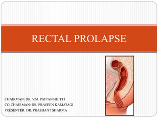
  • 1. CHAIRMAN: DR. V.M. PATTANSHETTI CO-CHAIRMAN: DR. PRAVEEN KAMATAGI PRESENTER: DR. PRASHANT SHARMA RECTAL PROLAPSE
  • 2. HISTORICAL PERSPECTIVE • Evidence of rectal prolapse (rectal procidentia) exists from ancient times. • The first described mortality from rectal prolapse was in the 4th century. • The initiating cause of prolapse is elusive. The path to rectal prolapse generally occurs slowly over a period of years. • Suggested contributing factors include standing, constipation, colic, coughing, sneezing, weak anal sphincters and pelvic floor musculature, an abnormal cul-de-sac, and distal rectal intussusception. • The proposed treatments have changed significantly over time as well. • Treatments such as cauterizing the rectal mucosa, binding patient’s legs together, and applying trusses have been replaced with variations of resection and rectopexy today.
  • 3. INTRODUCTION • Rectal prolapse is an uncommon, disabling condition that has long fascinated surgeons. • Few clinical disorders have generated such a large number of surgical procedures, with varying degrees of surgical outcomes, as has rectal prolapse. • Confusing terminology is a major problem in the study of rectal prolapse and the terms that must be distinguished are: 1. Mucosal Prolapse 2. Internal Intussusception (Occult Prolapse) 3. Complete Rectal Prolapse (Procidentia)
  • 4. • MUCOSAL PROLAPSE is caused by a looseness or breaking down of the connective tissue between the submucosa of the rectum and anal canal and the underlying muscle. •This usually starts in the anal canal and, in its earliest form, is represented by prolapsing hemorrhoids. •With progression, more anal canal mucosa and distal rectal mucosa protrude, leading to the characteristic picture of linear mucosal furrows and absence of the perianal sulcus. •Mucosal prolapse doesn’t progress to complete prolapse and is best considered part of the spectrum of hemorrhoidal disease.
  • 5. •INTERNAL INTUSSUSCEPTION of the rectum is a distinct clinical entity that may represent the precursor of a complete rectal prolapse. It can only be diagnosed reliably by obtaining a defecating proctogram. Patients with internal intussusception shouldn’t undergo operation as the basic underlying abnormalities in pelvic floor function cannot be addressed by surgery •COMPLETE RECTAL PROLAPSE is defined as protrusion of the full thickness of the rectal wall through the anal orifice. •It is the entity to which the various surgical procedures are directed.
  • 7. ANAL CANAL STRUCTURE, ANUS, AND ANAL VERGE •The anus or anal orifice is an anteroposterior cutaneous slit, that along with the anal canal remains virtually closed at rest, as a result of tonic circumferential contraction of the sphincters and the presence of anal cushions. • The edge of the anal orifice, the anal verge or margin (Ano-cutaneous line of Hilton), marks the lowermost edge of the anal canal and is sometimes the level of reference for measurements taken during sigmoidoscopy. • Others favor the dentate line as a landmark because it is more precise. The difference between the anal verge and the dentate line is usually 1–2 cm. • The epithelium distal to the anal verge acquires hair follicles, glands, including apocrine glands, and other features of normal skin, and is the source of perianal hidradenitis suppurativa, (inflammation of the apocrine glands).
  • 8. ANATOMIC VS. SURGICALANAL CANAL Two definitions are found describing the anal canal:: • The “anatomic” or “embryologic” anal canal is only 2.5 cm long, extending from the anal verge to the dentate line, the level that corresponds to the proctodeal membrane. • The “surgical” or “functional” anal canal is longer, extending for approximately 4.0 cm (in men) from the anal verge to the anorectal ring (levator ani).
  • 9. • The anorectal ring is at the level of the distal end of the ampullary part of the rectum and forms the anorectal angle, and the beginning of a region of higher intraluminal pressure. Therefore, this definition correlates with digital, manometric, and sonographic examinations.
  • 10. ANATOMIC RELATIONS OF THE ANAL CANAL • Posteriorly, the anal canal is related to the coccyx and Anteriorly to the perineal body and the lowest part of the posterior vaginal wall in the female, and to the urethra in the male. • The ischium and the ischiorectal fossa are situated on either side. • The fossa ischiorectal contains fat and the inferior rectal vessels and nerves, which cross it to enter the wall of the anal canal.
  • 11. MUSCLES OF THE ANAL CANAL  The muscular component of the mechanism of continence can Be stratified into three functional groups: 1. Lateral compression from the pubococcygeus, 2. Circumferential closure from the Internal and external anal sphincter, and 3. Angulation from the Puborectalis
  • 12. INTERNALANAL SPHINCTER • The internal anal sphincter represents the distal 2.5- to 4.0-cm condensation of the circular muscle layer of the rectum. • As a consequence of both intrinsic myogenic and extrinsic autonomic neurogenic properties, the internal anal sphincter is a smooth muscle in a state of continuous maximal contraction, and represents a natural barrier to the involuntary loss of stool and gas. • • The lower rounded edge of the internal anal sphincter can be felt on physical examination, about 1.2 cm distal to the dentate line. • The groove between the internal and external anal sphincter, the intersphincteric sulcus, can be visualized or easily palpated. • Endosonographically, the internal anal sphincter is a 2 to 3 mm thick circular band and shows a uniform hypoechogenicity.
  • 13. EXTERNALANAL SPHINCTER • The external anal sphincter is the elliptical cylinder of striated muscle that envelops the entire length of the inner tube of smooth muscle, but it ends slightly more distal than the internal anal sphincter. • The deepest part of the external anal sphincter is intimately related to the pubo- rectalis muscle, which can actually be considered a component of both the levator ani and the external anal sphincter muscle complexes. • Endosonographically, the puborectalis and the external anal sphincter, despite their mixed linear echogenicity, are both predominantly hyperechogenic, with a mean thickness of 6 mm (range, 5–8 mm). • In the male, the upper half of the external anal sphincter is enveloped anteriorly by the conjoined longitudinal muscle, whereas the lower half is crossed by it. • In the female, the entire external anal sphincter is encapsulated by a mixture of fibers derived from both longitudinal and internal anal sphincter muscles.
  • 14.  The automatic continence mechanism is formed by the resting tone, maintained by the internal anal sphincter, magnified by voluntary, reflex, and resting external anal sphincter contractile activities. • In response to conditions of threatened incontinence, such as increased intra-abdominal pressure and rectal distension, the external anal sphincter and puborectalis reflexively and voluntarily contract further to prevent fecal leakage.  The external anal sphincter and the pelvic floor muscles, unlike other skeletal muscles, which are usually inactive at rest, maintain unconscious resting electrical tone through a reflex arc at the cauda equina level.
  • 15. CONJOINED LONGITUDINAL MUSCLE • Whereas the inner circular layer of the rectum gives rise to the internal anal sphincter, the outer longitudinal layer, at the level of the anorectal ring, mixes with fibers of the levator ani muscle to form the conjoined longitudinal muscle. • This muscle descends between the internal and external anal sphincter, and ultimately some of its fibers, referred to as the corrugator cutis ani muscle, traverse the lowermost part of the external anal sphincter to insert into the perianal skin. • Possible functions of the conjoined longitudinal muscle include: 1. Attaching the Anorectum to the pelvis; and 2. Acting as a Skeleton that supports and binds the internal and external sphincter complex together.
  • 16.
  • 17. EPITHELIUM OF THE ANAL CANAL • The lining of the anal canal consists of an upper mucosal (endoderm) and a lower cutaneous (ectoderm) segment. • The dentate (pectinate) line is the “saw-toothed” junction between these two distinct origins of venous and lymphatic drainage, nerve supply, and epithelial lining. • Above this level, the intestine is innervated by the sympathetic and parasympathetic systems, with venous, arterial, and lymphatic drainage to and from the hypogastric vessels. • Distal to the dentate line, the anal canal is innervated by the somatic nervous system, with blood supply and drainage from the inferior hemorrhoidal system. These differences are important when the classification and treatment of hemorrhoids are considered. • The pectinate or dentate line corresponds to a line of anal valves that represent remnants of the proctodeal membrane. Above each valve, there is a little pocket known as an Anal Sinus or Crypt. These crypts are connected to a variable number of glands, in average 6 (range, 3–12).
  • 18. • More than one gland may open into the same crypt, whereas half the crypts have no communication. • The anal gland ducts, in an outward and downward route, enter the submucosa; two- thirds enter the internal anal sphincter, and half of them terminate in the intersphincteric plane. • Obstruction of these ducts, presumably by accumulation of foreign material in the crypts, may lead to perianal abscesses and fistulas.
  • 19. • Cephalad to the dentate line, 8–14 longitudinal folds, known as the rectal columns (Columns of Morgagni), have their bases connected in pairs to each valve at the dentate line. • At the lower end of the columns are the anal papillae. The mucosa in the area of the columns consists of several layers of cuboidal cells and has a deep purple color because of the underlying internal hemorrhoidal plexus.  This 0.5- to 1.0 cm strip of mucosa above the dentate line is known as the Anal Transition or Cloacogenic Zone. • Cephalad to this area, the epithelium changes to a single layer of columnar cells and macroscopically acquires the characteristic pink color of the rectal mucosa.  The cutaneous part of the anal canal consists of modified squamous epithelium that is thin, smooth, pale, stretched, and devoid of hair and glands.
  • 20. RECTUM • Both proximal and distal limits of the rectum are controversial: the rectosigmoid junction is considered to be at the level of the third sacral vertebra by anatomists but at the sacral promontory by surgeons, and likewise, the distal limit is regarded to be the dentate line by anatomists and the muscular anorectal ring by surgeons. • The rectum measures 12–15 cm in length and has three lateral curves: the upper and lower are convex to the right and the middle is convex to the left. These curves correspond intraluminally to the folds or Valves of Houston.
  • 21.  Although the rectal valves do not contain all muscle wall layers from a clinical point of view, they are a good location for performing a rectal biopsies, because they are readily accessible with minimal risk for perforation. • The two left-sided folds are usually noted at 7–8 cm and at 12–13 cm, respectively, and the one on the right is generally at 9–11 cm. The middle valve (Kohlrausch’s plica) is the most consistent in presence and location and corresponds to the level of the anterior peritoneal reflection.
  • 22. • The rectum is characterized by its wide, easily distensible lumen, and the absence of taeniae, epiploic appendices, haustrations, or a well defined mesentery. • The word “mesorectum” has gained widespread popularity among surgeons to address the perirectal areolar tissue, which is thicker posteriorly, containing terminal branches of the inferior mesenteric artery and enclosed by the fascia propria.  The “mesorectum” may be a metastatic site for a rectal cancer and is removed during surgery for rectal cancer without neurologic sequelae, because no functionally significant nerves pass through it. • The upper third of the rectum is anteriorly and laterally invested by peritoneum; the middle third is covered by peritoneum on its anterior aspect only. • Finally, the lower third of the rectum is entirely extraperitoneal, because the anterior peritoneal reflection occurs at 9.0–7.0 cm from the anal verge in men and at 7.5–5.0 cm from the anal verge in women.  Valve of Houston: Rectum  (Spiral) Valve of Heister: Cystic Duct  Valve of Hasner: Nasolacrimal Duct
  • 23. ANATOMIC RELATIONS OF THE RECTUM • The rectum occupies the sacral concavity and ends 2–3 cm anteroinferiorly from the tip of the coccyx. At this point, it angulates backward sharply to pass through the levators and becomes the anal canal. • Anteriorly, in women, the rectum is closely related to the uterine cervix and posterior vaginal wall; in men, it lies behind the bladder, vas deferens, seminal vesicles, and prostate. Posterior to the rectum lie the median sacral vessels and the roots of the sacral nerve plexus.
  • 24. FASCIAL RELATIONSHIPS OF THE RECTUM • The parietal endo-pelvic fascia lines the walls and floor of the pelvis and continues on the internal organs as a visceral pelvic fascia. • The lateral ligaments or stalks of the rectum are distal condensations of the pelvic fascia that form a roughly triangular structure with a base on the lateral pelvic wall and an apex attached to the lateral aspect of the rectum. • The lateral stalks are comprised essentially of connective tissue and nerves, and the middle rectal artery does not traverse them. Branches, however, course through in approximately 25% of cases. Consequently, division of the lateral stalks during rectal mobilization is associated with a 25% risk for bleeding.  One theoretical concern in ligation of the stalks is leaving behind lateral mesorectal tissue, which may limit adequate lateral or mesorectal margins during cancer surgery. • The presacral fascia is a thickened part of the parietal endopelvic fascia that covers the concavity of the sacrum and coccyx, nerves, the middle sacral artery, and presacral veins. Operative dissection deep to the presacral fascia may cause troublesome bleeding from the underlying presacral veins.
  • 25.  Presacral hemorrhage occurs as frequently as 4.6–7.0% of resections for rectal neoplasms, and despite its venous nature, can be life threatening. This is a consequence of two factors:  The difficulty in securing control because of retraction of the vascular stump into the sacral foramen, and  The high hydrostatic pressure of the presacral venous system. • The rectosacral fascia is an anteroinferiorly directed thick fascial reflection from the presacral fascia at the S-4 level to the fascia propria of the rectum just above the anorectal ring.  The rectosacral fascia, classically known as the Fascia of Waldeyer, is an important landmark during posterior rectal dissection.  The Visceral Pelvic Fascia of Denonvilliers is a tough fascial investment that separates the extraperitoneal rectum anteriorly from the prostate and seminal vesicles or vagina.
  • 26.
  • 27. ARTERIAL SUPPLY OF THE RECTUM AND ANAL CANAL • The superior hemorrhoidal/rectal artery is the continuation of the inferior mesenteric artery, once it crosses the left iliac vessels. •The artery descends in the sigmoid mesocolon to the level of S-3 and then to the posterior aspect of the rectum. •In 80% of cases, it bifurcates into right, usually wider, and left terminal branches; multiple branches are present in 17%. These divisions, once within the submucosa of the rectum, run straight downward to supply the lower rectum and the anal canal. • The superior and inferior hemorrhoidal/rectal arteries represent the major blood supply to the anorectum. In addition, it is also supplied by the internal iliac arteries.  The contribution of the middle hemorrhoidal artery varies with the size of the superior hemorrhoidal artery; this may explain its controversial anatomy. Some authors report absence of the middle hemorrhoidal artery in 40–88% whereas others identify it in 94– 100% of specimens.
  • 28.  The middle hemorrhoidal artery is more prone to be injured during low anterior resection, when anterolateral dissection of the rectum is performed close to the pelvic floor and the prostate and seminal vesicles or upper part of the vagina are being separated. • The anorectum has a profuse intramural anastomotic network, which probably accounts for the fact that division of both superior and middle hemorrhoidal arteries does not result in necrosis of the rectum. • The paired inferior hemorrhoidal arteries are branches of the internal pudendal artery, which in turn is a branch of the internal iliac artery.
  • 29.
  • 30. VENOUS DRAINAGE AND LYMPHATIC DRAINAGE OF THE RECTUM AND ANAL CANAL • The anorectum also drains, via middle and inferior hemorrhoidal/rectal veins, to the internal iliac vein and then to the inferior vena cava.  The external hemorrhoidal plexus, situated subcutaneously around the anal canal below the dentate line, constitutes when dilated the external hemorrhoids. • The internal hemorrhoidal plexus is situated submucosally, around the upper anal canal and above the dentate line. The internal hemorrhoids originate from this plexus. • Lymph from the upper two-thirds of the rectum drains exclusively upward to the inferior mesenteric nodes and then to the paraaortic nodes. • Lymphatic drainage from the lower third of the rectum occurs not only cephalad, along the superior hemorrhoidal and inferior mesentery arteries, but also laterally, along the middle hemorrhoidal vessels to the internal iliac nodes.
  • 31. • In the anal canal, the dentate line is the landmark for two different systems of lymphatic drainage: above, to the inferior mesenteric and internal iliac nodes, and below, along the inferior rectal lymphatics to the superficial inguinal nodes, or less frequently along the inferior hemorrhoidal artery.  In the female, drainage at 5 cm above the anal verge in the lymphatic may also spread to the posterior vaginal wall, uterus, cervix, broad ligament, fallopian tubes, ovaries, and cul-de-sac, and at 10 cm above the anal verge, spread seems to occur only to the broad ligament and cul-de-sac.
  • 32.
  • 33. INNERVATION OF THE RECTUM AND ANAL CANAL • Innervation of the Rectum – The sympathetic supply of the rectum and the left colon arises from L-1, L-2, and L-3. – Two main hypogastric nerves, on either side of the rectum, carry sympathetic innervation from the hypogastric plexus to the pelvic plexus. – The parasympathetic fibers to the rectum and anal canal emerge through the sacral foramen and are called the nervi erigentes (S-2, S-3, and S-4). – The periprostatic plexus, a subdivision of the pelvic plexus situated on Denonvilliers’ fascia, supplies the prostate, seminal vesicles, corpora cavernosa, vas deferens, urethra, ejaculatory ducts, and bulbourethral glands. – Sexual function is regulated by cerebrospinal, sympathetic, and parasympathetic components. Erection of the penis is mediated by both parasympathetic (arteriolar vasodilatation) and sympathetic inflow (inhibition of vasoconstriction). – All pelvic nerves lie in the plane between the peritoneum and the endopelvic fascia and are in danger of injury during rectal dissection.
  • 34.  Permanent bladder paresis occurs in 7–59% of patients after abdomino-perineal resection of the rectum; the incidence of impotence is reported to range from 15 to 45%, and that of ejaculatory dysfunction from 32 to 42%.  The overall incidence of sexual dysfunction after proctectomy has been reported to reach 100% when wide dissection is performed for malignant disease.  Dissections performed for benign conditions are undertaken closer to the bowel wall, thus reducing the possibility of nerve injury.
  • 35. – Trauma to the autonomic nerves may occur at several points. During high ligation of the inferior mesenteric artery, close to the aorta, the sympathetic preaortic nerves may be injured. – Division of both superior hypogastric plexus and hypogastric nerves may occur also during dissection at the level of the sacral promontory or in the presacral region.  In such circumstances, sympathetic denervation with intact nervi-erigentes results in retrograde ejaculation and bladder dysfunction. – The nervi erigentes are located in the posterolateral aspect of the pelvis, and at the point of fusion with the sympathetic nerves are closely related to the middle hemorrhoidal artery.  Injury to these nerves will completely abolish erectile function. – The pelvic plexus may be damaged either by excessive traction on the rectum, particularly laterally, or during division of the lateral stalks when this is performed close to the lateral pelvic wall.
  • 36. – Finally, dissection near the seminal vesicles and prostate may damage the periprostatic plexus, leading to a mixed parasympathetic and sympathetic injury. This can result in erectile impotence as well as a flaccid, neurogenic bladder. – Sexual complications after rectal surgery are readily evident in men but are probably underdiagnosed in women.
  • 37. • ANAL CANAL – The internal anal sphincter is supplied by sympathetic (L-5) and parasympathetic nerves (S-2, S-3, and S-4) following the same route as the nerves to the rectum. – The external anal sphincter is innervated on each side by the inferior rectal branch of the pudendal nerve (S-2 and S-3) and by the perineal branch of S-4.  Despite the fact that the puborectalis and external anal sphincter have somewhat different innervations, these muscles seem to act as an indivisible unit. – After unilateral transection of a pudendal nerve, external anal sphincter function is still preserved because of the crossover of the fibers at the spinal cord level.  Anal sensation is carried in the inferior rectal branch of the pudendal nerve and is thought to have a role in maintenance of anal continence.
  • 38. PELVIC FLOOR MUSCULATURE  The muscles within the pelvis can be divided into three categories (1) the anal sphincter complex; (2) pelvic floor muscles; and (3) muscles that line the sidewalls of the osseous pelvis.
  • 39. LEVATOR ANI • The levator ani muscle, or pelvic diaphragm, is the major component of the pelvic floor. It is a pair of broad, symmetric sheets composed of three striated muscles: ileococcygeus, pubococcygeus, and puborectalis. • The pelvic floor is “incomplete” in the midline where the lower rectum, urethra, and either the dorsal vein of the penis in men, or the vagina in women, pass through it. This defect is called the levator hiatus and consists of an elliptic space situated between the two pubococcygeus muscles. • The puborectalis muscle is a strong, U-shaped loop of striated muscle that slings the anorectal junction to the posterior aspect of the pubis.
  • 40. THE ANORECTAL RING AND THE ANORECTALANGLE • Two anatomic structures of the junction of the rectum and anal canal are related to the puborectalis muscle: the anorectal ring and the anorectal angle. The anorectal ring, a term coined by Milligan and Morgan, is a strong muscular ring that represents the upper end of the sphincter, more precisely the puborectalis, and the upper border of the internal anal sphincter, around the anorectal junction.  It is of clinical relevance, because division of this structure during surgery for abscesses or fistula inevitably results in fecal incontinence.  The anteriorly directed pull of the puborectalis contributes to the angulation between the rectum and anal canal, the anorectal angle.
  • 41. • The Anorectal Angle is thought to be the result of the anatomic configuration of the U- shaped sling of puborectalis muscle around the anorectal junction.  Whereas the anal sphincters are responsible for closure of the anal canal to retain gas and liquid stool, the puborectalis muscle and the anorectal angle are designed to maintain gross fecal continence.
  • 43.  The normal function of the anorectum represents a complex interaction between anatomical, neurological, sensory, myogenic and hormonal factors.  Failure or weakness of any one part or combination of parts of these factors may lead to symptoms of many common diseases seen in colorectal practice.  The anorectal physiology laboratories are designed to provide informations about the function of the neurological, sensory and anotomical components of anorectal function.
  • 44. MECHANISM OF DEFECATION • The development of a mass peristaltic wave by the left colon results in the delivery of stool to the lower sigmoid colon and rectum. Once the stool content arrived in the rectum several physiological events take place. 1. There is reflex relaxation of the internal sphincter and contraction of the distal external sphincter. This is called Rectoanal Inhibitory Reflex (RAIR). 2. As the result of the reflex relaxation of the internal sphincter the content of the rectum comes in contact with the highly sensitive lower end of the anal canal and the nature of the content either solid, liquid or gas is determined accurately. 3. This increase in the volume of the rectum leads to an increase in the rectal pressure that is maintained for 1-2 minutes before the intra-rectal pressure returns to normal by process called accommodation which allows the relaxation of the rectum to accommodate the increased rectal volume. 4. As the rectal volume increases there is a gradual rise in the rectal pressure and at a particular pressure the urge to defecate occurs.
  • 45.  If the urge occurs at a socially acceptable time to proceed with defecation or if the accommodation has reaches its upper limit the process of defecation occurs and the individual will normally assume a squatting or sitting position.  By Valsalva maneuver the intra-abdominal and intra-rectal pressures are raised. The anorectal angle is straightened the entire sphincter mechanism relaxes and the pelvic floor forms funnel shape.  Both straining and recto-sigmoid contraction aids in emptying of the rectum.  If this process is accompanied by further mass peristalsis the entire left colon may be evacuated. If not the evacuation may occur in a piecemeal fashion.  Once the rectum is empty the external sphincter and puborectalis transiently contract (the closing reflex).  Following defecation, sphincter pressure, intra-abdominal pressure, return to their normal resting pressure and the anorectal angle returns to its position.
  • 46.  Constipation manifested as outlet obstruction may occur when any portion of this process is interrupted.  Such a situation may occur in the following conditions: 1. Failure of puborectalis to relax (Paradoxical puborectalis, Anismus, levator syndrome). 2. Abnormal high resting pressure (Nut-cracker anus). 3. Excessive accommodation (Mega rectum). 4. External compression of the rectum (Enterocele). 5. Incomplete evacuation (Larger rectocele). 6. Intussusception (Overt or Covert prolapse). 7. Excessive pelvic descent (descending Perineal syndrome). Whether the perineal descent is the result of chronic straining due to outlet obstruction or a primary cause of the constipation is debatable. 8. Constipation can also be a manifestation of dysfunctional colon mobility.
  • 47. ALTERED ANORECTAL PHYSIOLOGY • Altered anorectal physiology may lead to two important conditions, constipation and incontinence. I. In the case of constipation quality of feces, hard or soft, type (excessive straining or failure of recognize a call to stool) and frequency may help direct to the initial evaluation. II. In the case of incontinence frequency and type of incontinence either to gas, liquid or solid is important for the evaluation. Factors that contribute to anal continence are: 1. The anal canal high pressure zone 2. Anal canal length 3. Anorectal angle 4. Rectal compliance 5. Anorectal sensation 6. Anal cushions 7. Stool consistency and frequency.
  • 48.  The anal canal high pressure zone is due to internal anal sphincter, external anal sphincter and the puborectalis muscle.  The internal anal sphincter is responsibles for 85 percent of the resting pressure. The external sphincter and puborectal muscle are responsible for squeeze pressure of the anal canal. The anal canal pressure can be elevated voluntarily for about one minute only. The same action of these muscles is responsible for the anal canal length.  The anorectal angle is not constant and it varies according to the strain and increase in intra-abdominal pressure.  When the puborectalis muscle relaxes the angle is widened and defecation occurs. The rectal compliance plays a roll in maintenance of continence by the accommodation reflex.  In disease like ulcerative colitis and irradiation proctitis the rectum becomes less compliant and the ability to stretch the rectal wall may be decreased. As a result, the rectal pressure remains high and when further increase in pressure occurs, the continence becomes dependent upon external anal sphincter and puborectalis muscle which can last for one minute only.
  • 49. • The consistency of the stool is an important factor for continence. Solid feces can be controlled and regulated even by weak sphincters and fluid stools can not be controlled even by a normal sphincter. • The sensory components of the mechanism of continence are critical in allowing a satisfactory reaction time to read to a bolus of stool or gas entering the rectum.  Sensation is associated with the sampling reflex. This reflex results in the transient relaxation of the internal sphincter with contraction of distal external sphincter, allowing for discrimination of the contents in the upper anal canal and rectum as either gas or liquid or solid.
  • 50. BASIC ANORECTAL PHYSIOLOGY—LABORATORY TESTS In clinical practice, the basic tests which are useful include: 1. Anorectal manometry (ARM) 2. Anorectal electromyography (EMG) 3. Dynamic proctography (DPG) 4. Colon transit time (CTT) 5. Trans anal ultrasound (TAUS) 6. Estimation of neurological function (ENF).  It is important to select carefully those tests that will provide useful clinical information for an individual patient.
  • 51. A. ANORECTAL MANOMETRY • Anorectal manometry is an objective method for assessing anal muscular tone, rectal compliance, and anorectal sensation and of verifying the integrity of the rectoanal inhibitory reflex (RAIR). • It is a useful investigation in patients with fecal incontinence to identify the presence of sensory or muscular defects as well as to find out the functional weakness of internal and external anal sphincter. • It may be helpful in evaluation of Chronic Constipation and Hirschsprung's disease. • It helps to determine whether sphincter weakness or dysfunction is likely to be the cause of symptoms. Other tests such as electromyography, ultrasound or other radiological study are needed to determine the underlying pathology. • The most basic components of anorectal manometry are pressure transuding catheter and a recorder. • Different types of transuding catheters are available such as water perfused catheter, micro transducer and balloon catheters.
  • 52. • Regardless which catheter are used, manometric examination measure a resting pressure (RP) a squeeze pressure (SP) and the rectoanal inhibitory reflex (RAIR) and the anal canal length. • Resting pressure and squeeze pressure are obtained by measuring the pressure at multiple points in the ano-rectum. This can be done by pulling the catheter out at 0.5 cm intervals and recording pressure at each point. • The maximum squeeze pressure is twice the resting pressure and it indicates the function of external sphincter. • The anal canal length is determined by measuring from the point at which the pressure suddenly rises higher than the basal rectal tone to that where the pressure suddenly drops off at or near the anal verge.
  • 53. • The anal canal length is generally shorter in incontinence patients. The anal canal length is usually increased after post anal repair which predict the return of continence.  The rectoanal inhibitory reflex (RAIR) is usually initiated by instillation of air or water into the balloon in the rectum. This causes a reflex contraction of the external anal sphincter with synchronous relaxation of the internal anal sphincter. This reflex is absent in Hirschsprung's disease and some cases of scleroderma.  Chronically relaxed internal sphincter (low resting pressure) and failure to further relax may be associated with fecal impaction and overflow incontinence. • To measure the rectal sensation and compliance an intra-rectal balloon is connected to a pressure monitor is slowly filled with warm saline and the individual is asked to note when distention is first feel (minimum perceived or threshold volume) and when the further distention can not be tolerable (maximum tolerable volume). By recording the intrarectal pressure at various volumes a compliance curve can he obtained. The maximum tolerable volume is more useful in biofeedback treatment for anal incontinence.
  • 54. B. AMBULATORY ANORECTAL MANOMETRY • In an effort to observe the anorectal continence mechanism in a more suitable environment ambulatory anorectal manometry has been developed. • Usually a micro transducer catheter used from which constant recordings are made. Surface electromyography can also be coupled with it.
  • 55. C. BALLOON RETENTION AND EXPULSION TEST • The ability of the rectum to retain or expel an intra rectal balloon can be evaluated. The intra rectal balloon is filled with water at 60 cc per minute till it is expelled or maximum tolerable volume is reached. • Water filled balloon stimulate like the solid or semisolid stool rather than free water. Some research people use air filled rectal balloon at the end of the standard anorectal manometry study. • This test helps to discriminate which patient truly have anismus and will benefit from bio- feedback therapy.
  • 56. D. RECTAL TONE MEASUREMENT • Recently a technique is described for measuring rectal tone (as opposed to compliance) • Changes in the tone are measured by an electronic barostat that measures change in the volume in an intrarectal balloon maintained at a constant pressure.  Profound changes have been recorded in rectal tone that where not detected manometrically. E. SALINE INFUSION TEST • This is an investigation done for the evaluation of fluid incontinent individuals. • Isotonic saline 1500 cc at body temperature is infused per anum at 60 cc per minute and the time of first leak and the total volume of leak are noted.  The time of first leak is significally lower in incontinent patients.
  • 57. ELECTROPHYSIOLOGICAL TESTS • Anal electromyography (EMG) is the recording of the electrical activity from the muscle fibers of the external sphincter and puborectalis complex during rest, during maximum squeeze, during defecations and in response to various reflexes. • It has been used for sphincter mapping in patients with faecal incontinence and also is of use in assessing the function of the pelvic floor in individuals with constipation. • Anorectal EMG consists of two basic evaluations, assess the conduction time and the assessment of pelvic floor activity.
  • 58.  To assess the conduction time in the Pudendal nerve from Alcock's canal to the end organ in the external sphincter and this test is called as Pudendal Nerve Terminal Motor Latency (PNTML). • Normal conduction occurs in 2.0 + 0.2 milli seconds. Prolonged times suggest injury to the large fast conducting fibres. • In patients with neuropathtic incontinence PNTML is measured by using a specially designed glove with a stimulating electrode on the finger tip and a recording electrode at the base of the finger. • The stimulating electrode is used intra rectally to stimulate the pudendal nerve as it exits the Alcock's canal and the recording electrode documents the response in the external anal sphincter.
  • 59. • Prolonged PNTML is found in several disorders associated with difficult vaginal delivery or chronic straining at stool and fecal incontinence. Severe constipation, prolapse rectum, uterine prolapse and solitary rectal ulcer syndrome can also prolong PNTML.
  • 60.  EMG can also be used for the activity of the external sphincter by inserting electrode in all four quadrants to detect areas of electrically silent, which indicate a lack of muscle at that spot. • Single fiber EMC shows density of muscle fibers innervated by individual axon in the muscle suspected to he damaged by a neurogenic process.  A raised fiber density indicates reinnervation of the muscle.
  • 61. DYNAMIC PROCTOGRAPHY (DPG) (DEFECOGRAPHY) • Dynamic Proctography (DPG) is used to evaluate function of the pelvic floor at the time of defecation. • DPG has many variations in name as well as the procedures. Videoproctography, Defecography, Defecating Scintigraphy, all accomplish the same essential assessment of the ano-rectal angle, ano-rectal function, pelvic floor descent and the length of the anal canal.  Patients with colonic inertia often undergo defecography in order to exclude pelvic floor dysfunction. • DPG is performed by thick mixture of barium contrast and methyl cellulose in the rectum. Additional liquid contrast is placed in the vagina generally the patient sits on a radiolucent toilet seat when the test is done under videofluroscopic monitoring. • This allows the evaluation to occur during three phases – Rest, Evacuation and Recovery. • The technique can be enhanced with the use of 'Triple Contrast'. This technique combines the use of the standard DPG with the contrast in the small bowel.
  • 62. • The 3rd element of the triple contrast is contrast in the urinary bladder so that its interaction with the pelvic floor at the time of straining can also be evaluated. This tripple contrast DPG gives the most thorough evaluation of pelvic floor at the time of defecation. • DPG can demonstrate paradoxical contraction of the puborectalis, internal intussusception, enterocele, rectocele, pelvic floor hernia and incomplete evacuation.
  • 63. TRANSANAL ULTRASONOGRAM (TAUS) • This test is useful in the assessment of fecal incontinence.  This provides the most accurate and complete evaluation for detecting the anal sphincter defect. • Ultrasonography is a safe test, without risk of radiation exposure. Therefore, it is ideal for children and pregnant women. It is also a more cost-effective and safer technique. • The problem of differentiating local recurrence from that of postoperative changes can be minimize by performing a base line endorectal ultrasonography 3 months after surgery by which time most acute inflammatory reaction has decreased. • Generally a probe with 7-10 mhz, 360 degree rotating transducer is used. • In TUS the internal sphincter is visualized as a hypoechogenicity band around the anal canal and the external sphincter as a band of mixed echogenicity.  Defects in either band can be readily identified and measured.
  • 64. PRESENTATION AND WORK-UP • Patients with rectal prolapse will often report a history of constipation and even incontinence. Women are 6 times more likely than men to suffer from prolapse. • The primary presenting complaint is often of a protuberance or bulge from the anus, which is often mistaken for hemorrhoidal disease. It may spontaneously reduce or remain prolapsed, causing considerable distress. • Other complaints may be a feeling of incomplete evacuation of stool, bleeding, mucus discharge, urinary incontinence, and pain. • The prolapse may be apparent on initial exam; other times it will need to be elicited by asking the patient to perform a Valsalva. At times, it is even necessary to examine the patient after having them strain while squatting on the commode. • Once the prolapse is produced, it is important to differentiate true full-thickness rectal prolapse with concentric mucosal rings from simple hemorrhoidal disease, which is identified by radial folds of hemorrhoidal tissue.
  • 65.
  • 66. • Colonoscopy is required in all patients considered candidates for operative repair of rectal prolapse to rule out a neoplastic lead point. • On endoscopic examination, the rectal mucosa in a patient with chronic recurrent prolapse will show signs of circumferential erythema. • Other diagnostic tests include anal manometry, endorectal ultrasound, and defecography.  Defecography is valuable in occult, intermittent prolapse to document marked mobility of the rectosigmoid junction away from the normal point of fixation at the sacral promontory.
  • 67. ACUTE TREATMENT • The immediate management of obvious prolapse is reduction. Before any attempts at reducing the prolapse, it is of utmost importance to fully assess the viability of the rectum. • Ischemia or frank necrosis may be present in the neglected or demented patient. If significant full-thickness ischemia is present, reduction should be avoided and operative resection will likely be required. • The majority of patients with viable prolapse will respond to continuous, steady pressure in the office setting or emergency room. At times, intravenous sedation or even general anesthesia in the operating room may be necessary for full reduction. • Applying table salt or sugar to the mucosa can reduce the swelling of the incarcerated rectum and aid in reduction. • Another technique described for the stubborn prolapse involves encompassing the rectum with an elastic compression wrap. • Hyaluronidase injection into the prolapsed rectum has also been described to aid in reduction.
  • 68. TRANSABDOMINAL OPTIONS • Abdominal approaches to definitive repair of prolapse consistently show lower recurrence rates compared with perineal approaches. • The transabdominal approach is the preferred repair in patients who can tolerate a transabdominal operation. • Ideally, the operation should be limited in morbidity and mortality, have a low recurrence rate, and address the patient’s associated constipation or incontinence, if present. • Care must also be given to evaluating the patient for the presence of concurrent anterior compartment prolapse to assess the feasibility of repair of these prolapsing organs as well in a combined operation. • There are two primary technical components in the abdominal approach to rectal prolapse: rectopexy and resection. • The goal of rectopexy is to mobilize the rectum out of the pelvis to straighten the low rectum in the curve of the sacrum and secure it proximally at the sacral promontory so that prolapse cannot recur.
  • 69. • In 1963, Ripstein described the posterior rectopexy: a posterior rectal dissection to the level of the coccyx and a sling of fascia lata around the rectum anchored to the proximal sacrum. • Modifications using biologic and synthetic meshes have come and gone over time. • Rectopexy can even be performed without the use of mesh by securing peritoneal wings of the rectum to the sacral promontory with suture or tacks. This technique, however, results in slightly higher recurrence rates than rectopexy with mesh. • The extent of rectal mobilization required during rectopexy has also been an issue of debate. • Circumferential mobilization may decrease the rate of prolapse recurrence; however, a known drawback of rectopexy is constipation, and division of the anterolateral ligaments (parasympathetic innervations) during dissection.
  • 70. • The most recent addition to the abdominal operation for rectal prolapse is the Ventral Rectopexy that involves mobilization of the anterior portion of the rectum without a posterior or lateral dissection. • Mesh shaped in the form of a “hockey stick” is then sutured from the anterior rectal wall to the sacral promontory and covered with peritoneum.  This technique reduces the need for extensive posterior dissection. Some authors report lower constipation rates, while maintaining recurrence rates similar to posterior Rectopexy.
  • 71. • The use of laparoscopy in the repair of rectal prolapse has been studied extensively. The integrity of the repair has not been compromised with the laparoscopic approach, as recurrence rates are similar.  The short-term postoperative benefits of laparoscopic surgery are realized in patients with rectal prolapse, including  Decreased postoperative pain,  Shorter hospital length of stay, and  Quicker return of bowel function.  These recovery benefits of laparoscopy may also allow more high-risk patients to be approached transabdominally, who may otherwise have only been considered for the less durable perineal approaches.
  • 72. PERINEAL APPROACHES • While abdominal approaches for rectal prolapse have the lowest recurrence rates with better overall functional outcomes, the majority of patients with prolapse are elderly. • Many of these patients have significant comorbidities that limit their ability to tolerate an abdominal approach. • Additionally, though a much smaller cohort, young patients, especially males, may consider a perineal approach to limit the added risk of sexual dysfunction. • Up to 60% of patients suffering from rectal prolapse will undergo a perineal approach. • When rectal prolapse recurs after an abdominal approach, a second abdominal approach can be technically difficult. In these circumstances, a perineal approach may be a viable option. • However, a perineal resection-rectopexy can spell doom if the blood supply of the rectosigmoid was previously divided, and the perineal resection leaves an ischemic segment of rectum in place.
  • 73. PERINEAL PROCTOSIGMOIDECTOMY (ALTEMEIER PROCEDURE) • Perineal proctosigmoidectomy for rectal prolapse was originally introduced by Mikulicz in 1899 and popularized by Altemeier et al. • The procedure can be performed in lithotomy or prone jack-knife position. Anesthetic options include regional or general anesthesia. • The operation is begun by applying gentle retraction to the rectum so that the redundant rectum is externalized, recreating the prolapse. • The anal canal is held open with a Lone Star retractor (Lone Star Company, Frisco, Texas).
  • 74. • The dentate line is identified. An injection of dilute epinephrine solution is administered circumferentially 1 to 2 cm proximal to the dentate line followed by a full-thickness, circumferential rectal incision with electrocautery. • The avascular intersphincteric plane is developed up to the pelvis, and the distal rectum is pulled from the pelvis. • Using either suture ligation or an energy device, the vascular supply of the exposed bowel is then ligated as the dissection is continued proximally.
  • 75. • The redundant rectum and sigmoid colon are extracted to the level of the left colon, and the bowel is divided under mild stretch at the point of planned coloanal anastomosis. • The more bowel removed, the better. A posterior rectopexy with a postanal levatorplasty has been shown to improve anal continence. • The rectopexy is performed by using a nonabsorbable suture to fix the posterior surface of the nonprolapsed bowel to the pre-sacrococcygeal fascia. The normal anorectal angle is reconstructed with a posterior levator-plasty to restore the anorectal angle.
  • 76. • A hand-sewn colo-anal anastomosis is performed between left colon and dentate line.A stapled anastomosis can be performed using two purse strings. However, some authors report a higher probability of recurrence with a stapled anastomosis. • Ultimately, patients selected for a perineal procto-sigmoidectomy should expect an improved continence compared with times of prolapse and decreased constipation with overall improved quality of life. • However, a disadvantage of the perineal proctosigmoidectomy is the increased recurrence rates of 12% to 24% compared with the abdominal approach. This can be reduced by removing all of the redundant distal colon up to the left colon.
  • 77. ANORECTAL MUCOSECTOMY WITH MUSCULAR PLICATION (DELORME PROCEDURE) • In 1900, Delorme described the anorectal mucosectomy with muscular plication for the treatment of rectal prolapse.  A significant advantage of the Delorme procedure is that no full-thickness bowel resection with anastomosis is performed. Similar to the Altemeier procedure, the bowel is completely prolapsed and dilute epinephrine solution is injected in a submucosal plane. • Using electrocautery, a circumferential submucosal incision is then made 1 cm proximal to the dentate line. Being careful not to violate the muscular layer of the rectal wall, a submucosal dissection is performed and extended proximally. • A mucosectomy twice the length of the prolapse is performed. • After the mucosal sleeve is excised, longitudinal plicating sutures are placed in rows along the length of the rectal wall musculature 1 cm apart. • The sutures are not tied until all the sutures have been placed. As the longitudinal sutures are tied around the circumference of the rectum, the cut edge of the mucosa is brought to the level of the anal canal.
  • 78. • Finally, the proximal and distal mucosal edges are re-approximated using absorbable sutures at the dentate line. • The muscular plication and mucosal anastomosis can be alternatively performed using a circular stapler. • In patients with a patulous anus on physical examination and in efforts to improve continence, a posterior levatorplasty can also be performed. • The Delorme procedure for rectal prolapse offers patients a safe procedure, with reported low morbidity of 9.5% but reported recurrence rates of 12% to 31%.  Significant preexisting long-term constipation has been associated with higher recurrence rates, while pelvic floor repair with levator-plasty has been associated with reduced recurrence rates.  Patients may complain of the urge to defecate because of the reefed muscle in the anal canal.
  • 79. Anorectal mucosectomy with muscular plication (Delorme procedure).
  • 80. THIERSCH PROCEDURE • Anal encirclement for the treatment of rectal prolapse was introduced in 1891 by Thiersch.  In the original description, Thiersch used a silver wire to encircle the anus in an effort to provide a mechanical replacement of the relaxed sphincter.  The procedure does not correct the prolapse but simply mechanically supports the anal sphincter function by narrowing the anal canal and preventing external rectal prolapse. • The procedure can be performed quite quickly. With the patient in prone jackknife or lithotomy position, in reference to the center of the anus, two lateral perianal skin incisions are made 180 degrees apart. • The incisions are connected subcutaneously by tunneling through the ischiorectal fossa and perineal body. Next, a piece of polypropylene mesh or silicone roll 1.5 cm in height is tunneled circumferentially around the entire anus.  “The presence of a foreign body would incite an inflammatory reaction and create an adhesive circumferential band to create support of the perineum.”
  • 81. • The mesh is tightened to a point that allows only the passage of one finger into the anal canal, and the mesh is sutured to itself at this level of tension. • Reported complications include recurrence rates of up to 75%, anal stenosis, fecal impaction, and even erosion of the mesh or Silastic product into the anorectum.  Because of the high recurrence rates and reported complications, indications for the procedure are very narrow but include patients with mild to moderate rectal prolapse and significant life-threatening comorbidities, with anticipated short life expectancy, that prevent them from either abdominal or resective perineal approaches.
  • 82. COMPARISON BETWEEN THE DIFFERENT MODALITIES • Historically, elderly patients with significant comorbidities were only candidates for perineal approaches. • The perineal approaches traditionally rendered less durable long-term control of prolapse compared to abdominal approaches.  Comparing the Altemeier procedure to Delorme procedure, a randomized controlled study showed there were fewer recurrences after the Altemeier procedure (24% vs. 31%), but functional outcomes were similar.  With abdominal rectopexy, there is no significant difference in terms of recurrence between the methods used for fixation.  In investigating the degree of rectal mobilization necessary, circumferential mobilization rather than preservation of the lateral ligaments has been associated with decreased recurrent prolapse. However, circumferential mobilization is also associated with increased postoperative constipation.
  • 83. • In comparison of resection rectopexy to rectopexy alone, lower rates of both constipation and recurrence were encountered with resection rectopexy. • Unfortunately, resection is associated with a finite anastomotic leak rate. Although it has been a long-held belief that perineal approaches are associated with a significantly higher recurrence rate than abdominal approaches, a 2015 Cochrane review of 15 randomized controlled trials and over 1000 patients failed to display superiority of any approach in terms of recurrence rates or quality of life.  Therefore the surgeon’s experience and judgment are paramount to the choice of operative approach based on patient characteristics, the clinical situation, and expertise.
  • 84. MANAGEMENT OF RECURRENCE • It is important to understand patient and technical factors that are associated with recurrence patterns. • During perineal proctosigmoidectomy, stapled anastomosis, shorter specimen length, and severe preexisting constipation were all associated with an increased risk of recurrence. • A postanal repair and levatorplasty were associated with lower recurrence rates. • During abdominal rectopexy, variables identified as related to recurrence included incontinence, constipation, the extent of rectal mobilization, and whether sigmoid resection was performed in the setting of constipation. • As mentioned previously, circumferential rectal mobilization was associated with decreased recurrence. • Overall, a high recurrence rate after either approach is likely due to technical failures such as inadequate fixation or incomplete mobilization.
  • 85. • Poor decision making and failure to recognize preexisting conditions such as colonic dysmotility in a patient undergoing rectopexy without resection will ultimately render suboptimal results. • Repair of recurrent rectal prolapse should be undertaken only after consideration of several complexities that guide choice of approach. • Traditionally, if a perineal resection was initially performed, repeat perineal resection is often the safest route to avoid compromising the remaining proximal blood supply. • Similarly, a recurrence after abdominal resection rectopexy should not be approached with a perineal resection, as there is great risk for rectal ischemia after ligating both the proximal and then distal rectal blood supply. However, a perineal proctosigmoidectomy is a very reasonable option if abdominal rectopexy without resection was performed as the index procedure.
  • 86. • A second abdominal approach is often a technically difficult procedure in the pelvis that can increase the potential for added patient morbidity. • Similarly, it is reasonable to consider abdominal rectopexy without resection after a recurrence from a perineal resection.  Therefore, in the case of recurrent prolapse, the index procedure often dictates the reoperative surgical approach. Mesh suspension may be considered if tissues were inadequate for pexy at the initial operation.
  • 87. INTERNAL INTUSSUSCEPTION (INTERNAL RECTAL PROLAPSE) • Internal rectal intussusception is described as internal telescoping of the rectum that does not result in protrusion from the anal canal. When the rectum protrudes from the anal canal, rectal prolapse occurs. • The Oxford grading system has been established to grade the severity of rectal prolapse based on evacuation proctography. • While internal intussusception has been identified in asymptomatic individuals and has historically been treated as a normal variant, evidence now suggests that patients with internal intussusception can suffer from a spectrum of disorders ranging from obstructed defecation syndrome to fecal incontinence. As such, detailed physical examination and often extensive diagnostic imaging work-up are required.
  • 88. HISTORY • Women with internal rectal intussusception commonly suffer from a range of symptoms. • Historically, symptoms of internal intussusception included only symptoms of obstructive defecation; however, evidence now suggests that fecal incontinence also frequently plays a significant role in symptomatology. • In fact, fecal incontinence can be the predominant complaint in patients with high- grade internal intussusception. In females with internal intussusception, fecal incontinence was reported in 56%, while complaints of incomplete evacuation (45%), straining at defecation (34%), and digital assistance during defecation (34%) were also commonly reported. • The majority of patients had complaints of urge incontinence, while passive incontinence and incontinence to flatus were reported less often. During the initial clinical encounter of a patient with pelvic defecatory dysfunction, it is necessary to quantify the severity of symptoms. • The subsequent success of therapy can be gauged by comparison of function before and after treatment using grading scales, which include Wexner constipation and incontinence score, fecal incontinence severity index score, and quality-of-life.
  • 89. PATHOPHYSIOLOGY • Initial evidence pointed toward changes in the rectoanal inhibitory reflex (RAIR) caused by the intussusceptum as the source of incontinence. • Reducing the intussusceptum with rectopexy, the RAIR would return to normal, and continence would be restored. • Upon evaluation with anal manometry, initial results suggested a significant reduction in the average maximum resting pressure (MRP) as the grade of rectal intussusception increased. This predicted the role of incontinence was mainly through a reduction in internal anal sphincter tone induced by the inhibitory reflex as the rectum stretched the anal canal. • Subsequent follow-up data, however, showed that the MRP between patients with and without intussusception did not differ, suggesting that the reduction of MRP is not permanent. • Furthermore, urge incontinence associated with high-grade internal intussusception was significantly associated with a decrease in rectal sensory volume thresholds. One explanation for this observation involves the intussusceptum inciting a chronic irritation on the rectal wall, rendering a reduction in wall compliance with subsequent urge incontinence.
  • 90. • . Unlike incontinence, constipation severity does not increase with increasing Oxford grades of internal intussusception. • Further research is necessary to understand the relationship between internal rectal intussusception, the development of defecatory disorders, and the need for surgical repair.  The majority of mild to moderate intussusception seen on defecography is asymptomatic, and symptoms can be alleviated with the administration of dietary fiber supplements.
  • 91. PHYSICAL EXAM  Performing a digital rectal examination (DRE) is a necessary initial screening tool in the work-up of patients presenting with defecatory dysfunction. • Not only does the surgeon gain insight on anal sphincter pressures, but DRE also shows high positive predictive values in detecting patients with other forms of outlet obstruction such as paradoxical puborectalis contraction (PPC) or increased perineal descent (IPD). • Forward motion of the puborectalis shelf during attempts by the patient to push the examining finger out indicates paradoxical contraction or nonrelaxation (PPC). By having a high index of suspicion for PPC and IPD, physicians can appropriately guide therapy. • A sign of intussusception during proctoscopy and anoscopy includes invagination of the circumference of the rectal wall. Low-grade intussusception remains in the rectal vault while high-grade intussusception prolapses into the anal canal without external prolapse. • Valsalva maneuver can be helpful during examination to assist in diagnosis.
  • 92. PHYSIOLOGIC TESTING AND DIAGNOSTIC IMAGING • Advanced physiologic and imaging procedures in the evaluation of patients suffering from defecatory dysfunction are often required. • Anal ultrasound can evaluate for sphincter injury or sphincter defects responsible for fecal incontinence. When fecal incontinence is not explained by history of anal obstetric trauma, fistulotomy, abnormal anal manometry, or ultrasound, further work-up is necessary. • Most centers do not routinely perform evacuation defecography in patients with fecal incontinence; however, defecography is now often used in the work-up for intussusception and quite valuable in this role. • Physicians must remember the most common symptom in patients suffering from internal intussusception is fecal incontinence. • Using defecography, physicians have established a grading system for internal intussusception.
  • 93. • The Oxford grading scale was created to assist physicians in quantifying the severity of intussusception. • The system grades intussusception according to the lowest extent reached by the apex of the prolapse, in relation to the anal canal or a rectocele.  A rectocele is present in up to 89% of patients with internal intussusception. • Evidence supports the higher the Oxford grade of prolapse, the more significant the symptoms of urge incontinence. • The presence of intussuscepted rectum in the upper anal canal results in mucus drainage per anum.
  • 94. TREATMENT • Intervention and treatment of internal rectal intussusception is afforded in two realms: Biofeedback Therapy And Surgery. • Conservative measures of fiber supplementation, laxatives, and stool softeners should be trialed prior to any operative approach to management. • As with other forms of obstructed defecation and pelvic floor dysfunction, the cornerstone of therapy in internal intussusception is biofeedback and pelvic floor physiotherapy, from which up to 78% of patients benefit. • Biofeedback is administered by a physical therapist trained specifically in the technique, with a goal of reliable, reproducible relaxation of the puborectalis muscle. • A high degree of efficacy can be achieved with as few as five 1-hour sessions performed in an outpatient setting. • Surgery as a primary modality of treatment of internal intussusception has been met with incoming and outgoing tides of enthusiasm. This was especially the case with the introduction of the Stapled Trans-Anal Rectal Resection (STARR) procedure.
  • 95. • At its initial introduction, the procedure was reported to result in excellent outcomes with respect to improvement in constipation while yielding no untoward effects on rectal anatomy or physiology. • Subsequent reports revealed reason for concern in light of severe complications such as rectovaginal fistula, fecal incontinence, and rectal stricture.  While still a viable choice for internal intussusception, STARR is best suited for highly selected patients who have failed medical management in conjunction with biofeedback. • A similar wave of enthusiasm surrounds the performance of laparoscopic ventral mesh rectopexy for internal intussusception. • Originally described by D’Hoore et al., the procedure is now used in both full- thickness rectal prolapse and internal intussusception.
  • 96. • The rectum is mobilized in the ventral distribution without dividing the lateral rectal attachments or performing a presacral dissection. • A ventral mesh is insinuated into the rectovaginal septum after dissection of the rectovaginal plane. The mesh is affixed to the rectum, vaginal apex, and then to the sacral promontory. • Care is taken to implant the mesh below a reapproximated peritoneum so as to avoid contact with bowel other than the rectum. • The reported benefits over a standard rectopexy or resection rectopexy are a reduced rate of induced constipation, avoidance of anastomotic complication, and avoidance of presacral bleeding. • The laparoscopic ventral rectocolpopexy affords a favorable continence profile in correcting the prolapse while subjecting the patient to less risk than a rectopexy inclusive of posterior rectal mobilization.
  • 97. CONCLUSION  Abdominal approaches for the surgical management of rectal prolapse have increasingly been used.  However, recent evidence supports that perineal approaches may offer patients acceptable outcomes not clearly inferior to abdominal approaches.  A major consideration for the approach taken during the index operation, or in the setting of recurrence, should then focus on the surgeon’s comfort level and expertise combined with the patient’s surgical history and comorbid conditions.  Additionally, while biofeedback and fiber therapy remain the cornerstone of treatment in patients with internal rectal intussusception, isolated internal rectal intussusception can now be considered for surgical therapy.