
Recommended
Recommended
More Related Content
What's hot
What's hot (20)
Similar to Cornea -immune mediated disorders
Similar to Cornea -immune mediated disorders (20)
Recently uploaded
Recently uploaded (20)
Cornea -immune mediated disorders
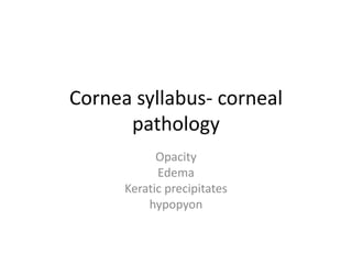
- 1. Cornea syllabus- corneal pathology Opacity Edema Keratic precipitates hypopyon
- 2. Corneal ulcer • Bacterial • Viral • Fungal • Pasasitic
- 3. Immunologically mediated corneal diseases Phlylenctnular keratitis Chronic serpenginous ulcer Interstitial keratitis Disciform keratitis
- 4. Miscelaneous Vitamin A deficiency Exposure keratitis Neurotropic keratopathy Photophthalmia
- 5. Corneal degeneration dystrophy and ectatic conditions • Eyebanking including eye donation and keratoplasty (basic) • Basic refractive surgeries
- 6. sclera • Scleritis • Episcleritis including DD, investigation and treatment • Staphyloma • Blue sclera
- 7. Immunologically mediated corneal diseases • Phlylenctnular keratitis • Chronic serpenginous ulcer • Interstitial keratitis • Disciform keratitis
- 8. Phylectenular keratoconjuncitivitis • Phylecten (bleb of vessels) • Symptoms • Phylectenulosis • Endogenous Tb allergen, tonsils and adenoids • Limbal location • Grey nodule, yellowish ulcer • Infiltration • Corneal margin
- 11. Fate of a phylecten • Absorption but no scar • Staphylococcol secondary bacterial infection • Treatment – phlecten stage and ulcer stage • Topical steroids: treatment of choice
- 12. Chronic serpenginous ulcer (rodent ulcer, mooren’s ulcer) • Definition : mostly immunomediated, autoimmune • Etiology: collagenolytic enzyme release • Severe pain • Start at corneal margin, spread circumferentially
- 13. • It commences as one or more grey infiltrates, which break down, forming small ulcers that spread and sooner or later coalesce. • The ulcer undermines the epithelium and superficial stromal lamellae at the advancing border, forming a whitish overhanging edge which is characteristic, while the base quickly becomes vascularized. • It rarely perforates, but progresses with intermissions for months until eventually a thin nebula is formed over the whole cornea and sight is greatly diminished. • Bilateral involvement with severe pain and relentless progression (‘malignant’) is more common in young adults, while a milder, usually unilateral, less painful form is seen in elderly patients.
- 14. Mooren’s ulcer
- 16. Progressive form and non-progressive form • Diagnosis: PUK like, overhanging edge • Diagnosis of exclusion • Treatment: • Peritomy • topical steroids • Contact lens • AMT • Conjunctival hooding • Lamellar keratoplasty • Scleral patch grafts • Medical management
- 17. Interstitial keratitis • Stromal type due to infection of allergy • Measels, typhoid, syphillis, TB or idiopathic • Syndromic: Cogans syndrome deafness vertigo tinnitus • Syphilitic (leutic) interstitial keratitis: inherited and delayed • Affects children and adults
- 19. Radiating lines and Ground glass appearance
- 20. Signs • Stromal haziness • Neovascularisation • Salmon patch: scarlet color • Spontaneous resolution • Cloudiness disappears • Vessels obliterated (ghost vessels) • Uveitis, cyclitis, choroiditis, KPs are seen • Bilateral • Acute stage 6 weeks
- 21. Diagnosis • Evidence of congential syphilis • Positive serological reaction
- 22. Treatment • Penicillin • Lubricants • Topical steroids • Cycloplegics for uveitis • Later stages: corneal grafting • Good prognosis
- 23. Disciform keratitis • Seen in adults • Unilateral • Virus etiology • Herpes ? • Immune mediated response • Not due to direct invasion • Similar to syphilitic interstitial keratitis
- 24. Clinical features • Central greyish disc stromal • Corenal thickening • DM folds • Immune ring of wessliy. • Corneal anaesthetic • No ulcer • Vision impaired • Uveitis associated
- 25. Disciform keratitis: slit lamp view
- 26. Bilateral circumscribed disc of stromal keratitis showing increased corneal thickness in the slit section and keratic precipitates at the back of cornea (arrows).
- 28. Treatment • Difficult • Topical steroids • Systemic antiviral