Presentation on how to chat with PDF using ChatGPT code interpreter
Food theorymay2013.withfigsnopage1
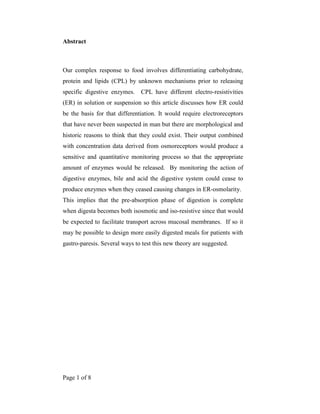
1. Page 1 of 8
Abstract
Our complex response to food involves differentiating carbohydrate,
protein and lipids (CPL) by unknown mechanisms prior to releasing
specific digestive enzymes. CPL have different electro-resistivities
(ER) in solution or suspension so this article discusses how ER could
be the basis for that differentiation. It would require electroreceptors
that have never been suspected in man but there are morphological and
historic reasons to think that they could exist. Their output combined
with concentration data derived from osmoreceptors would produce a
sensitive and quantitative monitoring process so that the appropriate
amount of enzymes would be released. By monitoring the action of
digestive enzymes, bile and acid the digestive system could cease to
produce enzymes when they ceased causing changes in ER-osmolarity.
This implies that the pre-absorption phase of digestion is complete
when digesta becomes both isosmotic and iso-resistive since that would
be expected to facilitate transport across mucosal membranes. If so it
may be possible to design more easily digested meals for patients with
gastro-paresis. Several ways to test this new theory are suggested.
2. Page 2 of 8
Bioimpedance background
Food has resistance to electrical current which is used to monitor gastric filling and emptying by
the epigastric impedance method.1.2
Fig 1 shows a trace when that method monitored gastric filling
when a volunteer drank 500ml of 10% glucose solution. There was a positive increase because
10% glucose solution in de-ionised water is more resistant (or impedic with alternating current)
than the body norm, or iso-impedic point. By contrast the deflection would be negative when the
subject drinks a liquid such as salty tomato juice which is conductive, or sub-iso-impedic, showing
what a wide range of effect can exist in common fluids. The decline from peak has been shown to
mirror gastric emptying. 1.2
Figure 1: A diagram of an Epigastric Impedance Trace.
There is a wide range of impedances in biological fluids. Fig 2 shows that de-ionised water has a
specific impedance about 18.31 Mega ohm-cm, which is 3 orders of magnitude greater than
tapwater at about 20 Kilo Ω-cm (3)
which shows the effect of a small concentration of ionized
particles such as Cl
-
. Normal saline and 10% glycerin solution are another 2 orders less impedic at
approximately 80–110 Ω-cm, while the protein fibrinogen at physiological concentrations is
approximately 309 Ω-cm. (4)
The value is affected by the bulk and ionized state of the molecules in
solution or suspension, so large molecule lipids (L) without charges will have higher impedances
than smaller molecule carbohydrate (C) or proteins (P) that usually have ionized radicals on their
surfaces. Hence, if the digestive system does use this characteristic of food it has a wide range in
which to work.
3. Page 3 of 8
Figure 2 De-ionised water and normal saline have specific impedances that are orders of
magnitude apart.
The wide range of resistivity in
physiological fluids
1
10
100
1000
10000
100000
1000000
10000000
100000000
Ultra Pure
Water
Tapwater
Normal Saline
.cm-1
Electro-Receptors?
To do that would require electro-receptors, which seems to be a remote possibility because they
have never been identified. However, they have never been specifically excluded or, it seems
even sought because nobody has suspected their existence. There is an interesting parallel in, of
all species, the monotreme mammal the duck-billed platypus. That animal was known to detect
its prey of small crustacean and worms in muddy water, at night and often with its eyes closed,
so how did it do it? Eventually someone recognized the similarity to the behaviour of fish and
sharks that track their prey by electroreceptors that detect the minute electrical currents emitted
by the muscles of their prey. Proske et al5
described how monotreme electroreceptor structure
gave no clue to its function when they wrote:
“The ability of animals to be able to detect weak electric fields in their environment was
recognized only relatively recently, perhaps because we, ourselves, are unaware of
4. Page 4 of 8
any but the strongest fields. Thus, the ampullae of Lorenzini in elasmobranch fish were first
thought to be mechanoreceptors, then thermoreceptors and chemo-receptors. It was not until
the late 1950s that convincing behavioural evidence was provided for an electro-
receptive role. At about the same time the tuberous receptors of mormyrid and gymnotid fish
were recognized as electroreceptors. It was not until the 1980s that electroreceptors were
described in urodele amphibians and monotremes.”
Figure 3. The structure of platypus electroreceptors.
Platypus bill's two kinds of pore. The electrosensitive pore (far left) is the opening of a mucus
gland duct while the touch-sensitive pore on the far right contains a pushrod device that triggers
nerves when compressed by a mechanical wave in the water or when it comes into contact with an
object (Australian Geographic).
Drawings of electroreceptors as in Fig 3 show a central, mucus-filled compartment that provides
a conductive pathway down to the nerve endings at its base. The resemblance of this
compartment to human mucus glands was noted by Andres et al (1991)6
who wrote: “Monotreme
electroreceptors have similar structures to classic, human duodenal mucus glands and were even
designated “sensory mucous glands”. Therefore it seems possible that human mucus-producing
glands in the duodenal mucosa, commonly called Brunner’s glands, could also have an
electroreceptor function.
5. Page 5 of 8
Osmoreceptors?
Osmoreceptors are long established and their output influences production of gastric secretions
and pancreatic digestive peptides.7-12
but they could not distinguish between CP and L at equal
concentrations, unlike the situation when osmolarity is combined with resistivity as Fig 4
attempts to portray.
Figure 4. Gastric hydrochloric acid would cause different effects on the impedance and
osmolarity of carbohydrate, protein and lipid suspensions...
Adding Hydrochloric Acid
Pure water
Zero osmolarity
Oils and Fats
Protein CHO
Osmolarity
Conductive Impedic
?
Some ionisation and molecular splitting? ?
?
Effects of acid and enzymes
Digestion itself would cause changes in the osmo-electrical nature of the gastric contents and the
proposed system could monitor them, providing a feedback loop by which the need for more
enzyme would be identified. Equally, when enzyme or acid increments cause no change it would
be a signal to cease producing them. Fig 4 attempts to illustrate the effect of acid separating
CPL but clearly specific CPL enzymes would have more specific effects, such as lipase
increasing the osmolarity of lipids by fragmenting molecules. Bile would reduce lipid resistivity
by adding charged particles to the lipid during micelle formation, and since micelles are
aggregated lipid molecules it would also reduce osmolarity, but it would not affect carbohydrate.
6. Page 6 of 8
The pattern of gastric emptying includes a sampling period.
It is possible that gastric emptying includes a sampling period when electro and osmo receptor
data are processed. The plateau seen in Fig 1 during the decline from peak impedance may
represent such a period. It has appeared frequently in traces from individual subjects although
group data tended to revert towards the classic mono-exponential mean of textbooks. It was
detected by epigastric impedance partly by its continuous record of gastric volume, unlike a
periodic sampling method such as scintigraphy where sampling occurs at intervals of comparable
duration to the plateau itself. A second reason is that epigastric impedance normally monitors
liquid test meals, partly because liquid specific impedance is more easily manipulated and
measured than solid meals and liquids provide more uniform and interpretable profiles.
A mechanical aspect of this plateau was shown to me by Professor Heading in Edinburgh in a
video of ultrasound images of gastric emptying from a volunteer who had swallowed water laced
with biscuit-crumbs. They shone like stars in a black background and it was surprising to see
them moving repeatedly back and forth between the antrum and duodenum in an oddly
purposeful way as though some kind of sampling was occurring.
Discussion
We make a complex response to food, yet there is no convincing theory of its orchestration
despite decades of research into many aspects of it, such as the digestive peptides. We do not
know what triggers specific responses to CPL or why the responses cease as digestion is
completed. Such a deep mystery requires a radical solution and to propose the existence of
human electroreceptors is certainly that. Unlikely as they must seem, there is a precedent in the
work of Andres et al 6
who pointed out that electroreceptors in monotreme mammals were not
suspected for decades despite the fact that their structure was well known. Clearly it is feasible, if
currently improbable, that mucus glands of the human duodenum have the required sensors at
their depths but they have been credited with functions concerned with mucus production.
Proske et al suggested that that this was partly due to a general lack of awareness of the functions
of electroreceptors and their extraordinary sensitivity in particular. Platypus and echidna use
them to detect the minute currents emitted by the muscles of river crustaceans and worms.
Electroreceptors in elasmobranch species are amongst the most sensitive receptors known. For
example, sharks that can detect 0.01 microvolt/cm(14, 15)
which has been illustrated by the
Reefquest Shark protection organization’s website as “the equivalent to the electric field of a
flashlight battery connected to electrodes some 10,000 miles (16,000 kilometres) apart in the
ocean.(16)
In freshwater fish species electroreceptors are reported to detect 0.1-10 mv/cm.(14)
7. Page 7 of 8
The parallel in human digestion is the lack of awareness of the possible advantages of
electroreceptor data, particularly of a highly sensitive nature; the main one being quantification
of the CPL contents of digesta so that responses are also accurately quantified. Not only would
that minimise wasteful production of enzymes but it would enable responses to small amounts,
so making the most advantage of the food available.
This becomes possible when electroreceptor data is combined with osmolarity. An appealing
aspect of this theory is that it enhances the role of osmoreceptors. Their output on concentration
without knowing the CPL content of the suspension could only trigger a non-specific response.
Likewise, electroreceptor data alone cannot quantify CPL, but combined with concentration it
can do so.
An innovative hypothesis should suggest ways to test its assumptions and predictions and in this
case the list includes the following:
1. Measure resistivities of selected meals, including within simulated duodenal suspensions.
Differences should be large enough to be detected by electro-receptors and follow the
predicted pattern.
2. Examine duodenal mucous glands for nerve endings that could function as electro-
receptors.
3. Test the correlation between enzymatic and hormonal responses to test meals of
prescribed resistivity and CPL constitution. There should be smaller responses to meals
nearer to the iso-resistive point than those further from it.
4. Devise meals that could be taken by patients with gastroparesis to show whether
absorption is facilitated the nearer the meals are to isoresistive.
5. Measure the permeability of gut membranes to liquids of varying resistivity. Isoresistive
fluids should pass more easily through them than hyper or hypo resistive liquids
6. Assess the resistivity and osmotic changes caused by digestive enzymes acting on their
usual substrates, such as lipases on lipids. Do they cause a shift towards isoresistivity as
the theory predicts?
Conclusion
This theory, or series of speculations, describes a possible way for the human gut to respond
quantitatively to food. The wide range of electro-resistivity in carbohydrate, protein and lipid
solutions or suspensions offers a feasible way for the gut to distinguish them, particularly if they
change in consistent and distinctive ways in the presence of digestive enzymes.
Acknowledgement
8. Page 8 of 8
I thank Professor Nicholas Spyrou of the department of medical physics at Surrey University for
his inspiration and friendship over many years while we developed the gastric monitor, the basis
for inventing this hypothesis.
References
1. McClelland GR and Sutton JA (1985) Epigastric impedance; a non-invasive method for the assessment of
gastric emptying and motility. Gut, 26, 6, 607-614.
2. McClelland GR and Sutton JA (1986) A comparison of the gastric and central nervous system effects of
two substituted benzamides in normal volunteers. Br J Clin Pharmacol, 21, 503-509.
3. Iverson A (1964) The Measured Resistivity of Pure Water and Determination of the Limiting Mobility of
OH-
from 5 to 55°J. Phys. Chem., 68 (3), pp 515–521
4. Park S et al (2008). Electrical Characteristics of… Fluids for Endoscopic Mucosal Resection J Dig Dis Sci,
Vol 53, Number 6, 1573-2568.Sircus W (1958) Studies on the mechanism in the duodenum inhibiting
gastric secretion. Q J Exp Physiol 43:114.
5. Proske U Gregory J. E. and A. Iggo Sensory receptors in monotremes. Phil.Trans. R. Soc. Lond. B (1998)
353, 1187-1198Martin, R. A. 2003. www.elasmo-research.org/copyright.htm (C)
6. Andres, K. H., von During, M., Iggo, A. & Proske, U. 1991 The anatomy and ¢ne structure of the
echidnaTachyglossus aculeatus snout with respect to its different trigeminal sensory receptors including the
electroreceptors. Anat. Embryol. 184, 371-393.
7. Ward AS, Wilkins RA, Cockel R, et al (1969) Duodenal inhibition of gastric secretion by osmotic agents in
normal subjects and patients with duodenal ulcer. Gut, 10: 1020.
8. Knightly JJ, Vanamee P, Lawrence W Jr, (1960) The effect of intra-duodenal hypertonic glucose on biliary
and pancreatic secretion. Surg. Forum 11:371.
9. Lawrence W Jr, Khemntigan A, Hudock J, et al (1961) The effect of intra-duodenal administration of
hypertonic glucose solution on external pancreatic secretion. Surgery 49: 666.
10. Schapiro H, Britt LG, Evans SR, Wilson H. (1967 The effect of infusion of hypertonic fluids into the upper
intestines on pancreatic secretion Am J Surg 113:65.
11. Dyck WP (1971) Influence of intra-jejunal glucose on pancreatic exocrine function in man.
Gastroenterology 60: 864.
12. Teichmann RK, Swierczek JS, Rayford PL et al (1979) Effect of duodenal osmolality on gastric and
secretin release and on gastric and pancreatic secretion. World J Surg 3, 623-630.
13. Bullock TH and Heiligenberg W (1986) Electroreception in Wiley Series in Neurobiology (Ed RG
Northcutt) New York, John Wiley & Sons.
14. Pettigrew JD, (1999) Electroreception in monotremes. J Experimental Biology, 202; 1447-1454.
15. Szabo G. (1965) Sense organs of the lateral line system in some electric fish of the Gymnotidae.
Gymnarchidae and Mormyridae. J Morphology 117, 229-250.
16. Reefquest shark website: www.reefquest.com