
Recommended
More Related Content
What's hot
What's hot (18)
Similar to Muscular system
Similar to Muscular system (20)
Recently uploaded
Recently uploaded (20)
Muscular system
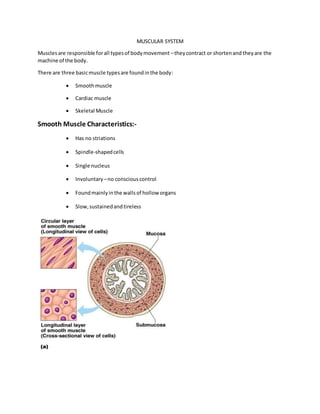
- 1. MUSCULAR SYSTEM Musclesare responsible forall typesof bodymovement –theycontract or shortenand they are the machine of the body. There are three basicmuscle typesare foundinthe body: Smoothmuscle Cardiac muscle Skeletal Muscle Smooth Muscle Characteristics:- Has no striations Spindle-shapedcells Single nucleus Involuntary –no consciouscontrol Foundmainlyinthe wallsof hollow organs Slow,sustainedandtireless
- 2. Cardiac Muscle Characteristics:- Has striations Usuallyhasa single nucleus Joinedtoanothermuscle cell atan intercalateddisc Involuntary Foundonlyinthe heart Steadypace! Skeletal muscles Classification Of Muscles:- A. ByFascicular Orientation 1.Parallel 2.Pennate 3 Spiral 4 Cruciate Parallel ( Relativetomuscle directionof pull):- (a) Quadrilateral- Quadratus - lumborum,Thyrohyoid (b)Longandstrap like- Sartorius
- 3. (c) Strap like withtendinousintersection- Rectusabdominis (d) Fusiform- Bicepsbrachii Pennate muscles:- (a) Unipennate –FlexorPollicislongus (b)Bipennate- Rectusfemoris,Dorsal interossei of hand (c )Multipennate - Deltoid (d)Circumpennate-Tibialisanterior Spiral Muscles:- Supinator Cruciate Muscles:- Sternocledomastoid,Masseter B. By Type Of Skeletal Muscle Fibre 1. Slowor Red fibresortype I fibres 2. Fast or White fibresortype IIfibres C. By Insertionnearoraway fromjoint 1. ShuntMuscle( AwayfromJoint) 2. Spurt Muscle ( NearJoint) Naming of skeletal Muscles:- Direction of muscle fibers Example: rectus (straight) Relative size of the muscle Example: maximus (largest) Location of the muscle Example: many muscles are named for bones (e.g., temporalis) Number of origins Example: triceps (three heads)
- 4. Location of the muscles origin and insertion Example: sterno (on the sternum) Shape of the muscle Example: deltoid (triangular) Action of the muscle Example: flexor and extensor (flexes or extends a bone) Nomenclature of Muscles:- On Basisof : 1.Shape of muscle Deltoid,Quadratus,Rhomboid,Lumbricals 2.Size Major , minor, longus, brevis 3. NumberOf Head Biceps,triceps, Quadricepsfemoris, Digastric 4. Position Supraspinatus,Infraspinatus,Abdominis, Oculi,oris 5.Depth External oblique,Internaloblique,FlexorD.Superficialis,FlexorD. Profundus 6. Attachment: Sternocledomastoid, coracobrachialis 7. Action: Flexor,Extensor,Abductor SKELETAL MUSCLES- NAME, ORIGIN, INSERTION & ACTION Musclesof Facial Expression(thatdonotworkby crossinga joint) ACTION ORIGIN INSERTION Orbicularisoculi Closeseye (squint), lowerseyebrows Frontal bone and maxilla Eyelid Orbicularisoris Closeslips(purses, protrudes) Maxillaandmandible Skinand muscle around mouth
- 5. Zygomaticusmajor Raisescornersof mouth (smile) Zygomaticbone Skinand muscle at corner of mouth Buccinator Compresscheeks (whistling,sucking) Mandible andmaxilla molarregions Orbicularisoris Frontalis Raise eyebrows Epicranial aponeurosis Skinof eyebrows Musclesthat act on the Jaw (formasticationandfacial expression) Musclesthat act on Neck(tomove head) Musclesthat act on Shoulder(tomove the arm) ACTION ORIGIN INSERTION Masseter Elevatesmandible (asin closingmouthwhile chewing) Zygomaticarch and bone Mandible ramusand angle Temporalis Elevatesmandible Temporal,frontal, parietal bones Coronoidprocessof mandible Platysma Pullslowerlipdown(as infrowning) Depresses mandible (opening mouthas when chewingorsurprised) Pectoralismuscle Mandible,skinof lower face ACTION ORIGIN INSERTION Sternocleidomastoid Sternocleidomastoid Both sides:flexesneck (as inlookingdown) One side:cockshead Manubrium, clavicle Mastoidprocessof temporal bone Spleniuscapitis Both sides:extends neck(as inlookingup) One side:cockshead C7 to T3 (variable) Nuchal line andmastoid process ACTION ORIGIN INSERTION Deltoid(anterior) Flexionandmedial rotationof humerusat shoulder Clavicle Deltoidtuberosityof humerus Deltoid(middle) Abductshumerusat Acromion Deltoidtuberosityof
- 6. Rotator Cuff Muscles(ALLthese small muscleshelptokeepheadof humerussnugin glenoidfossa) Musclesthat act on Scapula(to move shoulder) ACTION ORIGIN INSERTION Levatorscapulae Elevatesandretracts scapula(as inbringing shoulderupandin towardear) C1 to C4 Medial borderof scapula,superiorto spine Rhomboideusmajor Retracts scapula(asin pullingthe shoulders back) T2 to T5 Medial borderof scapula Serratusanterior Protracts scapula (whichpushesthe arm forwardinfront of the Ribs1 to 9 Anteriorsurface of vertebral borderof scapula shoulder(asinupward flap) humerus Deltoid(posterior) Extensionandlateral rotationof humerusat shoulder Scapularspine Deltoidtuberosity of humerus Pectoralismajor Flexion,adduction, medial rotationof humerusat shoulder(as inoverheadfistraise,or benchpressmotion) Sternumto rib7 Medial half of clavicle Intertuberculargroove of humerus Latissimusdorsi Extension,adduction, mediallyrotation humerusat shoulder (gymlat pull-downs) T7 to L5, ribs 10 to 12, iliaccrest Intertuberculargroove of humerus(muscle travelsunderaxillary region) ACTION ORIGIN INSERTION Subscapularis Mediallyrotates humerusat shoulder. Stabilizesshoulder. Subscapularfossaof scapula Lessertubercle of anteriorhumerus Supraspinatus Abductsarm at shoulder.Stabilizes shoulder. Supraspinousfossaof scapula Greatertubercle of top of humerus Infraspinatus Laterallyrotates humerusat shoulder. Stabilizesshoulder. Infraspinousfossaof scapula Middle partof greater tubercle of posterior humerus Teresminor Laterallyrotates humerusat shoulder. Stabilizesshoulder. Lateral borderof scapula Inferiorpartof greater tubercle of posterior humerus
- 7. scapula,as inpunching or hugging) Trapezius(upper) Elevatesscapula(asin shrugging) Occipital bone,C1-C7 Lateral 1/3 clavicle and acromionprocess Trapezius(middle) Retracts scapula(asin pullingshouldersback) T1 to T5 Spine of scapula Trapezius(lower) Retracts anddepresses scapula(as inpulling shouldersbackand down) T6 to T12 Medial 1/3 of scapula spine Musclesthat act on the Elbow(tomove the forearm) ACTION ORIGIN INSERTION Brachialis Flexeselbow(asin bringingaspoonto your mouth) Anteriordistal shaftof humerus Coronoidprocessof ulna Pronatorteres Pronates forearmat elbow(asinturninga doorknoblaterally) Medial epicondyle of humerus,coronoid processof ulna Lateral midshaftof radius Bicepsbrachii Flexeselbow,supinates forearm(asin scooping up waterinyour hand to drink) Long head: supraglenoidtubercle of scapula Short head:coracoid processof scapula Radial tuberosity Tricepsbrachii Extendsforearmat elbow(asinloweringa spoonback to the table) Long head: infraglenoid tubercle of scapula Lateral head: posterior proximal shaftof humerus Medial head: posterior distal shaftof humerus Olecranonof ulna Musclesthat act on Wrist (tomove the handand fingers) ACTION ORIGIN INSERTION Extensorcarpi radialis brevis Extendswrist,radially deviateswrist (abduction) Lateral epicondyle of humerus Metacarpal III(base) Flexorcarpi radialis Flexeswrist,radially deviateswrist (abduction) Medial epicondyle of humerus MetacarpalsII and III (base) Extensorcarpi ulnaris Extendswrist,ulnar deviateswrist (adduction) Lateral epicondyle of humerus,posterior shaftof ulna Metacarpal V (base)
- 8. Flexorcarpi ulnaris Flexeswrist,ulnar deviateswrist (adduction) Medial epicondyle of humerus Pisiform, hamate, metacarpal V (base) Extensordigitorum Extendsdigitsof hand, and wrist Lateral epicondyle of humerus Dorsal side of digits2 to 5 Flexordigitorum Flexesdigitsof the hand,and wrist Medial epicondyle of humerus Anteriorside of digits2 to 5 Musclesthat act on Vertebral Column ACTION ORIGIN INSERTION Spinalisthoracis One side:lateral flexion of vertebral column Both sides:extension and hyperextensionof vertebral column T11 to L2 T3 to T7 Rectusabdominis Compressesabdomen, flexesvertebral column Pubiscrestand symphysis Xiphoidprocess,costal cartilage 5 to 7 Musclesof Breathing(musclesthatworkonthe vertebral column) ACTION ORIGIN INSERTION External intercostals Pullsribsuptoward origin,elevatesribcage to cause inspiration Inferiorborderof rib above Superiorbordersof rib below Diaphragm Diaphragmdropsdown to enlarge thoracic cavity,causing inspiration Xiphoidprocess,ribs10 to 12, costal cartilage 5 to 9, lumbarvertebrae 1-5 Central tendon(in centerof the disc shapeddiaphragm) Musclesthat act at Hip(tomove leg) ACTION ORIGIN INSERTION Pectineus Flexeship,adductsleg Pubisandpubicramus From lessertrochanter to superiorpartof linea asperaof femur Adductorlongus Flexeship,adductsleg Superiorramusof pubis Lineaaspera(shorter insertionthanadductor magnus) Adductormagnus Extendship,adductsleg Inferiorischial ramus Lineaasperaof femur Gluteusmaximus Extendship(asin pushingdownwhile climbingastair),rotates leglaterally Ilium, sacrum, coccyx Gluteal tuberosityof femur Gracilis Adductslegat hip, Pubis Medial aspectof
- 9. flexesknee proximal tibia Tensorfasciae latae (tensesorstabilizeship and knee joints), abductslegat hip Iliaccrest nearanterior superiorspine Lateral condyle of tibia Psoasmajor Flexeship(asinlifting legto place on stair) Vertebral bodiesT12 to L5 Lessertrochanterof femur Sartorius Flexeshipandflexes knee,laterallyrotates thigh(intocrosslegged position) Anteriorsuperioriliac spine Medial aspectof tibial tuberosity Musclesthat Flex Knee (hamstringsworkasa group,as whenpullingbackinpreparationtokicka ball) ACTION ORIGIN INSERTION Bicepsfemoris Extendship,flexes knee,laterallyrotates leg Long head:Ischial tuberosity Short head:Posterior mid-shaftof femur Headof fibula Semimembranosus Extendship,flexesknee Ischial tuberosity Posteriormedial condyle of tibia Semitendinosus Extendship,flexesknee Ischial tuberosity Medial surface of proximal tibia(slightly anteriorto insertionof semimembranosus) Musclesthat ExtendKnee (quadricepsworkasa group,as whenkickingaball) ACTION ORIGIN INSERTION Vastuslateralis Extendsknee Greatertrochanter, lateral lipof linea aspera(wrapsaround to anteriorsurface) Tibial tuberosityand patella Vastusmedialis Extendsknee Intertrochantericline, medial lipof linea aspera(wrapsaround to anteriorsurface) Tibial tuberosityand patella Vastusintermedius Extendsknee Anteriorshaftof femur Tibial tuberosityand patella Rectusfemoris Extendsknee,flexeship (kickingaball) Anteriorinferioriliac spine of coxal bone Tibial tuberosityand patella Musclesthat act on Ankle (tomove foot)
- 10. ACTION ORIGIN INSERTION Soleus Plantarflexionatankle Proximal thirdof tibia and fibula Calcaneus Gastrocnemius Plantarflexionatankle, flexesknee Medial andlateral epicondylesof femur Calcaneus Tibialisanterior Dorsiflexionand inversionatankle Lateral condyle and antero-lateral tibia Medial cuneiform, undermetatarsal 1 Function of Skeletal Muscles:- Produce movement Maintainposture Stabilize joints Generate heat MicroscopicAnatomyof Skeletal Muscle:- Cellsare multinucleate Nuclei are justbeneaththe sarcolemma Sarcolemma– specializedplasmamembrane Sarcoplasmicreticulum –specializedsmoothendoplasmicreticulum Myofibril Bundlesof myofilaments Myofibrilsare alignedtogive distrinctbands I band= lightband
- 11. A band= dark band Sarcomere Contractile unitof a muscle fiber Organization of the sarcomere Thickfilaments=myosinfilaments Composedof the proteinmyosin Has ATPase enzymes Thinfilaments=actinfilaments Composedof the proteinactin MYOSIN:- Myosin filaments have heads (extensions, or cross bridges) Myosin and actin overlap somewhat
- 12. Nerve Stimulus to Muscles:- Skeletal muscles must be stimulated by a nerve to contract (motor neruron) Motor unit One neuron Muscle cells stimulated by that neuron Neuromuscular junction:- A neuromuscular junction (or myoneural junction) is a chemical synapse formed by the contact between a motor neuron and a muscle fiber. It is at the neuromuscular junction that a motor neuron is able to transmit a signal to the muscle fiber, causing muscle contraction. Neuromuscularjunctions –associationsite of nerve andmuscle Synapticcleft– gap betweennerveandmuscle Nerve andmuscle donot make contact
- 13. Areabetweennerve andmuscle isfilledwithinterstitialfluid Transmission of Nerve Impulse to Muscle Neurotransmitter–chemical releasedbynerve uponarrival of nerve impulse The neurotransmitterforskeletal muscleisacetylcholine Neurotransmitterattachestoreceptorsonthe sarcolemma Sarcolemmabecomes permeabletosodium(Na+ ) Sodiumrushingintothe cell generatesanactionpotential Once started,muscle contractioncannotbe stopped The Sliding Filament Theory of Muscle Contraction Activationbynerve causesmyosinheads(crossbridges)toattachto bindingsitesonthe thin filament Myosinheadsthenbindto the nextsite of the thinfilament Thiscontinuedactioncausesa slidingof the myosinalongthe actin The resultisthat the muscle isshortened(contracted)
- 14. Contraction of a Skeletal Muscle:- Muscle fibercontractionis“all or none” Withina skeletal muscle,notall fibersmaybe stimulatedduringthe same interval Differentcombinationsof muscle fibercontractionsmaygive differingresponses Gradedresponses –differentdegreesof skeletalmuscle shortening,rapidstimulus=constant contractionor tetanus Muscle ResponsetoStrong Stimuli:- Muscle force depends upon the number of fibers stimulated More fibers contracting results in greater muscle tension Muscles can continue to contract unless they run out of energy Energy for Muscle Contraction: Initially,musclesusedstoredATPforenergy Bondsof ATPare brokento release energy Only4-6 secondsworthof ATPis storedbymuscles Afterthisinitial time,otherpathwaysmustbe utilizedtoproduce ATP Direct phosphorylation :- Muscle cellscontaincreatine phosphate (CP) CP isa high-energymolecule AfterATPis depleted,ADPisleft CP transfersenergytoADP,toregenerate ATP CP suppliesare exhaustedinabout20 seconds Anaerobic glycolysis:- Reactionthatbreaksdownglucose withoutoxygen Glucose isbrokendownto pyruvicacidto produce some ATP Pyruvicacidis convertedtolacticacid
- 15. Aerobic Respiration:- Seriesof metabolicpathwaysthatoccurinthe mitochondria Glucose isbrokendownto carbondioxide andwater,releasingenergy Thisis a slowerreactionthatrequirescontinuousoxygen Muscle Fatigue and Oxygen Debt:- When a muscle is fatigued, it is unable to contract The common reason for muscle fatigue is oxygen debt Oxygen must be “repaid” to tissue to remove oxygen debt Oxygen is required to get rid of accumulated lactic acid Increasing acidity (from lactic acid) and lack of ATP causes the muscle to contract less Types of Muscle Contractions:- Isotonic contractions Myofilaments are able to slide past each other during contractions The muscle shortens Isometric contractions Tension in the muscles increases The muscle is unable to shorten MUSCLE TONE:- Some fibers are contracted even in a relaxed muscle Different fibers contract at different times to provide muscle tone The process of stimulating various fibers is under involuntary control Effects of Exercise onMuscle:- Increase in muscle size
- 16. Increase in muscle strength Increase in muscle efficiency Muscle becomes more fatigue resistant Muscles andBody Movements:- Movement is attained due to a muscle moving an attached bone Muscles are attached to at least two points Origin – attachment to a immoveable bone Insertion – attachment to an movable bone Types of Ordinary Body Movements:- Flexion – decreases angle of joint and brings two bones closer together Extension- opposite of flexion Rotation- movement of a bone in longitudinal axis, shaking head “no” Abduction/Adduction (see slides) Circumduction (see slides) Prime mover – muscle with the major responsibility for a certain movement Antagonist – muscle that opposes or reverses a prime mover Synergist – muscle that aids a prime mover in a movement and helps prevent rotation Fixators Disorders relating tothe Muscular System:- • Muscular Dystrophy: inherited, muscle enlarge due to increased fat and connective tissue, but fibers degenerate and atrophy • Duchenne MD: lacking a protein to maintain the sarcolemma • Myasthemia Gravis: progressive weakness due to a shortage of acetylcholine receptors Abnormal Contractions:-
- 17. • spasm – involuntary contraction of one muscle • cramp – painful spasm • tetanus – multiple spasms of skeletal muscles • tic – involuntary twiches of muscles, usually under voluntary control • tremor – rhythmical, involuntary contractions of opposite groups of muscles • fasciculations – involuntary, short twiches on motor unit visible under the skin • fibrilace – spontaneous contractions of fibres of one muscle that aren´t visible under the skin • fascia (= perimysium externum) – fibrous envelope of muscle or muscle group – barrier for spreading of inflammation in that specific area • osteofascial septum (= septum osteofasciale) – fascial divider from the superficial fascia to the periosteum separates the space for muscle groups – compartment (compartimentum) Enthesopathy- Ex; Tenniselbow