SlideShare a Scribd company logo
1 of 7
Download to read offline
The International Journal of Periodontics & Restorative Dentistry
© 2021 BY QUINTESSENCE PUBLISHING CO, INC. PRINTING OF THIS DOCUMENT IS RESTRICTED TO PERSONAL USE ONLY.
NO PART MAY BE REPRODUCED OR TRANSMITTED IN ANY FORM WITHOUT WRITTEN PERMISSION FROM THE PUBLISHER.
227
Volume 41, Number 2, 2021
Submitted April 23, 2020; accepted July 7, 2020.
©2021 by Quintessence Publishing Co Inc.
A postextraction socket is always open to different treatment possibilities.
A straightforward clinical classification may help evaluate which surgical
approach is best suited for the case being treated. Four different classes are
defined on the basis of the local anatomy of the site, available bone volume,
and soft tissue level. For every clinical situation, either immediate placement,
early placement, alveolar ridge preservation, or staged approach can be
selected as a treatment modality according to the classifications listed. Int J
Periodontics Restorative Dent 2021;41:227–232. doi: 10.11607/prd.5069
A Clinical Classification System for the
Treatment of Postextraction Sites
1ProEd, Institute for Professional Education in Dentistry, Torino, Italy.
2
Division of Periodontology, Harvard School of Dental Medicine, Boston, Massachusetts,
USA.
3Private Practice, Milan, Italy.
Correspondence to: Dr Daniele Cardaropoli, ProEd Institute, Corso Galileo Ferraris 148,
10129 Torino, Italy. Fax +39-011-323683. Email: d.cardaropoli@proed.it
It is uniformly accepted that the
healing dynamic of a postextraction
alveolus involves the succession of
biologic phenomena. It is initiated
with the stabilization of the blood
clot up to the deposition of a new
bone matrix,1,2
which assists bone
crest remodeling at sites with a loss
of vertical and horizontal volume.3,4
Over time, different classifica-
tions of postextraction sites have
been introduced, each considering
the timing of implant placement
from a strictly chronologic point of
view,5,6
the presence or absence of
the buccal hard and soft tissue,7–9
or the quantity of bone volume
available in relation to the anatomy
and the position of the root of the
teeth.10,11
However, none of the classi-
fications presently available take
into account the anatomy of the
postextraction alveolus and the po-
sition of the overlying soft tissues,
the volume of the bone crest, and
the presence of periapical lesions
and unfavorable anatomical struc-
tures. Furthermore, available classi-
fications do not suggest a possible
therapeutic option for each particu-
lar clinical condition.
Thus, a new classification of
postextraction sites is proposed
based on the anatomical evaluation
of the bone crest and gingival tis-
sues, linking the classifications with
appropriate therapeutic options.
Daniele Cardaropoli, DDS1
Myron Nevins, DDS2
Paolo Casentini, DDS3
© 2021 BY QUINTESSENCE PUBLISHING CO, INC. PRINTING OF THIS DOCUMENT IS RESTRICTED TO PERSONAL USE ONLY.
NO PART MAY BE REPRODUCED OR TRANSMITTED IN ANY FORM WITHOUT WRITTEN PERMISSION FROM THE PUBLISHER.
The International Journal of Periodontics  Restorative Dentistry
228
Extraction Site Treatment
Classes
Postextraction sockets are classified
in four different categories (Fig 1),
and relative surgical treatment op-
tions are suggested. The necessary
information is collected from a care-
ful clinical and radiologic analyses
using both standard intraoral (Fig 2)
and 3D radiographic (CBCT) images
(Fig 3).
Class I: Intact Extraction Site
with Favorable Anatomical
Conditions
Specific characteristics
Characteristics of this classification
include (1) buccal cortical bone that
is intact or affected by damage not
exceeding 20% of the extent of the
wall; (2) an optimum soft tissue level;
and (3) local bone anatomy that al-
lows an ideal tridimensional implant
position and good primary stability.
Surgical treatment techniques
Treatment techniques for this case
include (1) immediate implant place-
ment (potentially combined with
an immediate restoration, if proper
primary stability can be achieved);
(2) alveolar ridge preservation and
delayed implant placement (after 4
to 6 months); and (3) spontaneous
healing and early implant placement
(4 to 8 weeks after extraction) with
bone and/or soft tissue augmenta-
tion.
Fig 1  Schematic drawings of the four different classifications: (a) Class I; (b) Class II;
(c) Class III; (d) Class IV.
a
c
b
d
Fig 2  Clinical images corresponding to the four different classifications. (a) Class I. An
intact socket with a large amount of bone on the palatal side, suitable for immediate im-
plant placement. (b) Class II. An intact socket that apically presents direct communication
with a large nasopalatine canal, which prevents immediate implant placement. (c) Class III.
A compromised socket with the facial bone plate resorbed to almost 50%. (d) A severely
compromised socket, with the facial bone plate resorbed more than 50%, vertical resorp-
tion of the palatal wall, and presence of a periapical lesion.
a
c
b
d
© 2021 BY QUINTESSENCE PUBLISHING CO, INC. PRINTING OF THIS DOCUMENT IS RESTRICTED TO PERSONAL USE ONLY.
NO PART MAY BE REPRODUCED OR TRANSMITTED IN ANY FORM WITHOUT WRITTEN PERMISSION FROM THE PUBLISHER.
229
Volume 41, Number 2, 2021
Class II: Intact Extraction
Site with Partially Favorable
Anatomical Conditions
Specific characteristics
Characteristics of this classification
include (1) buccal cortical bone that
is intact or affected by damage not
exceeding 20% of the extent of the
wall; (2) an optimum soft tissue level;
and (3) difficulty achieving an ideal
implant position and satisfactory
primary stability due to the pres-
ence of a large periodontal defect,
large periapical lesion, and/or ana-
tomical structures limiting immedi-
ate placement (eg, maxillary sinus
floor, mandibular canal).
Surgical treatment techniques
Treatment techniques for this case
include (1) alveolar ridge preserva-
tion and delayed implant placement
(after 4 to 6 months); and (2) sponta-
neous healing and delayed implant
placement (3 to 4 months after ex-
traction) with bone tissue augmen-
tation.
Class III: Partially Compromised
Extraction Site with
Unfavorable Anatomical
Conditions
Specific characteristics
Characteristics of this classification
include (1) resorption of the buccal
cortical bone, totaling 20% to 50%
of the wall; (2) suboptimal soft tissue
level, soft tissues presenting inflam-
mation, and/or thin and scalloped
gingival phenotype; and (3) local
bone anatomy allowing an ideal
Fig 3  CBCT radiologic images correspond-
ing to the four different classifications.
(a) Class I. An intact facial bone plate and
a large amount of bone available apical
and palatal to the root apex. (b) Class II.
An intact and thick buccal bone plate with
a periapical lesion between the root apex
and the maxillary sinus floor, preventing
immediate implant placement. (c) Class III.
Resorption of the facial bone plate and a
large amount of bone available apical and
palatal to the root apex. (d) Class IV. Severe
bone resorption on both the buccal and
palatal sides, together with insufficient api-
cal bone volume.
a
c
b
d
© 2021 BY QUINTESSENCE PUBLISHING CO, INC. PRINTING OF THIS DOCUMENT IS RESTRICTED TO PERSONAL USE ONLY.
NO PART MAY BE REPRODUCED OR TRANSMITTED IN ANY FORM WITHOUT WRITTEN PERMISSION FROM THE PUBLISHER.
The International Journal of Periodontics  Restorative Dentistry
230
tridimensional implant positioning
and good primary stability.
Surgical treatment techniques
Treatment techniques for this case
include (1) alveolar ridge augmen-
tation, possibly with soft tissue
augmentation, and staged implant
placement after 6 months; and (2)
spontaneous healing and early im-
plant placement (4 to 8 weeks after
extraction) with bone and/or soft tis-
sue augmentation.
Class IV: Severely
Compromised Extraction Site
with Unfavorable Anatomical
Conditions
Specific characteristics
Characteristics of this classification
include (1) severely compromised
socket walls, particularly with the
buccal bone wall loss exceeding
50%; (2) suboptimal soft tissue level,
soft tissues presenting inflamma-
tion, or thin and scalloped gingival
phenotype; and (3) difficulty achiev-
ing an ideal implant position and
satisfactory primary stability due to
the presence of a large periodontal
defect, large periapical lesion, and/
or anatomical structures limiting im-
mediate placement (eg, maxillary
sinus floor, mandibular canal).
Surgical treatment techniques
Treatment techniques for this case
include (1) alveolar ridge augmen-
tation, possibly with soft tissue
augmentation, and staged implant
placement after 6 months; (2) spon-
taneous healing, bone augmenta-
tion after 4 to 8 weeks, and staged
implant placement after an addi-
tional 6 months; and (3) spontane-
ous healing and implant placement
after 3 to 4 months, performed si-
multaneously with bone augmenta-
tion.
Discussion
In the literature, there are different
classifications of postextraction sites
based on the timing of tooth extrac-
tion in relation to implant insertion5,6
or the presence of buccal hard and
soft tissues.7–9
Other papers have
called for a sagittal root position (in
relation to the bony housing) during
immediate implant placement in the
maxillary esthetic zone.10,11
The new classification system
presented herein creates a relation-
ship between the socket anatomy
and the soft tissue level and thick-
ness. The main evaluation is based
on the integrity of the alveolus and
gingival level. An intact socket is
defined as presenting three intact
bony walls and at least 80% of the
fourth bony wall.12
For each distinct
clinical situation, more treatment
options are proposed.
When a Class I case is diag-
nosed, the socket is basically in-
tact. The minimum bone volume
needed for implant primary stabil-
ity is related to the surgical skills of
the clinician and the characteristics
of the implant system used, as im-
mediate placement is a technique-
sensitive procedure.13
In the hori-
zontal dimension, creating a bone-
to-implant gap of at least 2 mm be-
tween the implant and the internal
surface of the facial bone wall is rec-
ommended in order to create a suf-
ficient space to be filled with a bone
substitute.14–16
In the vertical dimen-
sion, the implant shoulder should
be placed approximately 1 mm
apical to the midfacial bone crest
to compensate for marginal bone
remodeling.17,18
To optimize esthetic
outcomes, a flapless approach13,19,20
and the delivery of an immediate
restoration is always a suggested
option when possible.21,22
The litera-
ture remains controversial regarding
the need for soft tissue augmenta-
tion in cases of thin gingival pheno-
type.
Following a more prudent ap-
proach, alveolar ridge preservation
(intended as the preservation of the
ridge volume within the envelope
existing at the time of extraction23
)
can be selected as an alternative.
The majority of the original ridge
volume can be maintained when the
fresh alveolus is filled with a bone
graft at the time of tooth extrac-
tion.24,25
Usually, implant surgery can
be performed after 4 to 6 months
with results similar to that of im-
plants inserted in native bone.26
A third option is soft tissue heal-
ing and early placement. An open
flap raised the day of implant place-
ment allows for a contour augmen-
tation using guided bone regenera-
tion.27
For Class II cases, the socket is
still intact, but unfortunately the lo-
cal anatomy of the alveolar ridge
does not allow for an ideal implant
position or good primary stability,
and thus immediate implant place-
ment is not indicated. In this clinical
situation, alveolar ridge preserva-
tion with a staged implant place-
© 2021 BY QUINTESSENCE PUBLISHING CO, INC. PRINTING OF THIS DOCUMENT IS RESTRICTED TO PERSONAL USE ONLY.
NO PART MAY BE REPRODUCED OR TRANSMITTED IN ANY FORM WITHOUT WRITTEN PERMISSION FROM THE PUBLISHER.
231
Volume 41, Number 2, 2021
ment following 4 to 6 months of
healing is suggested. Socket preser-
vation can biologically compensate
for marginal bone remodeling due
to the healing dynamics of the fresh
extraction site.28
When spontaneous healing
is the selected approach, a mean
horizontal ridge width reduction of
3.8 mm and a mean vertical ridge
height reduction of 1.24 mm can be
anticipated.23
This will then require
additional bone augmentation the
day of implant placement.
Class III is characterized by a
compromised extraction socket.
Ridge augmentation (defined as the
increase of the ridge volume beyond
the skeletal envelope existing at the
time of extraction23
) is a valuable
option, with delayed implant place-
ment 6 months later. Alternatively,
early placement can be selected.
Class IV is the least favorable
clinical condition, with a severely
compromised socket and a poor
bone anatomy that is unideal for im-
plant positioning and primary stabil-
ity. In this situation, ridge augmen-
tation can be a suitable approach.
Alternatively, spontaneous healing
and bone augmentation after 4 to
8 weeks (the time needed for com-
plete soft tissue healing and the
resolution of any local infection) is
an option.29
An implant can then be
placed 6 months after augmenta-
tion. A third possibility is spontane-
ous healing and implant placement
after 3 to 4 months (the time needed
for bone tissue healing), performed
simultaneously with bone augmen-
tation.
Conclusions
The clinical classification of postex-
traction sites is a useful, straightfor-
ward, and didactic decisional tool
for evaluating the socket anatomy.
This classification helps determine
a more predictable treatment op-
tion in terms of the timing of implant
placement and the best surgical ap-
proach.
Acknowledgments
The authors declare no conflicts of interest.
References
1.	Cardaropoli G, Araújo M, Lindhe J. Dy-
namics of bone tissue formation in tooth
extraction sites. An experimental study
in dogs. J Clin Periodontol 2003;30:809–
818.
2.	 Trombelli L, Farina R, Marzola A, Bozzi L,
Liljenberg B, Lindhe J. Modeling and re-
modeling of human extraction sockets. J
Clin Periodontol 2008;35:630–639.
3.	 Araújo MG, Lindhe J. Dimensional ridge
alterations following tooth extraction.
An experimental study in the dog. J Clin
Periodontol 2005;32:212–218.
4.	Tan WL, Wong TL, Wong MC, Lang NP.
A systematic review of post-extractional
alveolar hard and soft tissue dimensional
changes in humans. Clin Oral Implants
Res 2012;23(suppl 5):s1–s21.
5.	 Wilson TG Jr, Weber HP. Classification of
and therapy for areas of deficient bony
housing prior to dental implant place-
ment. Int J Periodontics Restorative
Dent 1993;13:451–459.
6.	
Hämmerle CH, Chen ST, Wilson TG
Jr. Consensus statements and recom-
mended clinical procedures regarding
the placement of implants in extraction
sockets. Int J Oral Maxillofac Implants
2004;19(suppl):s26–s28.
7.	Elian N, Cho SC, Froum S, Smith RB,
Tarnow DP. A simplified socket classifica-
tion and repair technique. Pract Priced
Aesthet Dent 2007;19:99–104.
8.	Juodzbalys G, Sakavicius D, Wang HL.
Classification of extraction sockets
based upon soft and hard tissue compo-
nents. J Periodontol 2008;79:413–424.
9.	Caplanis N, Lozada JL, Kan JY. Extrac-
tion defect assessment, classification,
and management. J Calif Dental Assoc
2005;33:853–863.
10.	 Kan JY, Roe P, Rungcharassaeng K, et al.
Classification of sagittal root position in
relation to the anterior maxillary osseous
housing for immediate implant place-
ment: A cone beam computed tomogra-
phy study. Int J Oral Maxillofac Implants
2011;26:873–876.
11.	 Gluckman H, Pontes CC, Du Toit J. Ra-
dial plane tooth position and bone
wall dimensions in the anterior max-
illa: A CBCT classification for immedi-
ate implant placement. J Prosthet Dent
2018;120:50–56.
12.	Cardaropoli D, Tamagnone L, Roffredo
A, Gaveglio L, Cardaropoli G. Socket
preservation using bovine bone mineral
and collagen membrane: A randomized
controlled clinical trial with histologic
analysis. Int J Periodontics Restorative
Dent 2012;32:421–430.
13.	 Buser D, Chappuis V, Belser UC, Chen S.
Implant placement post extraction in es-
thetic single tooth sites: When immedi-
ate, when early, when late? Periodontol
2000 2017;73:84–102.
14.	Morton D, Chen ST, Martin WC, Levine
RA, Buser D. Consensus statements and
recommended clinical procedures re-
garding optimizing esthetic outcomes
in implant dentistry. Int J Oral Maxillofac
Implants 2014;29(suppl):s216–s220.
15.	
Cardaropoli D, Gaveglio L, Gherlone
E, Cardaropoli G. Soft tissue contour
changes at immediate implants: A
randomized controlled clinical study.
Int J Periodontics Restorative Dent
2014;34:631–637.
16.	Cardaropoli D, Tamagnone L, Roffredo
A, De Maria A, Gaveglio L. Preserva-
tion of peri-implant hard tissues follow-
ing immediate postextraction implant
placement. Part I: Radiologic evalua-
tion. Int J Periodontics Restorative Dent
2019;39:633–641.
17.	 Cardaropoli D, Tamagnone L, Roffredo A,
Gaveglio L. Soft tissue contour changes
at immediate postextraction single-
tooth implants with immediate restora-
tion: A 12-month prospective cohort
study. Int J Periodontics Restorative
Dent 201535:191–198.
© 2021 BY QUINTESSENCE PUBLISHING CO, INC. PRINTING OF THIS DOCUMENT IS RESTRICTED TO PERSONAL USE ONLY.
NO PART MAY BE REPRODUCED OR TRANSMITTED IN ANY FORM WITHOUT WRITTEN PERMISSION FROM THE PUBLISHER.
The International Journal of Periodontics  Restorative Dentistry
232
18.	Chen ST, Darby I. The relationship be-
tween facial bone wall defects and di-
mensional alterations of the ridge fol-
lowing flapless tooth extraction in the
anterior maxilla. Clin Oral Implants Res
2017;28:931–937.
19.	 Nobuto T, Suwa F, Kono T, et al. Micro-
vascular response in the periosteum
following mucoperiosteal flap surgery
in dogs: Angiogenesis and bone re-
sorption and formation. J Periodontol
2005;76:1346–1353.
20.	Blanco J, Carral C, Argibay O, Liñares
A. Implant placement in fresh extraction
sockets. Periodontol 2000 2019;79:151–
167.
21.	 Saito H, Chu SJ, Zamzok J, et al. Flapless
postextraction socket implant place-
ment: The effects of a platform switch-
designed implant on peri-implant soft
tissue thickness—A prospective study.
Int J Periodontics Restorative Dent
2018;38(suppl):s9–s15.
22.	Cardaropoli D, Tamagnone L, Roffredo
A, De Maria A, Gaveglio L. Preserva-
tion of peri-implant soft tissues follow-
ing immediate postextraction implant
placement. Part II: Clinical evaluation.
Int J Periodontics Restorative Dent
2019;39:789–797.
23.	Hämmerle CH, Araújo MG, Simion M;
Osteology Consensus Group 2011. Evi-
dence-based knowledge on the biology
and treatment of extraction sockets.
Clin Oral Implants Res 2012;23(suppl
5):s80–s82.
24.	
Vignoletti F, Matesanz P, Rodrigo D,
Figuero E, Martin C, Sanz M. Surgical
protocols for ridge preservation after
tooth extraction. A systematic review.
Clin Oral Implants Res 2012;23(suppl
5):s22–s38.
25.	Avila-Ortiz G, Elangovan S, Kramer KW,
Blanchette D, Dawson DV. Effect of al-
veolar ridge preservation after tooth ex-
traction: A systematic review and meta-
analysis. J Dent Res 2014;93:950–958.
26.	Cardaropoli D, Tamagnone L, Roffredo
A, Gaveglio L. Evaluation of dental im-
plants placed in preserved and non-
preserved postextraction ridges: A
12-month postloading study. Int J Peri-
odontics Restorative Dent 2015;35:677–
685.
27.	Buser D, Chappuis V, Kuchler U, et al.
Long-term stability of early implant
placement with contour augmentation.
J Dent Res 2013;92(12 suppl):s176–s182.
28.	Araújo MG, da Silva JCC, de Mendonça
AF, Lindhe J. Ridge alterations follow-
ing grafting of fresh extraction sockets
in man. A randomized clinical trial. Clin
Oral Implants Res 2015;26:407–412.
29.	Urban IA, Monje A, Lozada JL, Wang
HL. Long-term evaluation of peri-im-
plant bone level after reconstruction
of severely atrophic edentulous maxilla
via vertical and horizontal guided bone
regeneration in combination with sinus
augmentation: A case series with 1 to 15
years of loading. Clin Implant Dent Relat
Res 2017;19:46–55.
© 2021 BY QUINTESSENCE PUBLISHING CO, INC. PRINTING OF THIS DOCUMENT IS RESTRICTED TO PERSONAL USE ONLY.
NO PART MAY BE REPRODUCED OR TRANSMITTED IN ANY FORM WITHOUT WRITTEN PERMISSION FROM THE PUBLISHER.

More Related Content

What's hot

Complication & failure of dental implants / cosmetic dentistry training
Complication & failure of dental implants / cosmetic dentistry trainingComplication & failure of dental implants / cosmetic dentistry training
Complication & failure of dental implants / cosmetic dentistry trainingIndian dental academy
 
Applications Of Intra- Oral Scanners( IOS) In Crown And Bridge.pptx
Applications Of Intra- Oral Scanners( IOS) In Crown And Bridge.pptxApplications Of Intra- Oral Scanners( IOS) In Crown And Bridge.pptx
Applications Of Intra- Oral Scanners( IOS) In Crown And Bridge.pptxAhmed Ali
 
Dental Implant diagnosis/ practice dentistry
Dental Implant diagnosis/ practice dentistryDental Implant diagnosis/ practice dentistry
Dental Implant diagnosis/ practice dentistryIndian dental academy
 
Computer guided implant surgery
Computer guided implant surgeryComputer guided implant surgery
Computer guided implant surgeryUS Dental
 
Full Crown Preparation and Some Clinical Modification PREPARION
Full Crown Preparation  and Some Clinical   Modification PREPARIONFull Crown Preparation  and Some Clinical   Modification PREPARION
Full Crown Preparation and Some Clinical Modification PREPARIONddert
 
Interim fixed restorations
Interim fixed restorationsInterim fixed restorations
Interim fixed restorationsMahak Ralli
 
The socket shield technique at molar sites
The socket shield technique at molar sitesThe socket shield technique at molar sites
The socket shield technique at molar sitesNaveed AnJum
 
Failures of fixed restorations
Failures of fixed restorationsFailures of fixed restorations
Failures of fixed restorationsMaged El-Gendy
 
Hard Tissue Augmentation.pptx
Hard Tissue Augmentation.pptxHard Tissue Augmentation.pptx
Hard Tissue Augmentation.pptxRinisha Sinha
 
Implant placement protocol
Implant placement protocolImplant placement protocol
Implant placement protocolmarwan mohamed
 
Complet dentures trail denture
Complet dentures  trail dentureComplet dentures  trail denture
Complet dentures trail denturememoalawad
 
Dental implant in esthetic zone
Dental implant in esthetic zoneDental implant in esthetic zone
Dental implant in esthetic zoneIsraa Awadh
 
Imaging Modalities in Dental Implant
Imaging Modalities in Dental ImplantImaging Modalities in Dental Implant
Imaging Modalities in Dental Implantassem awad
 

What's hot (20)

Complication & failure of dental implants / cosmetic dentistry training
Complication & failure of dental implants / cosmetic dentistry trainingComplication & failure of dental implants / cosmetic dentistry training
Complication & failure of dental implants / cosmetic dentistry training
 
Pontics in fpd
Pontics in fpdPontics in fpd
Pontics in fpd
 
Applications Of Intra- Oral Scanners( IOS) In Crown And Bridge.pptx
Applications Of Intra- Oral Scanners( IOS) In Crown And Bridge.pptxApplications Of Intra- Oral Scanners( IOS) In Crown And Bridge.pptx
Applications Of Intra- Oral Scanners( IOS) In Crown And Bridge.pptx
 
CBCT in Dentistry
CBCT in DentistryCBCT in Dentistry
CBCT in Dentistry
 
Socket shield technique
Socket shield techniqueSocket shield technique
Socket shield technique
 
Dental Implant diagnosis/ practice dentistry
Dental Implant diagnosis/ practice dentistryDental Implant diagnosis/ practice dentistry
Dental Implant diagnosis/ practice dentistry
 
Computer guided implant surgery
Computer guided implant surgeryComputer guided implant surgery
Computer guided implant surgery
 
Cone beam ct
Cone beam ctCone beam ct
Cone beam ct
 
Full Crown Preparation and Some Clinical Modification PREPARION
Full Crown Preparation  and Some Clinical   Modification PREPARIONFull Crown Preparation  and Some Clinical   Modification PREPARION
Full Crown Preparation and Some Clinical Modification PREPARION
 
Interim fixed restorations
Interim fixed restorationsInterim fixed restorations
Interim fixed restorations
 
The socket shield technique at molar sites
The socket shield technique at molar sitesThe socket shield technique at molar sites
The socket shield technique at molar sites
 
Deep carious lesions
Deep carious lesionsDeep carious lesions
Deep carious lesions
 
Failures of fixed restorations
Failures of fixed restorationsFailures of fixed restorations
Failures of fixed restorations
 
Hard Tissue Augmentation.pptx
Hard Tissue Augmentation.pptxHard Tissue Augmentation.pptx
Hard Tissue Augmentation.pptx
 
Periodontal flap
Periodontal flapPeriodontal flap
Periodontal flap
 
Gnathology/endodontic courses
Gnathology/endodontic coursesGnathology/endodontic courses
Gnathology/endodontic courses
 
Implant placement protocol
Implant placement protocolImplant placement protocol
Implant placement protocol
 
Complet dentures trail denture
Complet dentures  trail dentureComplet dentures  trail denture
Complet dentures trail denture
 
Dental implant in esthetic zone
Dental implant in esthetic zoneDental implant in esthetic zone
Dental implant in esthetic zone
 
Imaging Modalities in Dental Implant
Imaging Modalities in Dental ImplantImaging Modalities in Dental Implant
Imaging Modalities in Dental Implant
 

Similar to A Clinical Classification System for the Treatment of Postextraction SitesArticle

Histomorfometria mandibular si
Histomorfometria mandibular siHistomorfometria mandibular si
Histomorfometria mandibular siLuis Muñoz
 
EAO Stockholm easygraft
EAO Stockholm easygraftEAO Stockholm easygraft
EAO Stockholm easygraftMinas Leventis
 
Socket Management in the Esthetic Zone
Socket Management in the Esthetic ZoneSocket Management in the Esthetic Zone
Socket Management in the Esthetic Zonetheaacd
 
Prosthodontic rehabilitation of maxillary defect in a patient
Prosthodontic rehabilitation of maxillary defect in a patientProsthodontic rehabilitation of maxillary defect in a patient
Prosthodontic rehabilitation of maxillary defect in a patientNishu Priya
 
Classification of alveolar bone width
Classification of alveolar bone widthClassification of alveolar bone width
Classification of alveolar bone widthNavneet Randhawa
 
1 mucoderm eao2011_puisys_mucosal tissue thickening
1 mucoderm eao2011_puisys_mucosal tissue thickening1 mucoderm eao2011_puisys_mucosal tissue thickening
1 mucoderm eao2011_puisys_mucosal tissue thickening巨 力
 
summaryofcbct-161216070820.pptx
summaryofcbct-161216070820.pptxsummaryofcbct-161216070820.pptx
summaryofcbct-161216070820.pptxpoojamuley7
 
CBCT in Implants- Summary
CBCT in Implants- SummaryCBCT in Implants- Summary
CBCT in Implants- Summaryghidalawand
 
Ridge Augmentation Procedures
Ridge Augmentation Procedures Ridge Augmentation Procedures
Ridge Augmentation Procedures حامد بكري
 
Immediate Implants Placed Into Infected Sockets: Clinical Update with 3-Year ...
Immediate Implants Placed Into Infected Sockets: Clinical Update with 3-Year ...Immediate Implants Placed Into Infected Sockets: Clinical Update with 3-Year ...
Immediate Implants Placed Into Infected Sockets: Clinical Update with 3-Year ...Abu-Hussein Muhamad
 
Craniomaxillofac trauma reconstruction bone graft in cranifacial surgery/oral...
Craniomaxillofac trauma reconstruction bone graft in cranifacial surgery/oral...Craniomaxillofac trauma reconstruction bone graft in cranifacial surgery/oral...
Craniomaxillofac trauma reconstruction bone graft in cranifacial surgery/oral...Indian dental academy
 
Craniomaxillofac trauma reconstr bone graft in cranifacial surgery
Craniomaxillofac trauma reconstr bone graft in cranifacial surgeryCraniomaxillofac trauma reconstr bone graft in cranifacial surgery
Craniomaxillofac trauma reconstr bone graft in cranifacial surgeryIndian dental academy
 
Disraction Osteogenesis.pptx
Disraction Osteogenesis.pptxDisraction Osteogenesis.pptx
Disraction Osteogenesis.pptxIbnSaad1
 

Similar to A Clinical Classification System for the Treatment of Postextraction SitesArticle (20)

3rd publication JCDR-8th name.pdf
3rd publication JCDR-8th name.pdf3rd publication JCDR-8th name.pdf
3rd publication JCDR-8th name.pdf
 
Histomorfometria mandibular si
Histomorfometria mandibular siHistomorfometria mandibular si
Histomorfometria mandibular si
 
Immediate implants
Immediate implants Immediate implants
Immediate implants
 
EAO Stockholm easygraft
EAO Stockholm easygraftEAO Stockholm easygraft
EAO Stockholm easygraft
 
ek1
ek1ek1
ek1
 
3RD PUBLICATION - JCDR - Dr. RAHUL VC TIWARI, SIBAR INSTITUTE OF DENTAL SCIE...
3RD  PUBLICATION - JCDR - Dr. RAHUL VC TIWARI, SIBAR INSTITUTE OF DENTAL SCIE...3RD  PUBLICATION - JCDR - Dr. RAHUL VC TIWARI, SIBAR INSTITUTE OF DENTAL SCIE...
3RD PUBLICATION - JCDR - Dr. RAHUL VC TIWARI, SIBAR INSTITUTE OF DENTAL SCIE...
 
Socket Management in the Esthetic Zone
Socket Management in the Esthetic ZoneSocket Management in the Esthetic Zone
Socket Management in the Esthetic Zone
 
Prosthodontic rehabilitation of maxillary defect in a patient
Prosthodontic rehabilitation of maxillary defect in a patientProsthodontic rehabilitation of maxillary defect in a patient
Prosthodontic rehabilitation of maxillary defect in a patient
 
Classification of alveolar bone width
Classification of alveolar bone widthClassification of alveolar bone width
Classification of alveolar bone width
 
1 mucoderm eao2011_puisys_mucosal tissue thickening
1 mucoderm eao2011_puisys_mucosal tissue thickening1 mucoderm eao2011_puisys_mucosal tissue thickening
1 mucoderm eao2011_puisys_mucosal tissue thickening
 
summaryofcbct-161216070820.pptx
summaryofcbct-161216070820.pptxsummaryofcbct-161216070820.pptx
summaryofcbct-161216070820.pptx
 
2
22
2
 
CBCT in Implants- Summary
CBCT in Implants- SummaryCBCT in Implants- Summary
CBCT in Implants- Summary
 
Ridge Augmentation Procedures
Ridge Augmentation Procedures Ridge Augmentation Procedures
Ridge Augmentation Procedures
 
Immediate Implants Placed Into Infected Sockets: Clinical Update with 3-Year ...
Immediate Implants Placed Into Infected Sockets: Clinical Update with 3-Year ...Immediate Implants Placed Into Infected Sockets: Clinical Update with 3-Year ...
Immediate Implants Placed Into Infected Sockets: Clinical Update with 3-Year ...
 
Craniomaxillofac trauma reconstruction bone graft in cranifacial surgery/oral...
Craniomaxillofac trauma reconstruction bone graft in cranifacial surgery/oral...Craniomaxillofac trauma reconstruction bone graft in cranifacial surgery/oral...
Craniomaxillofac trauma reconstruction bone graft in cranifacial surgery/oral...
 
Craniomaxillofac trauma reconstr bone graft in cranifacial surgery
Craniomaxillofac trauma reconstr bone graft in cranifacial surgeryCraniomaxillofac trauma reconstr bone graft in cranifacial surgery
Craniomaxillofac trauma reconstr bone graft in cranifacial surgery
 
Socket shield
Socket shield Socket shield
Socket shield
 
Disraction Osteogenesis.pptx
Disraction Osteogenesis.pptxDisraction Osteogenesis.pptx
Disraction Osteogenesis.pptx
 
Phd paper plain
Phd paper plainPhd paper plain
Phd paper plain
 

More from Nguyễn Thị Minh Hiền

More from Nguyễn Thị Minh Hiền (6)

Articulators.ppt
Articulators.pptArticulators.ppt
Articulators.ppt
 
Neurophysiology of LA and oral surgery.ppt
Neurophysiology of LA and oral surgery.pptNeurophysiology of LA and oral surgery.ppt
Neurophysiology of LA and oral surgery.ppt
 
dental caries.ppt
dental caries.pptdental caries.ppt
dental caries.ppt
 
Immediate placement and provisionalization of maxillary anterior single impla...
Immediate placement and provisionalization of maxillary anterior single impla...Immediate placement and provisionalization of maxillary anterior single impla...
Immediate placement and provisionalization of maxillary anterior single impla...
 
Side effects and_complications_of_dental_materials
Side effects and_complications_of_dental_materialsSide effects and_complications_of_dental_materials
Side effects and_complications_of_dental_materials
 
Articulator
ArticulatorArticulator
Articulator
 

Recently uploaded

Call Girls Rishikesh Just Call 9667172968 Top Class Call Girl Service Available
Call Girls Rishikesh Just Call 9667172968 Top Class Call Girl Service AvailableCall Girls Rishikesh Just Call 9667172968 Top Class Call Girl Service Available
Call Girls Rishikesh Just Call 9667172968 Top Class Call Girl Service AvailableSteve Davis
 
Circulatory Shock, types and stages, compensatory mechanisms
Circulatory Shock, types and stages, compensatory mechanismsCirculatory Shock, types and stages, compensatory mechanisms
Circulatory Shock, types and stages, compensatory mechanismsMedicoseAcademics
 
Call Girls Kathua Just Call 8250077686 Top Class Call Girl Service Available
Call Girls Kathua Just Call 8250077686 Top Class Call Girl Service AvailableCall Girls Kathua Just Call 8250077686 Top Class Call Girl Service Available
Call Girls Kathua Just Call 8250077686 Top Class Call Girl Service AvailableDipal Arora
 
ANATOMY AND PHYSIOLOGY OF RESPIRATORY SYSTEM.pptx
ANATOMY AND PHYSIOLOGY OF RESPIRATORY SYSTEM.pptxANATOMY AND PHYSIOLOGY OF RESPIRATORY SYSTEM.pptx
ANATOMY AND PHYSIOLOGY OF RESPIRATORY SYSTEM.pptxSwetaba Besh
 
Call Girls Wayanad Just Call 8250077686 Top Class Call Girl Service Available
Call Girls Wayanad Just Call 8250077686 Top Class Call Girl Service AvailableCall Girls Wayanad Just Call 8250077686 Top Class Call Girl Service Available
Call Girls Wayanad Just Call 8250077686 Top Class Call Girl Service AvailableDipal Arora
 
Call girls Service Phullen / 9332606886 Genuine Call girls with real Photos a...
Call girls Service Phullen / 9332606886 Genuine Call girls with real Photos a...Call girls Service Phullen / 9332606886 Genuine Call girls with real Photos a...
Call girls Service Phullen / 9332606886 Genuine Call girls with real Photos a...call girls hydrabad
 
👉 Chennai Sexy Aunty’s WhatsApp Number 👉📞 7427069034 👉📞 Just📲 Call Ruhi Colle...
👉 Chennai Sexy Aunty’s WhatsApp Number 👉📞 7427069034 👉📞 Just📲 Call Ruhi Colle...👉 Chennai Sexy Aunty’s WhatsApp Number 👉📞 7427069034 👉📞 Just📲 Call Ruhi Colle...
👉 Chennai Sexy Aunty’s WhatsApp Number 👉📞 7427069034 👉📞 Just📲 Call Ruhi Colle...rajnisinghkjn
 
7 steps How to prevent Thalassemia : Dr Sharda Jain & Vandana Gupta
7 steps How to prevent Thalassemia : Dr Sharda Jain & Vandana Gupta7 steps How to prevent Thalassemia : Dr Sharda Jain & Vandana Gupta
7 steps How to prevent Thalassemia : Dr Sharda Jain & Vandana GuptaLifecare Centre
 
💞 Safe And Secure Call Girls Coimbatore🧿 6378878445 🧿 High Class Coimbatore C...
💞 Safe And Secure Call Girls Coimbatore🧿 6378878445 🧿 High Class Coimbatore C...💞 Safe And Secure Call Girls Coimbatore🧿 6378878445 🧿 High Class Coimbatore C...
💞 Safe And Secure Call Girls Coimbatore🧿 6378878445 🧿 High Class Coimbatore C...dilbirsingh0889
 
Lucknow Call Girls Service { 9984666624 } ❤️VVIP ROCKY Call Girl in Lucknow U...
Lucknow Call Girls Service { 9984666624 } ❤️VVIP ROCKY Call Girl in Lucknow U...Lucknow Call Girls Service { 9984666624 } ❤️VVIP ROCKY Call Girl in Lucknow U...
Lucknow Call Girls Service { 9984666624 } ❤️VVIP ROCKY Call Girl in Lucknow U...Janvi Singh
 
Call 8250092165 Patna Call Girls ₹4.5k Cash Payment With Room Delivery
Call 8250092165 Patna Call Girls ₹4.5k Cash Payment With Room DeliveryCall 8250092165 Patna Call Girls ₹4.5k Cash Payment With Room Delivery
Call 8250092165 Patna Call Girls ₹4.5k Cash Payment With Room DeliveryJyoti singh
 
(RIYA)🎄Airhostess Call Girl Jaipur Call Now 8445551418 Premium Collection Of ...
(RIYA)🎄Airhostess Call Girl Jaipur Call Now 8445551418 Premium Collection Of ...(RIYA)🎄Airhostess Call Girl Jaipur Call Now 8445551418 Premium Collection Of ...
(RIYA)🎄Airhostess Call Girl Jaipur Call Now 8445551418 Premium Collection Of ...TanyaAhuja34
 
Call Girls Service Jaipur {9521753030 } ❤️VVIP BHAWNA Call Girl in Jaipur Raj...
Call Girls Service Jaipur {9521753030 } ❤️VVIP BHAWNA Call Girl in Jaipur Raj...Call Girls Service Jaipur {9521753030 } ❤️VVIP BHAWNA Call Girl in Jaipur Raj...
Call Girls Service Jaipur {9521753030 } ❤️VVIP BHAWNA Call Girl in Jaipur Raj...Janvi Singh
 
Difference Between Skeletal Smooth and Cardiac Muscles
Difference Between Skeletal Smooth and Cardiac MusclesDifference Between Skeletal Smooth and Cardiac Muscles
Difference Between Skeletal Smooth and Cardiac MusclesMedicoseAcademics
 
Bhawanipatna Call Girls 📞9332606886 Call Girls in Bhawanipatna Escorts servic...
Bhawanipatna Call Girls 📞9332606886 Call Girls in Bhawanipatna Escorts servic...Bhawanipatna Call Girls 📞9332606886 Call Girls in Bhawanipatna Escorts servic...
Bhawanipatna Call Girls 📞9332606886 Call Girls in Bhawanipatna Escorts servic...Dipal Arora
 
ANATOMY AND PHYSIOLOGY OF REPRODUCTIVE SYSTEM.pptx
ANATOMY AND PHYSIOLOGY OF REPRODUCTIVE SYSTEM.pptxANATOMY AND PHYSIOLOGY OF REPRODUCTIVE SYSTEM.pptx
ANATOMY AND PHYSIOLOGY OF REPRODUCTIVE SYSTEM.pptxSwetaba Besh
 
Cara Menggugurkan Kandungan Dengan Cepat Selesai Dalam 24 Jam Secara Alami Bu...
Cara Menggugurkan Kandungan Dengan Cepat Selesai Dalam 24 Jam Secara Alami Bu...Cara Menggugurkan Kandungan Dengan Cepat Selesai Dalam 24 Jam Secara Alami Bu...
Cara Menggugurkan Kandungan Dengan Cepat Selesai Dalam 24 Jam Secara Alami Bu...Cara Menggugurkan Kandungan 087776558899
 
Russian Call Girls In Pune 👉 Just CALL ME: 9352988975 ✅❤️💯low cost unlimited ...
Russian Call Girls In Pune 👉 Just CALL ME: 9352988975 ✅❤️💯low cost unlimited ...Russian Call Girls In Pune 👉 Just CALL ME: 9352988975 ✅❤️💯low cost unlimited ...
Russian Call Girls In Pune 👉 Just CALL ME: 9352988975 ✅❤️💯low cost unlimited ...chanderprakash5506
 
Lucknow Call Girls Just Call 👉👉8630512678 Top Class Call Girl Service Available
Lucknow Call Girls Just Call 👉👉8630512678 Top Class Call Girl Service AvailableLucknow Call Girls Just Call 👉👉8630512678 Top Class Call Girl Service Available
Lucknow Call Girls Just Call 👉👉8630512678 Top Class Call Girl Service Availablesoniyagrag336
 
❤️ Chandigarh Call Girls☎️98151-579OO☎️ Call Girl service in Chandigarh ☎️ Ch...
❤️ Chandigarh Call Girls☎️98151-579OO☎️ Call Girl service in Chandigarh ☎️ Ch...❤️ Chandigarh Call Girls☎️98151-579OO☎️ Call Girl service in Chandigarh ☎️ Ch...
❤️ Chandigarh Call Girls☎️98151-579OO☎️ Call Girl service in Chandigarh ☎️ Ch...Rashmi Entertainment
 

Recently uploaded (20)

Call Girls Rishikesh Just Call 9667172968 Top Class Call Girl Service Available
Call Girls Rishikesh Just Call 9667172968 Top Class Call Girl Service AvailableCall Girls Rishikesh Just Call 9667172968 Top Class Call Girl Service Available
Call Girls Rishikesh Just Call 9667172968 Top Class Call Girl Service Available
 
Circulatory Shock, types and stages, compensatory mechanisms
Circulatory Shock, types and stages, compensatory mechanismsCirculatory Shock, types and stages, compensatory mechanisms
Circulatory Shock, types and stages, compensatory mechanisms
 
Call Girls Kathua Just Call 8250077686 Top Class Call Girl Service Available
Call Girls Kathua Just Call 8250077686 Top Class Call Girl Service AvailableCall Girls Kathua Just Call 8250077686 Top Class Call Girl Service Available
Call Girls Kathua Just Call 8250077686 Top Class Call Girl Service Available
 
ANATOMY AND PHYSIOLOGY OF RESPIRATORY SYSTEM.pptx
ANATOMY AND PHYSIOLOGY OF RESPIRATORY SYSTEM.pptxANATOMY AND PHYSIOLOGY OF RESPIRATORY SYSTEM.pptx
ANATOMY AND PHYSIOLOGY OF RESPIRATORY SYSTEM.pptx
 
Call Girls Wayanad Just Call 8250077686 Top Class Call Girl Service Available
Call Girls Wayanad Just Call 8250077686 Top Class Call Girl Service AvailableCall Girls Wayanad Just Call 8250077686 Top Class Call Girl Service Available
Call Girls Wayanad Just Call 8250077686 Top Class Call Girl Service Available
 
Call girls Service Phullen / 9332606886 Genuine Call girls with real Photos a...
Call girls Service Phullen / 9332606886 Genuine Call girls with real Photos a...Call girls Service Phullen / 9332606886 Genuine Call girls with real Photos a...
Call girls Service Phullen / 9332606886 Genuine Call girls with real Photos a...
 
👉 Chennai Sexy Aunty’s WhatsApp Number 👉📞 7427069034 👉📞 Just📲 Call Ruhi Colle...
👉 Chennai Sexy Aunty’s WhatsApp Number 👉📞 7427069034 👉📞 Just📲 Call Ruhi Colle...👉 Chennai Sexy Aunty’s WhatsApp Number 👉📞 7427069034 👉📞 Just📲 Call Ruhi Colle...
👉 Chennai Sexy Aunty’s WhatsApp Number 👉📞 7427069034 👉📞 Just📲 Call Ruhi Colle...
 
7 steps How to prevent Thalassemia : Dr Sharda Jain & Vandana Gupta
7 steps How to prevent Thalassemia : Dr Sharda Jain & Vandana Gupta7 steps How to prevent Thalassemia : Dr Sharda Jain & Vandana Gupta
7 steps How to prevent Thalassemia : Dr Sharda Jain & Vandana Gupta
 
💞 Safe And Secure Call Girls Coimbatore🧿 6378878445 🧿 High Class Coimbatore C...
💞 Safe And Secure Call Girls Coimbatore🧿 6378878445 🧿 High Class Coimbatore C...💞 Safe And Secure Call Girls Coimbatore🧿 6378878445 🧿 High Class Coimbatore C...
💞 Safe And Secure Call Girls Coimbatore🧿 6378878445 🧿 High Class Coimbatore C...
 
Lucknow Call Girls Service { 9984666624 } ❤️VVIP ROCKY Call Girl in Lucknow U...
Lucknow Call Girls Service { 9984666624 } ❤️VVIP ROCKY Call Girl in Lucknow U...Lucknow Call Girls Service { 9984666624 } ❤️VVIP ROCKY Call Girl in Lucknow U...
Lucknow Call Girls Service { 9984666624 } ❤️VVIP ROCKY Call Girl in Lucknow U...
 
Call 8250092165 Patna Call Girls ₹4.5k Cash Payment With Room Delivery
Call 8250092165 Patna Call Girls ₹4.5k Cash Payment With Room DeliveryCall 8250092165 Patna Call Girls ₹4.5k Cash Payment With Room Delivery
Call 8250092165 Patna Call Girls ₹4.5k Cash Payment With Room Delivery
 
(RIYA)🎄Airhostess Call Girl Jaipur Call Now 8445551418 Premium Collection Of ...
(RIYA)🎄Airhostess Call Girl Jaipur Call Now 8445551418 Premium Collection Of ...(RIYA)🎄Airhostess Call Girl Jaipur Call Now 8445551418 Premium Collection Of ...
(RIYA)🎄Airhostess Call Girl Jaipur Call Now 8445551418 Premium Collection Of ...
 
Call Girls Service Jaipur {9521753030 } ❤️VVIP BHAWNA Call Girl in Jaipur Raj...
Call Girls Service Jaipur {9521753030 } ❤️VVIP BHAWNA Call Girl in Jaipur Raj...Call Girls Service Jaipur {9521753030 } ❤️VVIP BHAWNA Call Girl in Jaipur Raj...
Call Girls Service Jaipur {9521753030 } ❤️VVIP BHAWNA Call Girl in Jaipur Raj...
 
Difference Between Skeletal Smooth and Cardiac Muscles
Difference Between Skeletal Smooth and Cardiac MusclesDifference Between Skeletal Smooth and Cardiac Muscles
Difference Between Skeletal Smooth and Cardiac Muscles
 
Bhawanipatna Call Girls 📞9332606886 Call Girls in Bhawanipatna Escorts servic...
Bhawanipatna Call Girls 📞9332606886 Call Girls in Bhawanipatna Escorts servic...Bhawanipatna Call Girls 📞9332606886 Call Girls in Bhawanipatna Escorts servic...
Bhawanipatna Call Girls 📞9332606886 Call Girls in Bhawanipatna Escorts servic...
 
ANATOMY AND PHYSIOLOGY OF REPRODUCTIVE SYSTEM.pptx
ANATOMY AND PHYSIOLOGY OF REPRODUCTIVE SYSTEM.pptxANATOMY AND PHYSIOLOGY OF REPRODUCTIVE SYSTEM.pptx
ANATOMY AND PHYSIOLOGY OF REPRODUCTIVE SYSTEM.pptx
 
Cara Menggugurkan Kandungan Dengan Cepat Selesai Dalam 24 Jam Secara Alami Bu...
Cara Menggugurkan Kandungan Dengan Cepat Selesai Dalam 24 Jam Secara Alami Bu...Cara Menggugurkan Kandungan Dengan Cepat Selesai Dalam 24 Jam Secara Alami Bu...
Cara Menggugurkan Kandungan Dengan Cepat Selesai Dalam 24 Jam Secara Alami Bu...
 
Russian Call Girls In Pune 👉 Just CALL ME: 9352988975 ✅❤️💯low cost unlimited ...
Russian Call Girls In Pune 👉 Just CALL ME: 9352988975 ✅❤️💯low cost unlimited ...Russian Call Girls In Pune 👉 Just CALL ME: 9352988975 ✅❤️💯low cost unlimited ...
Russian Call Girls In Pune 👉 Just CALL ME: 9352988975 ✅❤️💯low cost unlimited ...
 
Lucknow Call Girls Just Call 👉👉8630512678 Top Class Call Girl Service Available
Lucknow Call Girls Just Call 👉👉8630512678 Top Class Call Girl Service AvailableLucknow Call Girls Just Call 👉👉8630512678 Top Class Call Girl Service Available
Lucknow Call Girls Just Call 👉👉8630512678 Top Class Call Girl Service Available
 
❤️ Chandigarh Call Girls☎️98151-579OO☎️ Call Girl service in Chandigarh ☎️ Ch...
❤️ Chandigarh Call Girls☎️98151-579OO☎️ Call Girl service in Chandigarh ☎️ Ch...❤️ Chandigarh Call Girls☎️98151-579OO☎️ Call Girl service in Chandigarh ☎️ Ch...
❤️ Chandigarh Call Girls☎️98151-579OO☎️ Call Girl service in Chandigarh ☎️ Ch...
 

A Clinical Classification System for the Treatment of Postextraction SitesArticle

  • 1. The International Journal of Periodontics & Restorative Dentistry © 2021 BY QUINTESSENCE PUBLISHING CO, INC. PRINTING OF THIS DOCUMENT IS RESTRICTED TO PERSONAL USE ONLY. NO PART MAY BE REPRODUCED OR TRANSMITTED IN ANY FORM WITHOUT WRITTEN PERMISSION FROM THE PUBLISHER.
  • 2. 227 Volume 41, Number 2, 2021 Submitted April 23, 2020; accepted July 7, 2020. ©2021 by Quintessence Publishing Co Inc. A postextraction socket is always open to different treatment possibilities. A straightforward clinical classification may help evaluate which surgical approach is best suited for the case being treated. Four different classes are defined on the basis of the local anatomy of the site, available bone volume, and soft tissue level. For every clinical situation, either immediate placement, early placement, alveolar ridge preservation, or staged approach can be selected as a treatment modality according to the classifications listed. Int J Periodontics Restorative Dent 2021;41:227–232. doi: 10.11607/prd.5069 A Clinical Classification System for the Treatment of Postextraction Sites 1ProEd, Institute for Professional Education in Dentistry, Torino, Italy. 2 Division of Periodontology, Harvard School of Dental Medicine, Boston, Massachusetts, USA. 3Private Practice, Milan, Italy. Correspondence to: Dr Daniele Cardaropoli, ProEd Institute, Corso Galileo Ferraris 148, 10129 Torino, Italy. Fax +39-011-323683. Email: d.cardaropoli@proed.it It is uniformly accepted that the healing dynamic of a postextraction alveolus involves the succession of biologic phenomena. It is initiated with the stabilization of the blood clot up to the deposition of a new bone matrix,1,2 which assists bone crest remodeling at sites with a loss of vertical and horizontal volume.3,4 Over time, different classifica- tions of postextraction sites have been introduced, each considering the timing of implant placement from a strictly chronologic point of view,5,6 the presence or absence of the buccal hard and soft tissue,7–9 or the quantity of bone volume available in relation to the anatomy and the position of the root of the teeth.10,11 However, none of the classi- fications presently available take into account the anatomy of the postextraction alveolus and the po- sition of the overlying soft tissues, the volume of the bone crest, and the presence of periapical lesions and unfavorable anatomical struc- tures. Furthermore, available classi- fications do not suggest a possible therapeutic option for each particu- lar clinical condition. Thus, a new classification of postextraction sites is proposed based on the anatomical evaluation of the bone crest and gingival tis- sues, linking the classifications with appropriate therapeutic options. Daniele Cardaropoli, DDS1 Myron Nevins, DDS2 Paolo Casentini, DDS3 © 2021 BY QUINTESSENCE PUBLISHING CO, INC. PRINTING OF THIS DOCUMENT IS RESTRICTED TO PERSONAL USE ONLY. NO PART MAY BE REPRODUCED OR TRANSMITTED IN ANY FORM WITHOUT WRITTEN PERMISSION FROM THE PUBLISHER.
  • 3. The International Journal of Periodontics Restorative Dentistry 228 Extraction Site Treatment Classes Postextraction sockets are classified in four different categories (Fig 1), and relative surgical treatment op- tions are suggested. The necessary information is collected from a care- ful clinical and radiologic analyses using both standard intraoral (Fig 2) and 3D radiographic (CBCT) images (Fig 3). Class I: Intact Extraction Site with Favorable Anatomical Conditions Specific characteristics Characteristics of this classification include (1) buccal cortical bone that is intact or affected by damage not exceeding 20% of the extent of the wall; (2) an optimum soft tissue level; and (3) local bone anatomy that al- lows an ideal tridimensional implant position and good primary stability. Surgical treatment techniques Treatment techniques for this case include (1) immediate implant place- ment (potentially combined with an immediate restoration, if proper primary stability can be achieved); (2) alveolar ridge preservation and delayed implant placement (after 4 to 6 months); and (3) spontaneous healing and early implant placement (4 to 8 weeks after extraction) with bone and/or soft tissue augmenta- tion. Fig 1  Schematic drawings of the four different classifications: (a) Class I; (b) Class II; (c) Class III; (d) Class IV. a c b d Fig 2  Clinical images corresponding to the four different classifications. (a) Class I. An intact socket with a large amount of bone on the palatal side, suitable for immediate im- plant placement. (b) Class II. An intact socket that apically presents direct communication with a large nasopalatine canal, which prevents immediate implant placement. (c) Class III. A compromised socket with the facial bone plate resorbed to almost 50%. (d) A severely compromised socket, with the facial bone plate resorbed more than 50%, vertical resorp- tion of the palatal wall, and presence of a periapical lesion. a c b d © 2021 BY QUINTESSENCE PUBLISHING CO, INC. PRINTING OF THIS DOCUMENT IS RESTRICTED TO PERSONAL USE ONLY. NO PART MAY BE REPRODUCED OR TRANSMITTED IN ANY FORM WITHOUT WRITTEN PERMISSION FROM THE PUBLISHER.
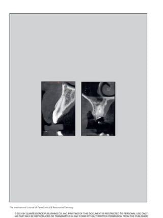
  • 4. 229 Volume 41, Number 2, 2021 Class II: Intact Extraction Site with Partially Favorable Anatomical Conditions Specific characteristics Characteristics of this classification include (1) buccal cortical bone that is intact or affected by damage not exceeding 20% of the extent of the wall; (2) an optimum soft tissue level; and (3) difficulty achieving an ideal implant position and satisfactory primary stability due to the pres- ence of a large periodontal defect, large periapical lesion, and/or ana- tomical structures limiting immedi- ate placement (eg, maxillary sinus floor, mandibular canal). Surgical treatment techniques Treatment techniques for this case include (1) alveolar ridge preserva- tion and delayed implant placement (after 4 to 6 months); and (2) sponta- neous healing and delayed implant placement (3 to 4 months after ex- traction) with bone tissue augmen- tation. Class III: Partially Compromised Extraction Site with Unfavorable Anatomical Conditions Specific characteristics Characteristics of this classification include (1) resorption of the buccal cortical bone, totaling 20% to 50% of the wall; (2) suboptimal soft tissue level, soft tissues presenting inflam- mation, and/or thin and scalloped gingival phenotype; and (3) local bone anatomy allowing an ideal Fig 3  CBCT radiologic images correspond- ing to the four different classifications. (a) Class I. An intact facial bone plate and a large amount of bone available apical and palatal to the root apex. (b) Class II. An intact and thick buccal bone plate with a periapical lesion between the root apex and the maxillary sinus floor, preventing immediate implant placement. (c) Class III. Resorption of the facial bone plate and a large amount of bone available apical and palatal to the root apex. (d) Class IV. Severe bone resorption on both the buccal and palatal sides, together with insufficient api- cal bone volume. a c b d © 2021 BY QUINTESSENCE PUBLISHING CO, INC. PRINTING OF THIS DOCUMENT IS RESTRICTED TO PERSONAL USE ONLY. NO PART MAY BE REPRODUCED OR TRANSMITTED IN ANY FORM WITHOUT WRITTEN PERMISSION FROM THE PUBLISHER.
  • 5. The International Journal of Periodontics Restorative Dentistry 230 tridimensional implant positioning and good primary stability. Surgical treatment techniques Treatment techniques for this case include (1) alveolar ridge augmen- tation, possibly with soft tissue augmentation, and staged implant placement after 6 months; and (2) spontaneous healing and early im- plant placement (4 to 8 weeks after extraction) with bone and/or soft tis- sue augmentation. Class IV: Severely Compromised Extraction Site with Unfavorable Anatomical Conditions Specific characteristics Characteristics of this classification include (1) severely compromised socket walls, particularly with the buccal bone wall loss exceeding 50%; (2) suboptimal soft tissue level, soft tissues presenting inflamma- tion, or thin and scalloped gingival phenotype; and (3) difficulty achiev- ing an ideal implant position and satisfactory primary stability due to the presence of a large periodontal defect, large periapical lesion, and/ or anatomical structures limiting im- mediate placement (eg, maxillary sinus floor, mandibular canal). Surgical treatment techniques Treatment techniques for this case include (1) alveolar ridge augmen- tation, possibly with soft tissue augmentation, and staged implant placement after 6 months; (2) spon- taneous healing, bone augmenta- tion after 4 to 8 weeks, and staged implant placement after an addi- tional 6 months; and (3) spontane- ous healing and implant placement after 3 to 4 months, performed si- multaneously with bone augmenta- tion. Discussion In the literature, there are different classifications of postextraction sites based on the timing of tooth extrac- tion in relation to implant insertion5,6 or the presence of buccal hard and soft tissues.7–9 Other papers have called for a sagittal root position (in relation to the bony housing) during immediate implant placement in the maxillary esthetic zone.10,11 The new classification system presented herein creates a relation- ship between the socket anatomy and the soft tissue level and thick- ness. The main evaluation is based on the integrity of the alveolus and gingival level. An intact socket is defined as presenting three intact bony walls and at least 80% of the fourth bony wall.12 For each distinct clinical situation, more treatment options are proposed. When a Class I case is diag- nosed, the socket is basically in- tact. The minimum bone volume needed for implant primary stabil- ity is related to the surgical skills of the clinician and the characteristics of the implant system used, as im- mediate placement is a technique- sensitive procedure.13 In the hori- zontal dimension, creating a bone- to-implant gap of at least 2 mm be- tween the implant and the internal surface of the facial bone wall is rec- ommended in order to create a suf- ficient space to be filled with a bone substitute.14–16 In the vertical dimen- sion, the implant shoulder should be placed approximately 1 mm apical to the midfacial bone crest to compensate for marginal bone remodeling.17,18 To optimize esthetic outcomes, a flapless approach13,19,20 and the delivery of an immediate restoration is always a suggested option when possible.21,22 The litera- ture remains controversial regarding the need for soft tissue augmenta- tion in cases of thin gingival pheno- type. Following a more prudent ap- proach, alveolar ridge preservation (intended as the preservation of the ridge volume within the envelope existing at the time of extraction23 ) can be selected as an alternative. The majority of the original ridge volume can be maintained when the fresh alveolus is filled with a bone graft at the time of tooth extrac- tion.24,25 Usually, implant surgery can be performed after 4 to 6 months with results similar to that of im- plants inserted in native bone.26 A third option is soft tissue heal- ing and early placement. An open flap raised the day of implant place- ment allows for a contour augmen- tation using guided bone regenera- tion.27 For Class II cases, the socket is still intact, but unfortunately the lo- cal anatomy of the alveolar ridge does not allow for an ideal implant position or good primary stability, and thus immediate implant place- ment is not indicated. In this clinical situation, alveolar ridge preserva- tion with a staged implant place- © 2021 BY QUINTESSENCE PUBLISHING CO, INC. PRINTING OF THIS DOCUMENT IS RESTRICTED TO PERSONAL USE ONLY. NO PART MAY BE REPRODUCED OR TRANSMITTED IN ANY FORM WITHOUT WRITTEN PERMISSION FROM THE PUBLISHER.
  • 6. 231 Volume 41, Number 2, 2021 ment following 4 to 6 months of healing is suggested. Socket preser- vation can biologically compensate for marginal bone remodeling due to the healing dynamics of the fresh extraction site.28 When spontaneous healing is the selected approach, a mean horizontal ridge width reduction of 3.8 mm and a mean vertical ridge height reduction of 1.24 mm can be anticipated.23 This will then require additional bone augmentation the day of implant placement. Class III is characterized by a compromised extraction socket. Ridge augmentation (defined as the increase of the ridge volume beyond the skeletal envelope existing at the time of extraction23 ) is a valuable option, with delayed implant place- ment 6 months later. Alternatively, early placement can be selected. Class IV is the least favorable clinical condition, with a severely compromised socket and a poor bone anatomy that is unideal for im- plant positioning and primary stabil- ity. In this situation, ridge augmen- tation can be a suitable approach. Alternatively, spontaneous healing and bone augmentation after 4 to 8 weeks (the time needed for com- plete soft tissue healing and the resolution of any local infection) is an option.29 An implant can then be placed 6 months after augmenta- tion. A third possibility is spontane- ous healing and implant placement after 3 to 4 months (the time needed for bone tissue healing), performed simultaneously with bone augmen- tation. Conclusions The clinical classification of postex- traction sites is a useful, straightfor- ward, and didactic decisional tool for evaluating the socket anatomy. This classification helps determine a more predictable treatment op- tion in terms of the timing of implant placement and the best surgical ap- proach. Acknowledgments The authors declare no conflicts of interest. References 1. Cardaropoli G, Araújo M, Lindhe J. Dy- namics of bone tissue formation in tooth extraction sites. An experimental study in dogs. J Clin Periodontol 2003;30:809– 818. 2. Trombelli L, Farina R, Marzola A, Bozzi L, Liljenberg B, Lindhe J. Modeling and re- modeling of human extraction sockets. J Clin Periodontol 2008;35:630–639. 3. Araújo MG, Lindhe J. Dimensional ridge alterations following tooth extraction. An experimental study in the dog. J Clin Periodontol 2005;32:212–218. 4. Tan WL, Wong TL, Wong MC, Lang NP. A systematic review of post-extractional alveolar hard and soft tissue dimensional changes in humans. Clin Oral Implants Res 2012;23(suppl 5):s1–s21. 5. Wilson TG Jr, Weber HP. Classification of and therapy for areas of deficient bony housing prior to dental implant place- ment. Int J Periodontics Restorative Dent 1993;13:451–459. 6. Hämmerle CH, Chen ST, Wilson TG Jr. Consensus statements and recom- mended clinical procedures regarding the placement of implants in extraction sockets. Int J Oral Maxillofac Implants 2004;19(suppl):s26–s28. 7. Elian N, Cho SC, Froum S, Smith RB, Tarnow DP. A simplified socket classifica- tion and repair technique. Pract Priced Aesthet Dent 2007;19:99–104. 8. Juodzbalys G, Sakavicius D, Wang HL. Classification of extraction sockets based upon soft and hard tissue compo- nents. J Periodontol 2008;79:413–424. 9. Caplanis N, Lozada JL, Kan JY. Extrac- tion defect assessment, classification, and management. J Calif Dental Assoc 2005;33:853–863. 10. Kan JY, Roe P, Rungcharassaeng K, et al. Classification of sagittal root position in relation to the anterior maxillary osseous housing for immediate implant place- ment: A cone beam computed tomogra- phy study. Int J Oral Maxillofac Implants 2011;26:873–876. 11. Gluckman H, Pontes CC, Du Toit J. Ra- dial plane tooth position and bone wall dimensions in the anterior max- illa: A CBCT classification for immedi- ate implant placement. J Prosthet Dent 2018;120:50–56. 12. Cardaropoli D, Tamagnone L, Roffredo A, Gaveglio L, Cardaropoli G. Socket preservation using bovine bone mineral and collagen membrane: A randomized controlled clinical trial with histologic analysis. Int J Periodontics Restorative Dent 2012;32:421–430. 13. Buser D, Chappuis V, Belser UC, Chen S. Implant placement post extraction in es- thetic single tooth sites: When immedi- ate, when early, when late? Periodontol 2000 2017;73:84–102. 14. Morton D, Chen ST, Martin WC, Levine RA, Buser D. Consensus statements and recommended clinical procedures re- garding optimizing esthetic outcomes in implant dentistry. Int J Oral Maxillofac Implants 2014;29(suppl):s216–s220. 15. Cardaropoli D, Gaveglio L, Gherlone E, Cardaropoli G. Soft tissue contour changes at immediate implants: A randomized controlled clinical study. Int J Periodontics Restorative Dent 2014;34:631–637. 16. Cardaropoli D, Tamagnone L, Roffredo A, De Maria A, Gaveglio L. Preserva- tion of peri-implant hard tissues follow- ing immediate postextraction implant placement. Part I: Radiologic evalua- tion. Int J Periodontics Restorative Dent 2019;39:633–641. 17. Cardaropoli D, Tamagnone L, Roffredo A, Gaveglio L. Soft tissue contour changes at immediate postextraction single- tooth implants with immediate restora- tion: A 12-month prospective cohort study. Int J Periodontics Restorative Dent 201535:191–198. © 2021 BY QUINTESSENCE PUBLISHING CO, INC. PRINTING OF THIS DOCUMENT IS RESTRICTED TO PERSONAL USE ONLY. NO PART MAY BE REPRODUCED OR TRANSMITTED IN ANY FORM WITHOUT WRITTEN PERMISSION FROM THE PUBLISHER.
  • 7. The International Journal of Periodontics Restorative Dentistry 232 18. Chen ST, Darby I. The relationship be- tween facial bone wall defects and di- mensional alterations of the ridge fol- lowing flapless tooth extraction in the anterior maxilla. Clin Oral Implants Res 2017;28:931–937. 19. Nobuto T, Suwa F, Kono T, et al. Micro- vascular response in the periosteum following mucoperiosteal flap surgery in dogs: Angiogenesis and bone re- sorption and formation. J Periodontol 2005;76:1346–1353. 20. Blanco J, Carral C, Argibay O, Liñares A. Implant placement in fresh extraction sockets. Periodontol 2000 2019;79:151– 167. 21. Saito H, Chu SJ, Zamzok J, et al. Flapless postextraction socket implant place- ment: The effects of a platform switch- designed implant on peri-implant soft tissue thickness—A prospective study. Int J Periodontics Restorative Dent 2018;38(suppl):s9–s15. 22. Cardaropoli D, Tamagnone L, Roffredo A, De Maria A, Gaveglio L. Preserva- tion of peri-implant soft tissues follow- ing immediate postextraction implant placement. Part II: Clinical evaluation. Int J Periodontics Restorative Dent 2019;39:789–797. 23. Hämmerle CH, Araújo MG, Simion M; Osteology Consensus Group 2011. Evi- dence-based knowledge on the biology and treatment of extraction sockets. Clin Oral Implants Res 2012;23(suppl 5):s80–s82. 24. Vignoletti F, Matesanz P, Rodrigo D, Figuero E, Martin C, Sanz M. Surgical protocols for ridge preservation after tooth extraction. A systematic review. Clin Oral Implants Res 2012;23(suppl 5):s22–s38. 25. Avila-Ortiz G, Elangovan S, Kramer KW, Blanchette D, Dawson DV. Effect of al- veolar ridge preservation after tooth ex- traction: A systematic review and meta- analysis. J Dent Res 2014;93:950–958. 26. Cardaropoli D, Tamagnone L, Roffredo A, Gaveglio L. Evaluation of dental im- plants placed in preserved and non- preserved postextraction ridges: A 12-month postloading study. Int J Peri- odontics Restorative Dent 2015;35:677– 685. 27. Buser D, Chappuis V, Kuchler U, et al. Long-term stability of early implant placement with contour augmentation. J Dent Res 2013;92(12 suppl):s176–s182. 28. Araújo MG, da Silva JCC, de Mendonça AF, Lindhe J. Ridge alterations follow- ing grafting of fresh extraction sockets in man. A randomized clinical trial. Clin Oral Implants Res 2015;26:407–412. 29. Urban IA, Monje A, Lozada JL, Wang HL. Long-term evaluation of peri-im- plant bone level after reconstruction of severely atrophic edentulous maxilla via vertical and horizontal guided bone regeneration in combination with sinus augmentation: A case series with 1 to 15 years of loading. Clin Implant Dent Relat Res 2017;19:46–55. © 2021 BY QUINTESSENCE PUBLISHING CO, INC. PRINTING OF THIS DOCUMENT IS RESTRICTED TO PERSONAL USE ONLY. NO PART MAY BE REPRODUCED OR TRANSMITTED IN ANY FORM WITHOUT WRITTEN PERMISSION FROM THE PUBLISHER.