Cytoplasm 2
•Download as DOC, PDF•
0 likes•42 views

- Coated vesicles, including clathrin-coated vesicles and coatomer-coated vesicles, transport molecules within cells through endocytosis and between organelles. - Lysosomes contain enzymes to degrade materials and form through the fusion of endosomes and lysosomal enzymes. Late endosomes transition to lysosomes after receiving lysosomal enzymes. - Peroxisomes contain enzymes for functions like fatty acid oxidation and are produced through the growth and division of preexisting peroxisomes.

Recommended
More Related Content
What's hot
What's hot (19)
Similar to Cytoplasm 2
Similar to Cytoplasm 2 (20)
More from Nawfal Aldujaily
More from Nawfal Aldujaily (20)
Recently uploaded
Recently uploaded (20)
Cytoplasm 2
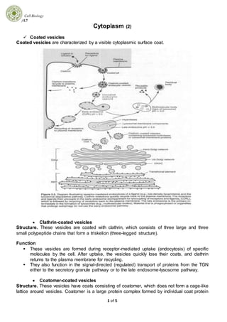
- 1. Cell Biology ) L7 1 of 5 Cytoplasm (2) Coated vesicles Coated vesicles are characterized by a visible cytoplasmic surface coat. Clathrin-coated vesicles Structure. These vesicles are coated with clathrin, which consists of three large and three small polypeptide chains that form a triskelion (three-legged structure). Function These vesicles are formed during receptor-mediated uptake (endocytosis) of specific molecules by the cell. After uptake, the vesicles quickly lose their coats, and clathrin returns to the plasma membrane for recycling. They also function in the signal-directed (regulated) transport of proteins from the TGN either to the secretory granule pathway or to the late endosome-lysosome pathway. Coatomer-coated vesicles Structure. These vesicles have coats consisting of coatomer, which does not form a cage-like lattice around vesicles. Coatomer is a large protein complex formed by individual coat protein
- 2. Cell Biology ) L7 2 of 5 subunits called COPs. Assembly of coatomer depends on the protein ADP-ribosylation factor (ARF), which binds guanosine triphosphate (GTP), becomes activated, and recruits coatomer subunits. ARF also helps to select the cargo molecules. Function Coatomer-coated vesicles mediate the continuous constitutive protein transport (default pathway; bulk flow) within the cell. Specific GTP binding proteins are present at each step of vesicle budding and fusion, and proteins called snares are believed to guide the vesicle movement. Vesicle v-snares bind to complimentary target t-snares. Coatomer-coated vesicles transport proteins from the RER to the ERGIC to the Golgi complex, from one Golgi cisterna to another, and from the TGN to the plasma membrane. Caveolin-coated vesicles. These coated vesicles are less common and less understood than those of the previous two categories. Structure. Caveolae are invaginations of the plasma membrane in endothelial cells and smooth muscle cells. They possess a distinct coat formed by the protein caveolin. Function. Caveolae have been associated with cell signaling and a variety of transport processes, such as transcytosis and endocytosis. Lysosomes Structure. Lysosomes are dense, membrane-bound organelles of diverse shape and size that function to degrade material. Lysosomes possess special membrane proteins and about 50 acid hydrolases, which are synthesized in the RER. ATP-powered proton pumps in the lysosome membrane maintain an acid pH (---- 5). Formation. Lysosomes are formed when sequestered material fuses with a late endosome and enzymatic degradation begins. Formation of a lysosome via one lysosomal pathway involves the following intermediates. o Early endosomes These irregular, peripherally located vesicles form part of the pathway for receptor- mediated endocytosis and contain receptor- ligand complexes. They are also known as the compartment for uncoupling of receptors and ligands (CURL). Their acidic interiors (pH 6) are maintained by ATP-driven proton pumps. The acidity aids in the uncoupling of receptors and ligands; receptors return to the plasma membrane and ligands move to a late endosome. o Late endosomes Late endosomes play a key role in a variety of lysosomal pathways and therefore are sometimes known as the intermediate compartment. These irregular vesicles (pH -,----- 5.5) located deep within the cell receive ligands via microtubular transport of vesicles from early endosomes. Late endosomes contain both lysosomal hydrolases and lysosomal membrane proteins; these are formed in the RER, transported to the Golgi complex for processing, and delivered in separate vesicles to late endosomes.1 Once late endosomes have received a full complement of lysosomal enzymes, they begin to degrade their ligands and are classified as lysosomes.
- 3. Cell Biology ) L7 3 of 5 Types of lysosomes . Lysosomes are named after the content of recognizable material; otherwise, the general term lysosome is used. Multivesicular bodies are formed by fusion of an early endosome containing endocytic vesicles with a late endosome. Phagolysosomes are formed by fusion of a phagocytic vacuole with a late endosome or a lysosome. *The terms primary and virgin lysosomes, formerly used for tiny vesicles believed to be lysosomes that have not yet engaged in digestive activity, are no longer used. Autophagolysosomes are formed by fusion of an autophagic vacuole with a late endosome or lysosome. Autophagic vacuoles are formed when cell components targeted for destruction become enveloped by smooth areas of membranes derived from the RER. Residual bodies are lysosomes of any type that have expended their capacity to degrade material. They contain undegraded material (e.g., lipofuscin and hemosiderin) and eventually may be excreted from the cell. Peroxisomes Structure. Peroxisomes (also known as microbodies) are membrane bound, spherical, or ovoid organelles. They originate from preexisting peroxisomes, which grow by importing specific cytosolic proteins that are recognized by receptor proteins (called peroxins) in the peroxisomal membrane. Then the peroxisome divides by fission; it has a life span of approximately 5-6 days. Function. Peroxisomes contain a variety of enzymes whose functions vary from the oxidation of long chain fatty acids, to the synthesis of cholesterol, to the detoxification of substances such as ethanol.
- 4. Cell Biology ) L7 4 of 5 B. Inclusions. Inclusions are accumulations of material that is not metabolically active. They usually are present in the cytosol only temporarily. o Glycogen appears as small clusters (or in hepatocytes as larger aggregates, known as rosettes) of electron-dense, 20- to 30-nm 13-particles, which are similar in appearance to, but larger than, ribosomes. Glycogen is not bound by a membrane but frequently lies close to the SER. Glycogen serves as a stored energy source that can be degraded to glucose, which enters the bloodstream to elevate blood sugar levels. o Lipid droplets vary markedly in size and appearance depending on the method of fixation and are not bound by a membrane. Lipid droplets are storage forms of triglycerides (an energy source) and cholesterol (used in the synthesis of steroids and membranes). o Lipofuscin appears as membrane-bound, electron-dense granular material varying greatly in size and often containing lipid droplets. Lipofuscin represents a residue of undigested material present in residual bodies. Because the amount of this material increases with age, it is called age pigment. It is most common in nondividing cells (e.g., cardiac muscle cells, neurons) but also is found in hepatocytes). o Centrosome Structure. The centrosome is located near the nucleus and contains two centrioles and a cloud of pericentriolar material. The centrioles exist as a pair of cylindrical rods (each 0.2 Rin wide and 0.5 long) oriented at right angles to one another. Each member of the pair is composed of nine triplets of microtubules (9 + 0 axoneme pattern) arranged radially in the shape of a pinwheel. The centrioles self-duplicate in the S phase of the cell cycle, as each parent centriole forms a procentriole at right angles to itself. Centrioles also form basal bodies, which appear identical to unpaired centrioles and which give rise to the axonemes of cilia and flagella. Function The centrosome is the major microtubule organizing center in the cell. The pericentriolar cloud of material contains hundreds of ring shaped structures composed of –y-tubulin, and each ring serves as a starting point for the polymerization of one microtubule. Centrioles play no role in nucleating microtubules, but help to maintain the organization of the centrosome. The centrosome itself is also duplicated during interphase (Sphase), then separates to form the poles of the mitotic spindle where microtubules originate and converge.
- 5. Cell Biology ) L7 5 of 5 C.Cytoskeleton Cytoskeleton. The cytoskeleton is the structural framework within the cytosol. It functions in maintaining cell shape, stabilizing cell attachments, facilitating endocytosis and exocytosis, and promoting cell motility. It includes the following major components: Microtubules Structure. Microtubules are straight, hollow tubules 25 nm in diameter and made of tubulin. They have a rigid wall composed of 13 protofilaments, each of which consists of a linear arrangement of tubulin dimers; each dimer consists of nonidentical a and 13 tubulin subunits. Microtubules are polar, with polymerization (assembly) and depolymerization (disassembly) occurring preferentially at the plus end as GTP is bound to tubulin dimers. Microtubules have microtubule-associated proteins (MAPs), which stabilize them and bind them to other cytoskeletal components and organelles; they also are associated with kinesin and cytoplasmic dynein, two force-generating proteins, which serve as "motors" for vesicle or organelle movement. Function. Microtubules maintain cell shape; aid in the transport of macromolecules within the cytosol; and promote the movement of chromosomes, cilia, and flagella. Microfilaments Structure. Microfilaments are also known as F actin or actin filaments. They are 7 nm in diameter and are composed of globular actin monomers (G actin) linked into a double helix. They display a polarity similar to that of microtubules; that is, their polymerization and depolymerization occurs preferentially at the plus end when ATP is bound by G actin. c. Many actin-binding proteins associate with microfilaments and modify their properties. d. Microfilaments are abundant at the periphery of the cell, where they are anchored to the plasma membrane via one or more intermediary proteins (e.g., a-actinin, vinculin, talin). Function. Microfilaments are involved in many cellular processes, such as establishing focal contacts between the cell and the extracellular matrix, locomotion of nonmuscle cells, formation of the contractile ring (in dividing cells), and the folding of epithelia into tubes during development. Intermediate filaments are 8 to 10 nm in diameter. They constitute a population of heterogeneous filaments that includes keratin, vimentin, desmin, glial fibrillary acid protein (GFAP), lamins, and neurofilaments. [Desmin and GFAP sometimes co-polymerize with vimentin and may be categorized as vimentin-like filaments.] In general, intermediate filaments provide mechanical strength to cells. They lack polarity and do not require GTP or ATP for assembly, which occurs along the entire length of the filament.