
Recommended
More Related Content
What's hot
What's hot (20)
Similar to 3RD WEEK OF DEVELOPMENT(4).pdf
Similar to 3RD WEEK OF DEVELOPMENT(4).pdf (20)
More from MujahedAldhabyani
More from MujahedAldhabyani (11)
Recently uploaded
Recently uploaded (20)
3RD WEEK OF DEVELOPMENT(4).pdf
- 1. 3RD WEEK OF DEVELOPMENT Ass.Prof.Dr. Saleh Nasser S. A. Alkardae Lecturer of Human Anatomy & Embryology Chairman of Anatomy Department, TUFOM_GUSTFOM
- 2. LEARNING OBJECTIVES At the end of the lecture, student should be able to • Define the gastrulation ( formation of three germ layers). • Discuss the development of primitive streak and related congenital anomalies. (Sacrococcygeal Teratoma) • Describe the development of notochordal process, notochord canal, prechordal plate and cloacal membrane. Ass.Prof.Dr. Saleh Nasser S. A. Alkardae
- 3. •GASTRULATION • DEVELOPMENT OF PRIMITIVE STREAK • DEVELOPMENT OF NOTOCHORD Ass.Prof.Dr. Saleh Nasser S. A. Alkardae
- 4. Development of embryo during third week • Is characterized by: • Appearance of primitive Streak. • Development of notochord. • Differentiation of three germ layers. Ass.Prof.Dr. Saleh Nasser S. A. Alkardae
- 5. GASTRULATION It is the sequence in the development of embryo which characterized by formation of third germ layer (Mesoderm) and indicates the beginning of 3rd week. Formative process by which the three germ layers are established.
- 6. • Bilaminar embryonic germ disc is converted into a trilaminar embryonic disc. • Extensive cell shape changes, rearrangement, movement, and changes in adhesive properties occur. • First sign of gastrulation is the formation of primitive streak . Ass.Prof.Dr. Saleh Nasser S. A. Alkardae
- 7. FORMATION OF THE 3 “GERM” LAYERS ❑Primitive streak (groove) on dorsal surface of epiblast ❑Gastrulation: invagination of epiblast cells ❑Days 14-15: they replace hypoblast becoming endoderm ❑Day 16: mesoderm (a new third layer) formed in between ❑Epiblast cells remaining on surface: ectoderm Ass.Prof.Dr. Saleh Nasser S. A. Alkardae
- 8. DEVELOPMENT OF PRIMITIVE STREAK Epibalstic cells proliferate and invaginate inside between epiblastic and hypoblastic layers. Due to proliferation and migration of epiblastic cells a linear thickening band will be developed in mid line on the dorsal aspect of embryonic disc in caudal part, this is the beginning of development of Primitive streak. The cells forming primitive streak are pluripotent. Ass.Prof.Dr. Saleh Nasser S. A. Alkardae
- 9. DEVELOPMENT OF PRIMITIVE NODE • Primitive streak develops into caudal 1/3 of embryo • The epiblastic cells proliferate continuously and they are moving inward and increasing in number in mid line. The collection of epiblastic cells which change their shape from columnar to polygonal cells called Mesenchymal cells and they are collected at the cranial end of Primitive streak called as Primitive node Ass.Prof.Dr. Saleh Nasser S. A. Alkardae
- 10. The moving surface cells first pile up to form a prominent bump known as the node.This occurs because the cells move along the top faster and sort of like crowds at a major concert.The node was discovered in mammals by Hensen and is appropriately named Hensen's node in rabbits and other organisms, but is only referred to as the node in humans. PRIMITIVE NODE Ass.Prof.Dr. Saleh Nasser S. A. Alkardae
- 11. • Gives rise to intraembryonic mesoderm (third layer of embryonic disc) • Formation of septum transversum • Formation of notochord • Determines the future craniocaudal axis of the embryo • Demarcates the embryo into left and right halves. Functions of Primitive Streak Ass.Prof.Dr. Saleh Nasser S. A. Alkardae
- 13. * Cells from the primitive streak move inwards to the ventral surface at the roof of yolk sac and displace the hypoblastic cells and form the Embryonic Endoderm. * The cells of Epiblast remain and form the Embryonic Ectoderm. * That means all three germs layers, Ectoderm, Mesoderm and Endoderm from Epiblastic cells. REPLACEMENT OF HYPO & EPIBLASTIC CELLS Ass.Prof.Dr. Saleh Nasser S. A. Alkardae
- 14. F A T E O F P R I M I T I V E S T R E A K * Primitive streak forms the embryonic mesoderm up to 4th week of embryo, after that the formation of mesoderm diminish and ultimately regresses completely. * Sometime the primitive streak persists and result in congenital anomalies called as Sacrococcygeal Teratoma Sacrococcygeal Teratoma ❖ Prevalence: 1 in 35,000 ❖ Gender: Female (80%) affected more as compare male. ❖ Morphology: Sacrococcygeal teratoma consists of cells which are derived from pluripotent primitive streak cells. ❖ These tumors contains tissues derived all three germ layers in incomplete stage of differentiation ❖ Can be diagnosed antenatally with of Ultrasonography. Ass.Prof.Dr. Saleh Nasser S. A. Alkardae
- 16. Situs Inversus Ass.Prof.Dr. Saleh Nasser S. A. Alkardae
- 17. What happens if there is “not enough” gastrulation? Caudal agenesis (sirenomelia) Premature regression of the primitive streak leads to widespread loss of trunk and lower limb mesoderm. VATeR association: Vertebral defects Anal atresia Tracheo-esophageal fistula Renal defects VACTeRL association: those above plus… Cardiovascular defects Limb (upper) defects Ass.Prof.Dr. Saleh Nasser S. A. Alkardae
- 18. If the primitive streak fails to regress, multipotent primitive streak cells can develop into multi- lineage tumors (containing ecto-, meso-, and endodermal tissues). What happens if there is “too much” gastrulation? Sacrococcygeal teratoma Ass.Prof.Dr. Saleh Nasser S. A. Alkardae
- 19. Ass.Prof.Dr. Saleh Nasser S. A. Alkardae
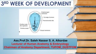
- 20. DEVELOPMENT NOTOCHORD • Prechordal plate, a small circular area of columnar endodermal cells where the ectoderm and endoderm are in contact. • The prechordal plate is the primordium of the oropharyngeal membrane which will lead to oral cavity Notochord is a midline cord of cells that develops in the region between the primitive streak and the precordial plate of embryonic disc. It develops from the primitive knot (Hensen’s node) of the primitive streak Ass.Prof.Dr. Saleh Nasser S. A. Alkardae
- 21. • The cells constantly migrating from the primitive streak and node and form a solid cord of Notochordal process which soon acquires lumen, called as Notochordal Canal Ass.Prof.Dr. Saleh Nasser S. A. Alkardae
- 22. • The Notochordal process elongates by invagination of cells from the primitive pit. The primitive pit extends into the notochordal process, forming a Notochordal canal. It is now a cellular tube that extends cranially from the primitive node to the prechordal plate. Ass.Prof.Dr. Saleh Nasser S. A. Alkardae
- 23. • The floor of the notochordal process fuses with the underlying embryonic endoderm. • The fused layers gradually undergo degeneration, resulting in the formation of openings in the floor of the notochordal process, which brings the notochordal canal into communication with the umbilical vesicle. Ass.Prof.Dr. Saleh Nasser S. A. Alkardae
- 24. • The openings rapidly become confluent and the floor of the notochordal canal disappears; the remains of the notochordal process form a flattened, grooved notochordal plate. • The proximal part of the notochordal canal persists temporarily as the neuroenteric canal, which forms a transitory communication between the amniotic and umbilical vesicle cavities. Ass.Prof.Dr. Saleh Nasser S. A. Alkardae
- 25. • The notochordal plate gets curved to form a tube. Ass.Prof.Dr. Saleh Nasser S. A. Alkardae
- 26. • Beginning at the cranial end of the embryo, the notochordal cells proliferate and the notochordal plate infolds to form the notochord tube. Ass.Prof.Dr. Saleh Nasser S. A. Alkardae
- 27. • The proliferation of cells of the tube converts it into a solid cord of cells to form definitive notochord. The endoderm is restored, and now the notochord is completely separated from the endoderm. Ass.Prof.Dr. Saleh Nasser S. A. Alkardae
- 28. 1) It forms the central axis of the developing embryo (embryonic disc). 2) It induces the formation of neural tube from the overlying ectoderm. 3) It provides central column around which vertebral bodies and intervertebral discs develop Functions of Notochord Ass.Prof.Dr. Saleh Nasser S. A. Alkardae
- 29. FATE OF NOTOCHORD ➢ The notochord extends from the oropharyngeal membrane to the primitive node. ➢ The notochord degenerates as the bodies of the vertebrae form, but small portions of it persist as the nucleus pulposus of each intervertebral disc. Ass.Prof.Dr. Saleh Nasser S. A. Alkardae
- 30. • The notochord functions as the primary inductor (signaling center) in the early embryo. • The developing notochord induces the overlying embryonic ectoderm to thicken and form the neural plate, the primordium of the central nervous system (CNS). NOTOCHORD ACT AS INDUCER Ass.Prof.Dr. Saleh Nasser S. A. Alkardae
- 31. In embryo the mesoderm intervene between ectoderm and endoderm but there three places where ectoderm and endoderm are direct in contact – At the oropharyngeal membrane cranially – In the median plane cranial to the primitive node, where the notochordal process is located – At the cloacal membrane caudally PLACES WHERE ECTODERM AND ENDODERM ARE DIRECT IN CONTACT Ass.Prof.Dr. Saleh Nasser S. A. Alkardae
- 32. FATEMAP ESTABLISHED DURING GASTRULATION Ass.Prof.Dr. Saleh Nasser S. A. Alkardae
- 33. REFERENCES * Keith L. Moore Developing Human 8th Edition Chapter-4 Pages 59 -65 THANK YOU