

Recommended
More Related Content
What's hot
What's hot (20)
Similar to Skeletal system anatomy physiology
Similar to Skeletal system anatomy physiology (20)
More from MuhammadUmer411
More from MuhammadUmer411 (6)
Recently uploaded
Recently uploaded (20)
Skeletal system anatomy physiology
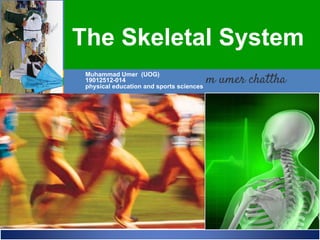
- 1. ELAINE N. MARIEB EIGHTH EDITION 5 Copyright © 2006 Pearson Education, Inc., publishing as Benjamin Cummings ESSENTIALS OF HUMAN ANATOMY & PHYSIOLOGY PART A Muhammad Umer (UOG) 19012512-014 physical education and sports sciences The Skeletal System
- 2. Divided into two divisions Axial skeleton Appendicular skeleton Parts of the skeletal system Bones (skeleton) Joints Cartilages Ligaments The Skeletal System
- 3. Functions of the Bones • Support of the body • Protection of soft organs • Movement due to attached skeletal muscles • Storage of minerals and fats • Blood cell formation
- 4. • The adult skeleton has 206 bones • Two basic types of osseous tissue Compact bone Is dense and looks smooth Homogenous Spongy bone Small needle-like pieces of bone Many open spaces Classification of Bones
- 5. Classification of Bones on the Basis of Shape
- 6. Long bones Typically longer than wide Have a shaft with heads at both ends Contain mostly compact bone • Examples: Femur, humerus Classification of Bones
- 7. Short bones Generally cube-shape Contain mostly spongy bone • Examples: Carpals, tarsals Classification of Bones
- 8. Flat bones Thin and flattened Usually curved Thin layers of compact bone around a layer of spongy bone • Examples: Skull, ribs, sternum Classification of Bones
- 9. Irregular bones Irregular shape Do not fit into other bone classification categories • Example: Vertebrae and hip Classification of Bones
- 10. Gross Anatomy of a Long Bone Diaphysis Shaft Composed of compact bone Epiphysis Ends of the bone Composed mostly of spongy bone
- 11. Structures of a Long Bone Periosteum Outside covering of the diaphysis Fibrous connective tissue membrane Sharpey’s fibers Secure periosteum to underlying bone Arteries Supply bone cells with nutrients
- 12. Articular cartilage Covers the external surface of the epiphyses Made of hyaline cartilage Decreases friction at joint surfaces Structures of a Long Bone
- 13. Medullary cavity Cavity of the shaft Contains yellow marrow (mostly fat) in adults Contains red marrow (for blood cell formation) in infants Structures of a Long Bone
- 14. Bone Markings • Surface features of bones • Sites of attachments for muscles, tendons, and ligaments • Passages for nerves and blood vessels • Categories of bone markings – Projections and processes – grow out from the bone surface – Depressions or cavities – indentations
- 17. • Osteon (Haversian System) – A unit of bone • Central (Haversian) canal – Opening in the center of an osteon – Carries blood vessels and nerves • Perforating (Volkman’s) canal – Canal perpendicular to the central canal – Carries blood vessels and nerves Microscopic Anatomy of Bone
- 18. Microscopic Anatomy of Bone
- 19. Microscopic Anatomy of Bone • Lacunae – Cavities containing bone cells (osteocytes) – Arranged in concentric rings • Lamellae – Rings around the central canal – Sites of lacunae
- 20. Microscopic Anatomy of Bone • Canaliculi – Tiny canals – Radiate from the central canal to lacunae – Form a transport system
- 21. Bone Formation, Growth and Remodeling • In embryos, the skeleton is primarily hyaline cartilage • During development, much of this cartilage is replaced by bone • Cartilage remains in isolated areas – Bridge of the nose – Parts of ribs – Joints
- 22. Bone Formation, Growth and Remodeling • Ossification – is the process of bone formation • It involves two major phases: – First, the hyaline cartilage model is completely covered with bone matrix by bone forming cells called osteoblasts. – Then, the enclosed hyaline cartilage model is digested away, opening up a medullar cavity within the newly formed bone.
- 23. Long Bone Formation and Growth
- 24. How do bones widen? • Osteoblasts in the periosteum add bone to the external face of the diaphysis as osteoclasts in the endosteum remove bone from the inner face of the diaphysis wall. • Appositional growth - The process by which the bones increase in diameter.
- 25. Formation and Growth of Long Bones
- 26. • Bones are remodelled continually in response changes in two factors: Calcium levels in the blood The pull of gravity and muscles on the skeleton.
- 27. • Osteocytes – Mature bone cells • Osteoblasts – Bone-forming cells • Osteoclasts – Bone-destroying cells – Break down bone matrix for remodeling and release of calcium • Bone remodeling is a process by both osteoblasts and osteoclasts Types of Bone Cells
- 28. • Is essential if bones are retain normal proportions and strength during long-bone growth as the body increases in size and weight. • Bones become thicker and form large projections to increase their strength in areas where bulky muscles are attached. Bone Remodeling
- 29. • A break in a bone • Types of bone fractures Closed (simple) fracture- break that does not penetrate the skin. Open (compound) fracture- broken bone penetrates through the skin • Bone fractures are treated by reduction or immobilization Realignment of the bone Bone Fractures
- 30. Bone Fractures
- 32. Type of Bone Fractures
- 33. Hematoma (blood-filled swelling) is formed. Break is splinted by fibrocartilage to form a callus. Fibrocartilage callus is replaced by a bony callus Bony callus is remodeled to form a permanent patch. Repair of Bone Fractures
- 34. 3. The bony callus forms As the more osteoblasts and osteoclasts migrate into the area and multiply, the fibrocartilage callus is gradually replaced by one made of spongy bone the bony callus. 4. Bone remodeling occurs Over the next few weeks or months, the bony callus is remodeled in response to the mechanical stressed placed on it and it forms a strong permanent “patch” at the fracture sight.
- 36. Axial Skeleton Forms the longitudinal axis of the body Divided into three parts: Skull Vertebral column Bony thorax
- 37. The Skull Two sets of bones Cranium Facial bones Bones are joined by sutures Only the mandible is attached by a freely movable joint.
- 38. Cranium The boxlike cranium is composed of eight large flat bones. Frontal Bone- forms the forehead, the bony projections under the eyebrows and the superior part of each eye’s orbit. Parietal Bones- form most of the superior and lateral walls of the cranium. They meet in the midline of the skull at the sagittal suture and form the coronal suture, where they meet the frontal bone.
- 39. Cranium Temporal Bone- it lies inferior to the parietal bones; they join them at the squamous sutures. Occipital Bone- the most posterior part of the cranium. It forms the floor and black wall of the skull. Sphenoid Bone- the butterfly-shaped sphenoid bone spans the width of the skull and forms part of the floor of the cranial cavity.
- 40. Cranium Ethmoid Bone- is very irregularly shaped and lies anterior to the sphenoid. It forms the roof of the nasal cavity and part of the medial walls of the orbits.