SlideShare a Scribd company logo
1 of 52
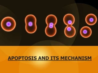
APOPTOSIS AND ITS MECHANISM
Table of content
 Introduction
 History
 Morphological changes
 Importance of apoptosis
 Biochemical changes
 Mechanism of apoptosis
 Assays of apoptosis
 Apoptosis in disease
 Conclusion
Introduction
• Release of enzymes in the cell that cause the degradation of the
cells own nuclear DNA, nuclear and cytoplasmic proteins leading
to cell death.
• The word Apoptosis means “Falling of”.
• Differs from necrosis as no swelling occurs
• Plasma membrane is altered and is called apoptotic bodies.
• Apoptotic bodies being highly “edible” are consumed easily by
Phagocytes.
History
• Term was given by Hippocrates
• Described by Rudolf Virchow
• Defined by Kerr in 1972
• In the 20th century the research is focused for clarification of
this method to cure diseases such as cancer, Alzheimer and
AIDS.
Morphological changes
• Apoptosis is characterized by:
– Mitochondrial leakage
– Activation of Caspases
– Shrinkage of the cell
– Membrane bound apoptotic bodies
– Chromatin condensation
– Phagocytosis by nearby cells
Importance of Apoptosis
• To eliminate harmful cells and cells that have lived
their usefulness.
– During growth old cells are replaced with new ones
– Unwanted cells are eliminated to prevent inflammation
and proliferation.
– Excess leukocytes left at the end of immune response
are eliminated.
– Lymphocytes that can cause auto-immune disease are
eliminated.
Cont.
• Occurs when cells are damaged especially
when damage is irreparable ( DNA or proteins
are damaged)
– Damage due to radiation and cytotoxic drugs.
– Misfolded proteins cause apoptosis
– Infection due to virus or infectious agents.
BIOCHEMICAL CHANGES IN
APOPTOSIS
Characteristic biochemical changes in cells undergoing
apoptosis:
I. Different proteins regulate these processes
II. ATP is required
III. DNA fragmentation occurs
IV. A series of reactions of activation of proteins take place.
Cont.
V. pH of the cell becomes acidic
VI. The apoptosis doesn’t effect adjacent cells
VII.Very rarely it requires medical treatment and
doesn’t case inflammation.
MECHANISM OF APOPTOSIS
Mechanism
• Apoptosis is regulated by biochemical pathways that control
the balance of death and survival inducing signals and
ultimately the activation of enzymes called Caspases.
• There are two different pathways for signaling apoptosis
– Mitochondrial Pathway (intrinsic pathway)
– Death Receptor Pathway (Extrinsic pathway)
• Both the pathways activate Caspases Proteins.
Intrinsic Pathway
 The intrinsic apoptosis pathway is activated by a
range of exogenous and endogenous stimuli,
such as DNA damage, ischemia, and oxidative
stress.
 It plays an important function in the elimination
of damaged cells.
DNA DAMAGE
Mitochondria along with
cytochrome-c also release a protein
called Smac/DIABLO.
It promote apoptosis and inhibit
anti-apoptosis proteins called
inhibitor of apoptosis proteins
(IAPs).
Cont.
• Entry of cytochrome-c into cytosol causes assembly of
Apoptosomes.
• Apaf-1 protein with cytochrome-c forms the seven
fold ring structure (apoptosomes)
• The attachment of caspases to the card domain of the
apoptosomes causes their activation.
Cont.
WORKING OF CASPASE-3
 Caspase-3 degraded the nuclease inhibitor and
consequently nuclease activated.
 Nuclease degraded the DNA inside nucleus.
 As a result cell cannot survive.
 Along caspase-9, caspase-2, caspase-8 and caspase-
10 initiate apoptosis.
 Caspase-3, caspase-6 and caspase-7 involved in
apoptosis.
 Caspase-6 cause disintegration of lamina and
cytoskeleton.
 P53 also activate protease for the degradation of
protein.
 All these incidents leads to cell death and
phagocytosis by macrophages.
Cont.
Components
• Death Receptors
• Death Ligands
• Adaptor Proteins
• Caspases
Death Receptors
• “Death receptors” that are members of the tumor
necrosis factor (TNF) receptor superfamily.
• Death receptors have a cytoplasmic domain of about
80 amino acids called the “death domain”.
• This death domain plays a critical role in transmitting
the death signal from the cell surface to the
intracellular signaling pathways.
Death Ligand
• The best characterized receptors & ligands corresponding death
receptors include:
Ligands Receptors
• FasR (CD95/APO1) FasL
• DR3 Apo3L
• DR4 (TRAIL-R1) Apo2L
• DR5 (TRAIL-R2) Apo2L
• TNFR1 TNF-α
• TNFR2 TNF-ß
Apoptotic Adaptor Proteins
• Apoptotic adaptor proteins play a critical role in regulating
pro- and anti-apoptotic signaling pathways Adaptor
proteins;
• FADD (Fas-associated death domain)
• TRADD (TNF receptor-associated death domain), are
recruited to ligand-activated, oligomerized death receptors
to mediate apoptotic signaling pathways.
Apoptosis inducing factor (AIF)
Assays of apoptosis
• Two different assay types are used to detect the
process of apoptosis
• The first assay detects initial events, whereas second
assay identifies the execution or terminal phase
• The apoptosis assays have been divided into six
different groups.
Cytomorphological altercation
• The observation of hematoxylin and eosin-stained tissue sections
with light microscopy allows the visualization of apoptotic cells.
• This method detects the cells in the later events of apoptosis, but
the cells in the early stage of apoptosis are not recognized.
• Transmission electron microscopy (TEM) is the gold standard for
the confirmation of apoptosis.
Cont.
• In TEM, cells undergoing apoptosis reveals several structural characteristics.
These characters include:
– electron-dense nucleus (marginalization of the nucleus in the early phase)
– nuclear fragmentation
– intact cell membrane even late in the cell disintegration phase
– disorganized cytoplasmic organelles
– large clear vacuoles
– phosphatidylserine at the cell surface.
• With the progression of apoptosis, these cells will lose the cell-to-cell
adhesions and will separate from neighboring cells.
• Eventually, the cell will fragment into apoptotic bodies with intact cell
membranes and will contain cytoplasmic organelles with or without nuclear
fragments.
DNA Fragmentation
• DNA Laddering technique can be used to detect apoptosis
– It involves the extraction of DNA from lysed cell homogenate separation by agarose gel
electrophoresis.
– The resulting bands of DNA form a DNA ladder that can be used to detect apoptosis in
tissues where the number of apoptic cells is high
– This technique can only detect apoptosis at the later stage.
• Another method include Terminal dUTP Nick end-labeling
(TUNEL).
– It detects the endonuclease cleavage products by enzymatically labelling the ends of
DNA strands.
– Terminal transferase is used to attach dUTP to the 3;-end of the DNA fragments.
– The dUTP is then labelled with variety of probes to allow detection by
light microscope, flourescence microscopy or flow cytometery.
– Although it is efficient but can give false result.
Cont.
• Another method include Terminal dUTP Nick end-labeling
(TUNEL).
– It detects the endonuclease cleavage products by enzymatically
labelling the ends of DNA strands.
– Terminal transferase is used to attach dUTP to the 3’-end of the
DNA fragments.
– The dUTP is then labelled with variety of probes to allow
detection by light microscope, fluorescence microscopy or flow
cytometery.
– Although it is efficient but can give false result.
Detection of Caspases
• More than 13 known caspases activity can be detected using various
types of assays.
• Immunoassays can detect cleaved substrates such as PARP and
known cell modifications such as phosphorylated histones.
• Western Blot, immunoprecipitation and immunohistochemistry
assays can be used to detect caspases activation.
• Real time PCR is also used to detect the expression of 112 genes
involved in apoptosis
• Microarrays are made to produce expression of genes that
encode factors involved in the regulation of programmed cell death.
Mitchondrial alterations
• Annexin V assays are used as early markers of apoptosis events.
• The translocation of phophatidylserine on the membrane surface is
an early event in apoptosis and can be detected by this assay.
• The cell is first bound to the FITC-labeled annexin V and
visualized with fluorescence microscopy.
• The integrity of membrane in apoptotic cell can also be detected
by Propidium iodide and trypan blue dyes.
• The dye may move inside the cell and then can also be visualized by
a light microscope.
Change in the Phosphatidylserine position
Detection in Whole Mounts
• Whole embryo or tissue can be visualized by Acridine orange,
Nile Blue Sulfate (NBS) and Neutral Red dyes.
• All the mentioned dyes are Acidophillic and concentrate in
regions where lysosomal and phagocytic activity is high.
• Drawback of this procedure is that Acridine orange is mutagenic
and toxic. Whereas NBS and Neutral Red do not penetrate deep
into tissues and can be lost during penetration.
Mitochondrial assays
• Mitochondria are important cellular organelles that maintain crucial
cellular energy balance, contain key regulators of cell death processes such
as apoptosis.
• Collapse of mitochondrial membrane potential is believed to coincide with
the permeabilization of the outer mitochondrial membrane, and release of
Cytochrome C and other pro-apoptotic proteins into the cytosol, which
then triggers the downstream events in the apoptotic cascade.
• The Laser scanning confocal microscopy (LSCM) creates thin optical
slices of living cells that are then used to monitor various mitochondrial
events in intact single cells throughout a period of time.
• Cytochrome c release from the mitochondria of living or fixed cells can
also be assayed using fluorescence and electron microscopy.
Apoptosis in Cancer
• Apoptosis is regulated by two major genes p53 & Bcl-2
• Tumor suppressor p53 controls senescence and apoptosis in
response to damage.
• Mutations or overexpression of these genes will result in cancer.
• In moat cases, Bcl-2 expression has seen elevated
• Most cancer cells are defective in apoptotic response.
Apoptosis in Neurodegenerative
Diseases
• Brain disorder that slowly destroys memory and thinking skills
• Apoptosis of hippocampal neurons in brain
1. Alzheimer's Disease:
2. Parkinson’s Disease:
• elevated activity of caspase-3 and increased expression of active caspase-3 in substantia
nigra.
• Apoptosis of the neurotransmitters Dopamine present in Substantia Nigra.
• Elevated levels of proapoptotic proteins, such as Bax, have also been seen in Parkinson’s
patients.
• rare, inherited disease that causes the progressive breakdown of nerve
cells in the brain.
• It usually effects person’s movement and thinking.
• In humans with HD, Caspases, among other proteins, cleave huntingtin
within the N-terminal region.
• Mutation in huntingtin may lead to apoptosis of neurons.
3. Huntington disease
Apoptosis & Necrosis
• Programmed Cell Death
• Induced by physiological stimuli
• No inflammation
• Shrinking of cytoplasm & condensation
of nucleus
• Blebbing of plasma membrane without
loss of integrity.
• ATP dependent pathways
Apoptosis Necrosis
• Premature cell death or tissue
• Induced by viruses, injury, infection
• Inflammation
• Swelling of cytoplasm and mitochondria
causing cell lysis.
• Loss of plasma membrane integrity
• No ATP is involved
• Programmed cell death which occurs in multicellular organisms.
• Characterized by specific morphological and biochemical features.
• Triggered by multi-signal pathways and regulated by extrinsic and
intrinsic ligands.
• Disordered apoptosis may lead to carcinogenesis and participates
in the pathogenesis of Alzheimer disease, Parkinson disease, AIDS
etc.
Conclusion
Apoptosis definition, mechanism , Apoptosis vs necrosis, Assays of apoptosis, and diseases related to Apoptosis

More Related Content

What's hot (20)

Apoptosis
Apoptosis Apoptosis
Apoptosis
 
Apoptosis
ApoptosisApoptosis
Apoptosis
 
Apoptosis
Apoptosis Apoptosis
Apoptosis
 
Programmed Cell death or Apoptosis
Programmed Cell death or ApoptosisProgrammed Cell death or Apoptosis
Programmed Cell death or Apoptosis
 
Apoptosis continued
Apoptosis continuedApoptosis continued
Apoptosis continued
 
Apoptosis, program cell death
Apoptosis, program cell deathApoptosis, program cell death
Apoptosis, program cell death
 
Apoptosis (presentation)
Apoptosis (presentation)Apoptosis (presentation)
Apoptosis (presentation)
 
Apoptosis
ApoptosisApoptosis
Apoptosis
 
Apoptosis (Programm of cell death)
Apoptosis (Programm of cell death)Apoptosis (Programm of cell death)
Apoptosis (Programm of cell death)
 
Apoptosis
ApoptosisApoptosis
Apoptosis
 
Adhesion molecules
Adhesion moleculesAdhesion molecules
Adhesion molecules
 
Apoptosis
ApoptosisApoptosis
Apoptosis
 
Cellular ageing
Cellular ageingCellular ageing
Cellular ageing
 
Apoptosis
ApoptosisApoptosis
Apoptosis
 
Apoptosis
ApoptosisApoptosis
Apoptosis
 
Apoptosis my ppt
Apoptosis my pptApoptosis my ppt
Apoptosis my ppt
 
Apoptosis
ApoptosisApoptosis
Apoptosis
 
Apoptosis
ApoptosisApoptosis
Apoptosis
 
Apoptosis ppt.
Apoptosis  ppt.Apoptosis  ppt.
Apoptosis ppt.
 
Apoptosis
ApoptosisApoptosis
Apoptosis
 

Similar to Apoptosis definition, mechanism , Apoptosis vs necrosis, Assays of apoptosis, and diseases related to Apoptosis

Apoptosis - programmed cell death that occurs in multicellular organisms
Apoptosis - programmed cell death that occurs in multicellular organismsApoptosis - programmed cell death that occurs in multicellular organisms
Apoptosis - programmed cell death that occurs in multicellular organismsMeethuRappai1
 
apopwqgdcbhuredgbcfyhhvhcctosis (1).pptx
apopwqgdcbhuredgbcfyhhvhcctosis (1).pptxapopwqgdcbhuredgbcfyhhvhcctosis (1).pptx
apopwqgdcbhuredgbcfyhhvhcctosis (1).pptxSarithaRani4
 
physiology of apoptosis first year mbbs medical.pdf
physiology of apoptosis first year mbbs medical.pdfphysiology of apoptosis first year mbbs medical.pdf
physiology of apoptosis first year mbbs medical.pdfDratoshKatiyar
 
Apoptosis / dental crown & bridge courses
Apoptosis / dental crown & bridge coursesApoptosis / dental crown & bridge courses
Apoptosis / dental crown & bridge coursesIndian dental academy
 
Apoptosis basics + microphotographs
Apoptosis basics + microphotographsApoptosis basics + microphotographs
Apoptosis basics + microphotographsashish223
 
Seminar- recent advances in apoptosis
Seminar- recent advances in apoptosisSeminar- recent advances in apoptosis
Seminar- recent advances in apoptosisEkta Jajodia
 
Intrinsic and Extrinsic Pathway of Apoptosis
Intrinsic and Extrinsic  Pathway of ApoptosisIntrinsic and Extrinsic  Pathway of Apoptosis
Intrinsic and Extrinsic Pathway of ApoptosisAnantha Kumar
 
Apoptosis roohit
Apoptosis roohitApoptosis roohit
Apoptosis roohitroohith
 
Apoptosis in health and diseases
Apoptosis in health and diseasesApoptosis in health and diseases
Apoptosis in health and diseasesmeenuev
 
Apoptosis.pptx
Apoptosis.pptxApoptosis.pptx
Apoptosis.pptxDrmustafa Ali
 
Cell death,regulators, apoptosis,necrosis,autophagy
Cell death,regulators, apoptosis,necrosis,autophagyCell death,regulators, apoptosis,necrosis,autophagy
Cell death,regulators, apoptosis,necrosis,autophagyDr.SIBI P ITTIYAVIRAH
 

Similar to Apoptosis definition, mechanism , Apoptosis vs necrosis, Assays of apoptosis, and diseases related to Apoptosis (20)

Apoptosis - programmed cell death that occurs in multicellular organisms
Apoptosis - programmed cell death that occurs in multicellular organismsApoptosis - programmed cell death that occurs in multicellular organisms
Apoptosis - programmed cell death that occurs in multicellular organisms
 
apopwqgdcbhuredgbcfyhhvhcctosis (1).pptx
apopwqgdcbhuredgbcfyhhvhcctosis (1).pptxapopwqgdcbhuredgbcfyhhvhcctosis (1).pptx
apopwqgdcbhuredgbcfyhhvhcctosis (1).pptx
 
Apoptosis
ApoptosisApoptosis
Apoptosis
 
physiology of apoptosis first year mbbs medical.pdf
physiology of apoptosis first year mbbs medical.pdfphysiology of apoptosis first year mbbs medical.pdf
physiology of apoptosis first year mbbs medical.pdf
 
Apoptosis / dental crown & bridge courses
Apoptosis / dental crown & bridge coursesApoptosis / dental crown & bridge courses
Apoptosis / dental crown & bridge courses
 
Apoptosis basics + microphotographs
Apoptosis basics + microphotographsApoptosis basics + microphotographs
Apoptosis basics + microphotographs
 
APOPTOSIS
APOPTOSISAPOPTOSIS
APOPTOSIS
 
Apoptosis
ApoptosisApoptosis
Apoptosis
 
Seminar- recent advances in apoptosis
Seminar- recent advances in apoptosisSeminar- recent advances in apoptosis
Seminar- recent advances in apoptosis
 
Intrinsic and Extrinsic Pathway of Apoptosis
Intrinsic and Extrinsic  Pathway of ApoptosisIntrinsic and Extrinsic  Pathway of Apoptosis
Intrinsic and Extrinsic Pathway of Apoptosis
 
Apoptosis roohit
Apoptosis roohitApoptosis roohit
Apoptosis roohit
 
Apoptosis
ApoptosisApoptosis
Apoptosis
 
Apoptosis in health and diseases
Apoptosis in health and diseasesApoptosis in health and diseases
Apoptosis in health and diseases
 
Slide share
Slide shareSlide share
Slide share
 
apoptosis.pptx
apoptosis.pptxapoptosis.pptx
apoptosis.pptx
 
APoptosis
APoptosisAPoptosis
APoptosis
 
Apoptosis.pptx
Apoptosis.pptxApoptosis.pptx
Apoptosis.pptx
 
Apoptosis
ApoptosisApoptosis
Apoptosis
 
Cell death,regulators, apoptosis,necrosis,autophagy
Cell death,regulators, apoptosis,necrosis,autophagyCell death,regulators, apoptosis,necrosis,autophagy
Cell death,regulators, apoptosis,necrosis,autophagy
 
Apoptosis
ApoptosisApoptosis
Apoptosis
 

Recently uploaded

BUMI DAN ANTARIKSA PROJEK IPAS SMK KELAS X.pdf
BUMI DAN ANTARIKSA PROJEK IPAS SMK KELAS X.pdfBUMI DAN ANTARIKSA PROJEK IPAS SMK KELAS X.pdf
BUMI DAN ANTARIKSA PROJEK IPAS SMK KELAS X.pdfWildaNurAmalia2
 
The dark energy paradox leads to a new structure of spacetime.pptx
The dark energy paradox leads to a new structure of spacetime.pptxThe dark energy paradox leads to a new structure of spacetime.pptx
The dark energy paradox leads to a new structure of spacetime.pptxEran Akiva Sinbar
 
FREE NURSING BUNDLE FOR NURSES.PDF by na
FREE NURSING BUNDLE FOR NURSES.PDF by naFREE NURSING BUNDLE FOR NURSES.PDF by na
FREE NURSING BUNDLE FOR NURSES.PDF by naJASISJULIANOELYNV
 
Bioteknologi kelas 10 kumer smapsa .pptx
Bioteknologi kelas 10 kumer smapsa .pptxBioteknologi kelas 10 kumer smapsa .pptx
Bioteknologi kelas 10 kumer smapsa .pptx023NiWayanAnggiSriWa
 
Speech, hearing, noise, intelligibility.pptx
Speech, hearing, noise, intelligibility.pptxSpeech, hearing, noise, intelligibility.pptx
Speech, hearing, noise, intelligibility.pptxpriyankatabhane
 
STOPPED FLOW METHOD & APPLICATION MURUGAVENI B.pptx
STOPPED FLOW METHOD & APPLICATION MURUGAVENI B.pptxSTOPPED FLOW METHOD & APPLICATION MURUGAVENI B.pptx
STOPPED FLOW METHOD & APPLICATION MURUGAVENI B.pptxMurugaveni B
 
Pests of Bengal gram_Identification_Dr.UPR.pdf
Pests of Bengal gram_Identification_Dr.UPR.pdfPests of Bengal gram_Identification_Dr.UPR.pdf
Pests of Bengal gram_Identification_Dr.UPR.pdfPirithiRaju
 
Base editing, prime editing, Cas13 & RNA editing and organelle base editing
Base editing, prime editing, Cas13 & RNA editing and organelle base editingBase editing, prime editing, Cas13 & RNA editing and organelle base editing
Base editing, prime editing, Cas13 & RNA editing and organelle base editingNetHelix
 
Functional group interconversions(oxidation reduction)
Functional group interconversions(oxidation reduction)Functional group interconversions(oxidation reduction)
Functional group interconversions(oxidation reduction)itwameryclare
 
THE ROLE OF PHARMACOGNOSY IN TRADITIONAL AND MODERN SYSTEM OF MEDICINE.pptx
THE ROLE OF PHARMACOGNOSY IN TRADITIONAL AND MODERN SYSTEM OF MEDICINE.pptxTHE ROLE OF PHARMACOGNOSY IN TRADITIONAL AND MODERN SYSTEM OF MEDICINE.pptx
THE ROLE OF PHARMACOGNOSY IN TRADITIONAL AND MODERN SYSTEM OF MEDICINE.pptxNandakishor Bhaurao Deshmukh
 
Dubai Calls Girl Lisa O525547819 Lexi Call Girls In Dubai
Dubai Calls Girl Lisa O525547819 Lexi Call Girls In DubaiDubai Calls Girl Lisa O525547819 Lexi Call Girls In Dubai
Dubai Calls Girl Lisa O525547819 Lexi Call Girls In Dubaikojalkojal131
 
User Guide: Magellan MX™ Weather Station
User Guide: Magellan MX™ Weather StationUser Guide: Magellan MX™ Weather Station
User Guide: Magellan MX™ Weather StationColumbia Weather Systems
 
Pests of soyabean_Binomics_IdentificationDr.UPR.pdf
Pests of soyabean_Binomics_IdentificationDr.UPR.pdfPests of soyabean_Binomics_IdentificationDr.UPR.pdf
Pests of soyabean_Binomics_IdentificationDr.UPR.pdfPirithiRaju
 
REVISTA DE BIOLOGIA E CIÊNCIAS DA TERRA ISSN 1519-5228 - Artigo_Bioterra_V24_...
REVISTA DE BIOLOGIA E CIÊNCIAS DA TERRA ISSN 1519-5228 - Artigo_Bioterra_V24_...REVISTA DE BIOLOGIA E CIÊNCIAS DA TERRA ISSN 1519-5228 - Artigo_Bioterra_V24_...
REVISTA DE BIOLOGIA E CIÊNCIAS DA TERRA ISSN 1519-5228 - Artigo_Bioterra_V24_...Universidade Federal de Sergipe - UFS
 
OECD bibliometric indicators: Selected highlights, April 2024
OECD bibliometric indicators: Selected highlights, April 2024OECD bibliometric indicators: Selected highlights, April 2024
OECD bibliometric indicators: Selected highlights, April 2024innovationoecd
 
Call Girls in Majnu Ka Tilla Delhi 🔝9711014705🔝 Genuine
Call Girls in Majnu Ka Tilla Delhi 🔝9711014705🔝 GenuineCall Girls in Majnu Ka Tilla Delhi 🔝9711014705🔝 Genuine
Call Girls in Majnu Ka Tilla Delhi 🔝9711014705🔝 Genuinethapagita
 
Davis plaque method.pptx recombinant DNA technology
Davis plaque method.pptx recombinant DNA technologyDavis plaque method.pptx recombinant DNA technology
Davis plaque method.pptx recombinant DNA technologycaarthichand2003
 
Best Call Girls In Sector 29 Gurgaon❤️8860477959 EscorTs Service In 24/7 Delh...
Best Call Girls In Sector 29 Gurgaon❤️8860477959 EscorTs Service In 24/7 Delh...Best Call Girls In Sector 29 Gurgaon❤️8860477959 EscorTs Service In 24/7 Delh...
Best Call Girls In Sector 29 Gurgaon❤️8860477959 EscorTs Service In 24/7 Delh...lizamodels9
 

Recently uploaded (20)

BUMI DAN ANTARIKSA PROJEK IPAS SMK KELAS X.pdf
BUMI DAN ANTARIKSA PROJEK IPAS SMK KELAS X.pdfBUMI DAN ANTARIKSA PROJEK IPAS SMK KELAS X.pdf
BUMI DAN ANTARIKSA PROJEK IPAS SMK KELAS X.pdf
 
The dark energy paradox leads to a new structure of spacetime.pptx
The dark energy paradox leads to a new structure of spacetime.pptxThe dark energy paradox leads to a new structure of spacetime.pptx
The dark energy paradox leads to a new structure of spacetime.pptx
 
FREE NURSING BUNDLE FOR NURSES.PDF by na
FREE NURSING BUNDLE FOR NURSES.PDF by naFREE NURSING BUNDLE FOR NURSES.PDF by na
FREE NURSING BUNDLE FOR NURSES.PDF by na
 
Bioteknologi kelas 10 kumer smapsa .pptx
Bioteknologi kelas 10 kumer smapsa .pptxBioteknologi kelas 10 kumer smapsa .pptx
Bioteknologi kelas 10 kumer smapsa .pptx
 
Speech, hearing, noise, intelligibility.pptx
Speech, hearing, noise, intelligibility.pptxSpeech, hearing, noise, intelligibility.pptx
Speech, hearing, noise, intelligibility.pptx
 
STOPPED FLOW METHOD & APPLICATION MURUGAVENI B.pptx
STOPPED FLOW METHOD & APPLICATION MURUGAVENI B.pptxSTOPPED FLOW METHOD & APPLICATION MURUGAVENI B.pptx
STOPPED FLOW METHOD & APPLICATION MURUGAVENI B.pptx
 
Pests of Bengal gram_Identification_Dr.UPR.pdf
Pests of Bengal gram_Identification_Dr.UPR.pdfPests of Bengal gram_Identification_Dr.UPR.pdf
Pests of Bengal gram_Identification_Dr.UPR.pdf
 
Base editing, prime editing, Cas13 & RNA editing and organelle base editing
Base editing, prime editing, Cas13 & RNA editing and organelle base editingBase editing, prime editing, Cas13 & RNA editing and organelle base editing
Base editing, prime editing, Cas13 & RNA editing and organelle base editing
 
Functional group interconversions(oxidation reduction)
Functional group interconversions(oxidation reduction)Functional group interconversions(oxidation reduction)
Functional group interconversions(oxidation reduction)
 
Hot Sexy call girls in Moti Nagar,🔝 9953056974 🔝 escort Service
Hot Sexy call girls in  Moti Nagar,🔝 9953056974 🔝 escort ServiceHot Sexy call girls in  Moti Nagar,🔝 9953056974 🔝 escort Service
Hot Sexy call girls in Moti Nagar,🔝 9953056974 🔝 escort Service
 
THE ROLE OF PHARMACOGNOSY IN TRADITIONAL AND MODERN SYSTEM OF MEDICINE.pptx
THE ROLE OF PHARMACOGNOSY IN TRADITIONAL AND MODERN SYSTEM OF MEDICINE.pptxTHE ROLE OF PHARMACOGNOSY IN TRADITIONAL AND MODERN SYSTEM OF MEDICINE.pptx
THE ROLE OF PHARMACOGNOSY IN TRADITIONAL AND MODERN SYSTEM OF MEDICINE.pptx
 
Dubai Calls Girl Lisa O525547819 Lexi Call Girls In Dubai
Dubai Calls Girl Lisa O525547819 Lexi Call Girls In DubaiDubai Calls Girl Lisa O525547819 Lexi Call Girls In Dubai
Dubai Calls Girl Lisa O525547819 Lexi Call Girls In Dubai
 
User Guide: Magellan MX™ Weather Station
User Guide: Magellan MX™ Weather StationUser Guide: Magellan MX™ Weather Station
User Guide: Magellan MX™ Weather Station
 
Pests of soyabean_Binomics_IdentificationDr.UPR.pdf
Pests of soyabean_Binomics_IdentificationDr.UPR.pdfPests of soyabean_Binomics_IdentificationDr.UPR.pdf
Pests of soyabean_Binomics_IdentificationDr.UPR.pdf
 
REVISTA DE BIOLOGIA E CIÊNCIAS DA TERRA ISSN 1519-5228 - Artigo_Bioterra_V24_...
REVISTA DE BIOLOGIA E CIÊNCIAS DA TERRA ISSN 1519-5228 - Artigo_Bioterra_V24_...REVISTA DE BIOLOGIA E CIÊNCIAS DA TERRA ISSN 1519-5228 - Artigo_Bioterra_V24_...
REVISTA DE BIOLOGIA E CIÊNCIAS DA TERRA ISSN 1519-5228 - Artigo_Bioterra_V24_...
 
REVISTA DE BIOLOGIA E CIÊNCIAS DA TERRA ISSN 1519-5228 - Artigo_Bioterra_V24_...
REVISTA DE BIOLOGIA E CIÊNCIAS DA TERRA ISSN 1519-5228 - Artigo_Bioterra_V24_...REVISTA DE BIOLOGIA E CIÊNCIAS DA TERRA ISSN 1519-5228 - Artigo_Bioterra_V24_...
REVISTA DE BIOLOGIA E CIÊNCIAS DA TERRA ISSN 1519-5228 - Artigo_Bioterra_V24_...
 
OECD bibliometric indicators: Selected highlights, April 2024
OECD bibliometric indicators: Selected highlights, April 2024OECD bibliometric indicators: Selected highlights, April 2024
OECD bibliometric indicators: Selected highlights, April 2024
 
Call Girls in Majnu Ka Tilla Delhi 🔝9711014705🔝 Genuine
Call Girls in Majnu Ka Tilla Delhi 🔝9711014705🔝 GenuineCall Girls in Majnu Ka Tilla Delhi 🔝9711014705🔝 Genuine
Call Girls in Majnu Ka Tilla Delhi 🔝9711014705🔝 Genuine
 
Davis plaque method.pptx recombinant DNA technology
Davis plaque method.pptx recombinant DNA technologyDavis plaque method.pptx recombinant DNA technology
Davis plaque method.pptx recombinant DNA technology
 
Best Call Girls In Sector 29 Gurgaon❤️8860477959 EscorTs Service In 24/7 Delh...
Best Call Girls In Sector 29 Gurgaon❤️8860477959 EscorTs Service In 24/7 Delh...Best Call Girls In Sector 29 Gurgaon❤️8860477959 EscorTs Service In 24/7 Delh...
Best Call Girls In Sector 29 Gurgaon❤️8860477959 EscorTs Service In 24/7 Delh...
 

Apoptosis definition, mechanism , Apoptosis vs necrosis, Assays of apoptosis, and diseases related to Apoptosis

  • 1. APOPTOSIS AND ITS MECHANISM
  • 2. Table of content  Introduction  History  Morphological changes  Importance of apoptosis  Biochemical changes  Mechanism of apoptosis  Assays of apoptosis  Apoptosis in disease  Conclusion
  • 3. Introduction • Release of enzymes in the cell that cause the degradation of the cells own nuclear DNA, nuclear and cytoplasmic proteins leading to cell death. • The word Apoptosis means “Falling of”. • Differs from necrosis as no swelling occurs • Plasma membrane is altered and is called apoptotic bodies. • Apoptotic bodies being highly “edible” are consumed easily by Phagocytes.
  • 4. History • Term was given by Hippocrates • Described by Rudolf Virchow • Defined by Kerr in 1972 • In the 20th century the research is focused for clarification of this method to cure diseases such as cancer, Alzheimer and AIDS.
  • 5. Morphological changes • Apoptosis is characterized by: – Mitochondrial leakage – Activation of Caspases – Shrinkage of the cell – Membrane bound apoptotic bodies – Chromatin condensation – Phagocytosis by nearby cells
  • 6. Importance of Apoptosis • To eliminate harmful cells and cells that have lived their usefulness. – During growth old cells are replaced with new ones – Unwanted cells are eliminated to prevent inflammation and proliferation. – Excess leukocytes left at the end of immune response are eliminated. – Lymphocytes that can cause auto-immune disease are eliminated.
  • 7. Cont. • Occurs when cells are damaged especially when damage is irreparable ( DNA or proteins are damaged) – Damage due to radiation and cytotoxic drugs. – Misfolded proteins cause apoptosis – Infection due to virus or infectious agents.
  • 8. BIOCHEMICAL CHANGES IN APOPTOSIS Characteristic biochemical changes in cells undergoing apoptosis: I. Different proteins regulate these processes II. ATP is required III. DNA fragmentation occurs IV. A series of reactions of activation of proteins take place.
  • 9. Cont. V. pH of the cell becomes acidic VI. The apoptosis doesn’t effect adjacent cells VII.Very rarely it requires medical treatment and doesn’t case inflammation.
  • 11. Mechanism • Apoptosis is regulated by biochemical pathways that control the balance of death and survival inducing signals and ultimately the activation of enzymes called Caspases. • There are two different pathways for signaling apoptosis – Mitochondrial Pathway (intrinsic pathway) – Death Receptor Pathway (Extrinsic pathway) • Both the pathways activate Caspases Proteins.
  • 12. Intrinsic Pathway  The intrinsic apoptosis pathway is activated by a range of exogenous and endogenous stimuli, such as DNA damage, ischemia, and oxidative stress.  It plays an important function in the elimination of damaged cells.
  • 14.
  • 15. Mitochondria along with cytochrome-c also release a protein called Smac/DIABLO. It promote apoptosis and inhibit anti-apoptosis proteins called inhibitor of apoptosis proteins (IAPs). Cont.
  • 16. • Entry of cytochrome-c into cytosol causes assembly of Apoptosomes. • Apaf-1 protein with cytochrome-c forms the seven fold ring structure (apoptosomes) • The attachment of caspases to the card domain of the apoptosomes causes their activation. Cont.
  • 17.
  • 18. WORKING OF CASPASE-3  Caspase-3 degraded the nuclease inhibitor and consequently nuclease activated.  Nuclease degraded the DNA inside nucleus.  As a result cell cannot survive.  Along caspase-9, caspase-2, caspase-8 and caspase- 10 initiate apoptosis.
  • 19.  Caspase-3, caspase-6 and caspase-7 involved in apoptosis.  Caspase-6 cause disintegration of lamina and cytoskeleton.  P53 also activate protease for the degradation of protein.  All these incidents leads to cell death and phagocytosis by macrophages. Cont.
  • 20. Components • Death Receptors • Death Ligands • Adaptor Proteins • Caspases
  • 21. Death Receptors • “Death receptors” that are members of the tumor necrosis factor (TNF) receptor superfamily. • Death receptors have a cytoplasmic domain of about 80 amino acids called the “death domain”. • This death domain plays a critical role in transmitting the death signal from the cell surface to the intracellular signaling pathways.
  • 22. Death Ligand • The best characterized receptors & ligands corresponding death receptors include: Ligands Receptors • FasR (CD95/APO1) FasL • DR3 Apo3L • DR4 (TRAIL-R1) Apo2L • DR5 (TRAIL-R2) Apo2L • TNFR1 TNF-Îą • TNFR2 TNF-ß
  • 23. Apoptotic Adaptor Proteins • Apoptotic adaptor proteins play a critical role in regulating pro- and anti-apoptotic signaling pathways Adaptor proteins; • FADD (Fas-associated death domain) • TRADD (TNF receptor-associated death domain), are recruited to ligand-activated, oligomerized death receptors to mediate apoptotic signaling pathways.
  • 24.
  • 25.
  • 26.
  • 27.
  • 28.
  • 29.
  • 30.
  • 31.
  • 33. Assays of apoptosis • Two different assay types are used to detect the process of apoptosis • The first assay detects initial events, whereas second assay identifies the execution or terminal phase • The apoptosis assays have been divided into six different groups.
  • 34. Cytomorphological altercation • The observation of hematoxylin and eosin-stained tissue sections with light microscopy allows the visualization of apoptotic cells. • This method detects the cells in the later events of apoptosis, but the cells in the early stage of apoptosis are not recognized. • Transmission electron microscopy (TEM) is the gold standard for the confirmation of apoptosis.
  • 35. Cont. • In TEM, cells undergoing apoptosis reveals several structural characteristics. These characters include: – electron-dense nucleus (marginalization of the nucleus in the early phase) – nuclear fragmentation – intact cell membrane even late in the cell disintegration phase – disorganized cytoplasmic organelles – large clear vacuoles – phosphatidylserine at the cell surface. • With the progression of apoptosis, these cells will lose the cell-to-cell adhesions and will separate from neighboring cells. • Eventually, the cell will fragment into apoptotic bodies with intact cell membranes and will contain cytoplasmic organelles with or without nuclear fragments.
  • 36. DNA Fragmentation • DNA Laddering technique can be used to detect apoptosis – It involves the extraction of DNA from lysed cell homogenate separation by agarose gel electrophoresis. – The resulting bands of DNA form a DNA ladder that can be used to detect apoptosis in tissues where the number of apoptic cells is high – This technique can only detect apoptosis at the later stage. • Another method include Terminal dUTP Nick end-labeling (TUNEL). – It detects the endonuclease cleavage products by enzymatically labelling the ends of DNA strands. – Terminal transferase is used to attach dUTP to the 3;-end of the DNA fragments. – The dUTP is then labelled with variety of probes to allow detection by light microscope, flourescence microscopy or flow cytometery. – Although it is efficient but can give false result.
  • 37.
  • 38. Cont. • Another method include Terminal dUTP Nick end-labeling (TUNEL). – It detects the endonuclease cleavage products by enzymatically labelling the ends of DNA strands. – Terminal transferase is used to attach dUTP to the 3’-end of the DNA fragments. – The dUTP is then labelled with variety of probes to allow detection by light microscope, fluorescence microscopy or flow cytometery. – Although it is efficient but can give false result.
  • 39.
  • 40. Detection of Caspases • More than 13 known caspases activity can be detected using various types of assays. • Immunoassays can detect cleaved substrates such as PARP and known cell modifications such as phosphorylated histones. • Western Blot, immunoprecipitation and immunohistochemistry assays can be used to detect caspases activation. • Real time PCR is also used to detect the expression of 112 genes involved in apoptosis • Microarrays are made to produce expression of genes that encode factors involved in the regulation of programmed cell death.
  • 41. Mitchondrial alterations • Annexin V assays are used as early markers of apoptosis events. • The translocation of phophatidylserine on the membrane surface is an early event in apoptosis and can be detected by this assay. • The cell is first bound to the FITC-labeled annexin V and visualized with fluorescence microscopy. • The integrity of membrane in apoptotic cell can also be detected by Propidium iodide and trypan blue dyes. • The dye may move inside the cell and then can also be visualized by a light microscope.
  • 42. Change in the Phosphatidylserine position
  • 43. Detection in Whole Mounts • Whole embryo or tissue can be visualized by Acridine orange, Nile Blue Sulfate (NBS) and Neutral Red dyes. • All the mentioned dyes are Acidophillic and concentrate in regions where lysosomal and phagocytic activity is high. • Drawback of this procedure is that Acridine orange is mutagenic and toxic. Whereas NBS and Neutral Red do not penetrate deep into tissues and can be lost during penetration.
  • 44. Mitochondrial assays • Mitochondria are important cellular organelles that maintain crucial cellular energy balance, contain key regulators of cell death processes such as apoptosis. • Collapse of mitochondrial membrane potential is believed to coincide with the permeabilization of the outer mitochondrial membrane, and release of Cytochrome C and other pro-apoptotic proteins into the cytosol, which then triggers the downstream events in the apoptotic cascade. • The Laser scanning confocal microscopy (LSCM) creates thin optical slices of living cells that are then used to monitor various mitochondrial events in intact single cells throughout a period of time. • Cytochrome c release from the mitochondria of living or fixed cells can also be assayed using fluorescence and electron microscopy.
  • 45.
  • 46. Apoptosis in Cancer • Apoptosis is regulated by two major genes p53 & Bcl-2 • Tumor suppressor p53 controls senescence and apoptosis in response to damage. • Mutations or overexpression of these genes will result in cancer. • In moat cases, Bcl-2 expression has seen elevated • Most cancer cells are defective in apoptotic response.
  • 47.
  • 48. Apoptosis in Neurodegenerative Diseases • Brain disorder that slowly destroys memory and thinking skills • Apoptosis of hippocampal neurons in brain 1. Alzheimer's Disease: 2. Parkinson’s Disease: • elevated activity of caspase-3 and increased expression of active caspase-3 in substantia nigra. • Apoptosis of the neurotransmitters Dopamine present in Substantia Nigra. • Elevated levels of proapoptotic proteins, such as Bax, have also been seen in Parkinson’s patients.
  • 49. • rare, inherited disease that causes the progressive breakdown of nerve cells in the brain. • It usually effects person’s movement and thinking. • In humans with HD, Caspases, among other proteins, cleave huntingtin within the N-terminal region. • Mutation in huntingtin may lead to apoptosis of neurons. 3. Huntington disease
  • 50. Apoptosis & Necrosis • Programmed Cell Death • Induced by physiological stimuli • No inflammation • Shrinking of cytoplasm & condensation of nucleus • Blebbing of plasma membrane without loss of integrity. • ATP dependent pathways Apoptosis Necrosis • Premature cell death or tissue • Induced by viruses, injury, infection • Inflammation • Swelling of cytoplasm and mitochondria causing cell lysis. • Loss of plasma membrane integrity • No ATP is involved
  • 51. • Programmed cell death which occurs in multicellular organisms. • Characterized by specific morphological and biochemical features. • Triggered by multi-signal pathways and regulated by extrinsic and intrinsic ligands. • Disordered apoptosis may lead to carcinogenesis and participates in the pathogenesis of Alzheimer disease, Parkinson disease, AIDS etc. Conclusion