
Recommended
More Related Content
What's hot
What's hot (20)
Similar to Anatomy of the Lumbar Region
Similar to Anatomy of the Lumbar Region (20)
Recently uploaded
Recently uploaded (20)
Anatomy of the Lumbar Region
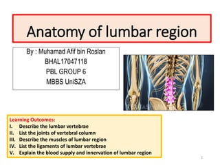
- 1. Anatomy of lumbar region By : Muhamad Afif bin Roslan BHAL17047118 PBL GROUP 6 MBBS UniSZA 1 Learning Outcomes: I. Describe the lumbar vertebrae II. List the joints of vertebral column III. Describe the muscles of lumbar region IV. List the ligaments of lumbar vertebrae V. Explain the blood supply and innervation of lumbar region
- 2. I. Lumbar vertebrae ▪ Body- kidney shaped body ▪ Pedicles- longer and wider than thoracic;oval shaped ▪ Spinous process- prominent posterior projection ▪ Transverse process- projects laterally from each pedicle provide attachment for intrinsic muscle and ligaments ▪ vertebral foramen- opening ▪ Pedicles- sides of vertebral arch ▪ Laminae- flat root plates,complete arch posteriorly 2 Posterior Posterior Anterior Anterior
- 3. II. Joints of vertebral column I. Joints of vertebral body: ▪ Symphyses ( secondary cartilaginous joint) 1. Annulus fibrosus fibrous ring consists of concentric lamellae of fibrocartilage forming circumference of IV disk ▪ Inserted into epiphyseal rims of the articular surface of vertebral bodies 2. Nucleus pulposuscore of the IV disk II. Joints of vertebral arch: 1. Zygapophysial joint(facet jt) ▪ Articulation: plane synovial joint between sup and inf articular process of adjacent vertebra ▪ Surrounded by thin joint capsule ▪ Permits gliding between articular process 3 Anterior Posterior Posterior
- 4. III. Muscles of the lumbar region Group of muscle Name of muscle Action Posterior Thoracolumbar fascia Ant layer- transmit tension produced by contraction of hip extensors to spinous process Post layer- actv by contraction of transversus abdominis Erector spinae: 1. Iliocostalis 2. Longissimus 3. Spinalis - Primary extensor of lumbar region when acting bilaterally - Unilaterally laterally flex the trunk and contribute to rotation Multifundus - Produce lumbar extension Lateral Quadratus lumborum - Stabilization in horizontal plane - Laterally flex the spine and control rotational motion Anterior Rectus abdominis - Flexor of trunk - Provide stability inacorset manner around trunk Abdominal wall - Stabilize lumbo-pelvic region Psoas major - Flexion of hip 4
- 5. POSTERIOR 5
- 6. IV. Lumbar ligaments ▪ Ligamentum flavum join laminae of adjacent vertebral arch ▪ Interspinal ligament connect adjoining spinous process,attaching from root to apex ▪ Supraspinous ligament connects the tip of spinous process from C7 to sacrum ▪ Anterior longitudinal ligament strong broad fibrous band that connects and cover anterolateral aspect of vertebral bodies and IV disc ▪ Posterior longitudinal ligament attached to the IV disc and posterior aspect of vertebra bodies from C2 to sacrum 6
- 7. V. Blood supply and innervation of lumbar vertebrae ▪ Blood supply: - Four paired of lumbar arteries that arise from post aspect of aorta ▪ Nerve supply: - Sinuvertebral nerve major sensory nerve - Innervates: 1. Post longitudinal ligament 2. Superficial layer of anullus fibrosus 3. Posterior vertebral periosteum 7
- 8. THANK YOU REFERENCES: 1. Keith L.Moore ,Arthur F.Daley ,Anne M.R Agur ;Clinically oriented anatomy sixth edition 2. www.slideshare.net/mobile/venus88/biomechanics-of-lumbar- spine 3. Atlas of human anatomy; F Netter 8