Dentin development
•Download as PPTX, PDF•
61 likes•7,355 views

dentin development, composition, physical charecters and chemical properties Dr. Mohana Pratima (MDS)

Recommended
More Related Content
What's hot
Viewers also liked
Viewers also liked (8)
Similar to Dentin development
Similar to Dentin development (20)
Recently uploaded
Recently uploaded (20)
Dentin development
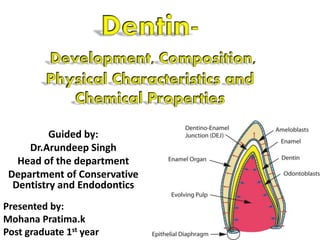
- 1. Guided by: Dr.Arundeep Singh Head of the department Department of Conservative Dentistry and Endodontics Presented by: Mohana Pratima.k Post graduate 1st year
- 2. • Definition. • Stages of tooth deveopment. • Dentinogenesis and mineralization. • Physical and chemical composition. • Types of dentin. • Theories of pain transmission. • Age and functional changes of dentin. • Clinical considerations. Dentinal carious zones Dentinal Hypersensitivity Dentin Bonding Agents Dentinogenesis Imperfecta Dentin Dysplasia Dens Evaginatus
- 3. Def: • A calcareous material similar to bone but harder and denser that composes the principal mass of a tooth, is formed by the odontoblasts of the surface of the dental papilla, and consists of a matrix containing minute parallel tubules which open into the pulp cavity and during life contain processes of the cells of the pulp - Merriam Webster • The formation of dentin, known as dentinogenesis begins prior to the formation of enamel and is initiated by the odontoblasts of the pulp. • Unlike enamel, dentin continues to form throughout life and can be initiated in response to stimuli, such as tooth decay or attrition.
- 6. Early bell stage Advanced bell stage Cervical loop
- 7. Dentinogenesis: • Begins at the cusp tip after odontoblasts have differentiated and begin collagen production • In odontoblasts differentiation, fibronectin, decorin, laminin, chondroiton sulfate may be involved. • Laminin alpha 2, subunit of laminin is essential for odontoblastic differentiation and to regulate the expression of dentin matrix proteins. • Factors like TGF, IGF, BMP help in organisation of odontoblast important for relocation of organelles. Orbans histology and embryology
- 8. • One of the key proteins involved in mineralization and secreted by the odontoblast is the Dentin phosphoprotein(DPP). • Binds to calcium, transports it to the mineralization front and controls the growth of apatite crystals. • Osteonectin: inhibits growth of apatite crystals, but promotes binding in collagen. • Osteopontin: a phosphoprotein, promotes mineralization. • Gla protein: acts as seeds or nucleators to attract and concentrate calcium. • Matrix vesicles are also involved in mineralization of mantle dentin. Orbans histology and embryology
- 9. • Initially daily increments of approximately 4um of dentin are formed. • This continuous until the crown is formed and the teeth erupt and move into occlusion. • Dentinogenesis is a two-phase sequence in that collagen matrix is first formed and then calcified. • Kroff’s fibers described the initial dentin deposition along the cusp tips. • Radicular dentin formation is slower when compared to coronal dentin, less mineralized with collagen fibers laid down parllel to CEJ. Orbans histology and embryology
- 11. Mineralization: • Earliest crystal deposition is in form of very fine plates of hydroxyapatite on the surfaces of the collagen fibrils and in the ground substances. • The apatite crystals found in dentin resemble those found in bone and cementum. Orbans histology and embryology
- 12. Provides the bulk and general form of tooth. Determines the shape of the crown. Physically & chemically the dentin closely resembles the bone. The main morphologic difference between bone & dentin is that some of the osteoblasts that form bone marrow enclosed within its matrix substance as osteocytes, whereas the dentin contains only the processes of the cells that form it. Orbans histology and embryology
- 13. Physical and chemical properties. Physical properties: • Teeth of young individuals- light yellowish in color, darkens with age. • Dentin is slight viscoelastic and is subjected to slight deformation. • Harder than bone but softer than enamel. • Dentin of primary teeth is less hard then permenant teeth. Chemical properties: • The lower content of mineral salts render it more radiolucent than enamel. It consistes of 35% of organic matter, water and 65% inorganic matter. Both are considered vital tissue because they contain because they contain living protoplasm. Orbans histology and embryology
- 14. Inorganic component: • Collagenous fibrils. • TypeI collagen is principal type of collagen found in dentine. • The protein of dentin matrix and bone are similar , but dentin sialoprotein and dentin phosphoprotein are present only in dentin. • Matrix also contains growth factors like TGF, FGF, IGF-s, BMP-s. • Matrix also plays an important role in dentin mineralization. • Hydroxyapatite. •Also contains small amounts of phosphates, carbonates, sulfates. • Organic and inorganic are substances can be seperated by either decalcification or inceneration Organic substances: Orbans histology and embryology
- 15. Structure • Tubules are found throughout normal dentin and are therefore its charectestic Dentinal tubules: • The course of dentinal tubules follows a gentle curve in the crown, so less in the root where it resembles gentle S shape. • These curvature are called primary curvatures. • Branches of the dentinal tubules near the terminals are referred to as terminal branches. • Over the entire lengths the tubules exhibit the minute, relatively Secondary curvature that are sinusoidal in shape Orbans histology and embryology
- 18. • The dentinal tubules have lateral branches throughout dentin, which are termed Canaliculi or microtubules. • A few dentinal tubules extend through the dentinoenamel junction into the enamel for several millimeters termed as enamel spindles. Orbans histology and embryology
- 19. Peritubular dentin: • The dentin that surrounds immediately the dentinal tubules is termed as peritubular dentin. • This dentin forms the walls of the tubules in all but the dentin near the pulp. • Highly mineralized then the dentin present between tubules. • Since the deposition occurs in the inner wall of the tubule rather then the outer wall, is called “intratubular dentin”. • The calcified tubules wall has inner organic lining termed the lamina limitans. • Between the odontoblastic process and the peritubular dentine, a space known as periodontoblastic space. • This space contains dentinal fluid. Orbans histology and embryology
- 21. Periodontoblastic space Odontoblastic process(tomes fibers) Dentinal tubules
- 22. Intertubular dentin: • The main body of dentine is composed of intertubular dentin. • Although it is highly mineralized, this matrix, like bone and cementum, is retained after decalcification. Predentin: • Located always adjacent to pulp tissue and is 2 to 6 um wide, depending on the extent of activity of odontoblast. • Not mineralized. • As the collagen fibers undergo mineralization at the predentin-dentin junction, the predentin becomes dentin and a new layer of predentin forms cicumpulpally. Orbans histology and embryology
- 24. Odontoblastic process: • Are the cytoplasmic extensions of the odontoblasts. • The odontoblast cells reside in the peripheral pulp at the pulp-dentin border and their process extend into dentinal tubules. • The process are larger in diameter near the pulp and taper further into dentin. • The odontoblast process divide near the DEJ and may extend into the enamel in the enamel spindles. Orbans histology and embryology
- 26. Types of dentine: Primary dentin Mantle dentin : • The name of the first-formed dentine in crown underlying DEJ. • Below DEJ is soft and provides cushioning effect to the tooth. • The fibrils are perpendicular to DEJ and organic matrix composed of large collagen fibrils. • The larger diameter collagen fibers are argyrophilic and are known as von Kroff’s fibers. • They contain mainly type III collagen. Circumpulpal dentin: • Forms the remaining primary dentin or bulk of the tooth. • Dentin that represents all the dentin formed before root completion. • May conatin slightly more mineral than mantle dentin. Orbans histology and embryology
- 27. Enamel Mantle dentin Circumpulpal dentin Irregular secondary (tertiary) dentin regular secondary dentin
- 30. Secondary dentin: • Is a narrow band of dentin bordering the pulp and representing that dentin formed after root completion. • This dentin contains fewer tubules than primary dentin. • Not formed uniformly and appears in greater amounts on the roof and floor of the coronal chamber, where it protects the pulp from exposure in older teeth. • Due to regular arrangement of dentinal tubules it is known as regular secondary dentin. Orbans histology and embryology
- 31. An accentuated calcio-traumatic band under the cavity represents an acute arrest in odontoblastic activity as a result of the operative procedure
- 32. Tertiary dentin: • It is reparative, response or reactive dentin. • This is localized formation of dentin on the pulp- dentin border, formed in reaction to trauma such as caries or restorative procedures Orbans histology and embryology
- 33. Incremental lines: • Appears as fine lines or striations in dentin. • These lines represent the daily rhythmic recurrent deposition of dentin matrix as well as a hesitation in the daily formative process. • The course of lines indicates the growth pattern of the dentin. • Incremental lines are accentuated because of disturbances in the matrix and mineralization process called contour lines(Owen) Orbans histology and embryology
- 34. • In deciduous teeth and in 1st permanent molars, where dentin is partly before and partly after birth, the prenatal and postnatal dentin are separated by an accentuated contour line termed as neonatal line seen in enamel and dentin. Orbans histology and embryology
- 35. Owencontourlines
- 36. Neonatal line
- 37. Interglobular dentin • Sometimes mineralization of dentin begins in small globular areas that fail to coalesce into homogenous mass. Results in zones of hypomineralization between the globules. • These zones are called globular dentin or interglobular space. Orbans histology and embryology
- 39. Granular layer: • When dry ground sections of the root dentin are visualized in transmitted light, a zone adjacent to cementum appears granular known as tomes’ granular layer. • This areas remain unmineralized like interglobular dentin. • Though Tomes granular layer and interglobular dentin have similarities in formation but differ in mineral content. • Tomes granular shows high calcium and phosphorous , while interglobular dentin showed higher content of sulfur. Orbans histology and embryology
- 40. Granular layer of Tomes Dentin (with tubules) cementum
- 41. Granular Layer of Tomes dentin cementum enamel
- 42. Innervation of dentin: • Nerve fibers shown to accompany 30% to 70% of odontoblastic process and these after referred to as intertubular nerve. • Synapse like relation between the process and nerve fibers were demonstrated. • These are the terminal process of myelinated nerve fibers if the dental pulp. Orbans histology and embryology
- 43. Direct Neural Stimulation •Which the nerves in dentin get stimulated. •The nerves in the dentinal tubules are not commanly seen and even present they do not extend beyond the inner dentin. Hydrodynamic theory •This fluid movement, either inward or outward stimulates pain mechanism in the tubules by the mechanical disturbances of the nerves closely associated with the odontoblasts and its process. •These endings may act as mechanoreceptors. Transduction theory •Presumes that odontoblast process is the primary structure excited by the stimulus and that impulse is transmitted to the nerve endings in the inner dentin. •No neurotransmitter vesicles in the odontoblast process to facilitate the synapse or synaptic specialization. Orbans histology and embryology
- 44. Permeability of dentin • Depends upon the patency of dentinal tubules. • Tubular occlusion, smear layer formation and lack of tubular communication between primary and irregular secondary dentin will result and reduce the permeability. • Reduction in dentin permeability would lessen the sensitivity of dentin. Orbans histology and embryology
- 45. Age and functional changes Vitality of dentin: • Dentin is laid down throughout the life although the teeth are erupted and functioning for a short time, dentinogenesis slows and further dentin formation is at much slow rate. • Pathologic effects of dental caries abrasion, attrition, or cutting of dentin of operative procedures cause changes in dentin. They are described as development of dead tracts, sclerosis and the addition of reparative dentin Orbans histology and embryology
- 46. Reparative dentin: • If by extensive abrasion, erosion, caries or operative procedures the odontoblastic process are exposed or cut, the odontoblasts die or survive. • If they survive the dentin that is produced is called reactionary or regenerated dentin. • Origin of new odontoblasts is from cells of cell rich zone . • The irregular nature of dentinal tubules, these types of dentin are referred as irregular secondary dentin. • It is believed that bacteria, living or dead or their toxic products as well as chemical substances from restorative materials migrate down the tubules to the pulp and stimulate pulpal response leading to reparative dentin formation Orbans histology and embryology
- 48. Dead tracts: • In dried ground sections of normal dentin the odontoblasts process disintegrate, and the empty tubules are filled with air. • They appear black in transmitted light and white in reflected light. • Where reparative dentin seals dentinal tubules at their pulpal ends are filled with fluid or gaseous substances. • These areas demonstrate decreased sensitivity and appear to a greater extent in older teeth. • These dead tracts are probably the initial step in the formation of sclerotic dentin. Orbans histology and embryology
- 50. Sclerotic dentin: • Also transparent dentin. • Stimuli not only induce additional formation of reparative dentin but also, lead to protective changes in the existing dentin. • The refractive indices of dentin in which tubules occluded are equalized, and such areas become transparent. • Seen mostly in elderly people especially in roots. And also in slow progression caries. • Mineral density is greater in this area of dentin. • Sclerotic dentin was harder than normal dentin, its fracture toughness reduced. • Tranparent - transmitted light • Dark- reflected light. Indicates location of dentin sclerosis Orbans histology and embryology
- 52. Zones of dentinal carious lesion Normal dentin: Deepest zone of carious dentin is normal with normal collagen, odontoblastic process and intertubular dentin. Subtransparent dentin: Intertubular dentin is demineralized, odontoblastic process are damaged. But no bacteria is found. Transparent dentin: Superficial to subtransparent. Softer than normal dentin. No bacteria so this layer is capable of remineralization. Turbid zone: Dentinal tubules are widened and distorted by bacterial penetration. Incapable of remineralization and must be removed before restoration. Operative dentistry-ramya raghu
- 53. Infected dentin: Decomposed dentin with destruction of dentinal tubules and collagen. High concentration of bacteria. Removed to prevent spread of infection. Narendranath operative dentistry and endodonticsOperative dentistry-ramya raghu
- 54. • Infected dentine is the outer layer and is softened and contaminated with bacteria. It is irreversibly denatured and not remineralized • Affected dentine has a demineralized phase, but not yet invaded by bacteria. It can be remineralized. • In clinical restorative treatment of dentine during cavity preparation it is infected dentine which is completely removed. The affected dentine, which may be remineralized after the completion of restorative treatment, is not removed and is preserved. 5 Affected & infected dentin Narendranath operative dentistry and endodontics
- 55. Caries-affected dentin interfaces showed a much thicker hybrid layer than was seen with non-carious dentin. Presumably, the thicker hybrid layer in caries- affected dentin may be due to the fact that caries-affected dentin is partially demineralized and more porous than non-carious dentin. Allow deeper penetration of the acid etchant, leading to a deeper demineralization with diffused monomer. 5 5Narendranath operative dentistry and endodontics
- 56. • The cells exposed dentin should not be insulted by bacterial toxins, strong drugs, undue operative trauma unnecessary thermal changes or irritating restorative materials • Rapid penetration and spread of caries in the dentin is the result of the tubule system in the dentin. • The enamel may be undermined at the DEJ, even when caries in the enamel is confined to a small surface area. • This is due to in part to the spaces created at the DEJ by enamel tufts, spindles and open and branched dentinal tubules. • The dentinal tubules provide a passage for invading bacteria and their products through either thin or thick dentinal layer. Orbans histology and embryology
- 57. Dentinal Hypersensitivity • Def: Short, sharp pain arising from exposed dentin in response to stimuli typically thermal, evaporative, tactile, osmotic or chemical and which cannot be ascribed to any other form of dental defect or pathology. Etiology: Enamel loss: • Occlusal wear • Tooth brush • Dietary erosion • Abfraction • Parafunctional habits Operative dentistry-ramya raghu
- 58. Cemental loss: • Gingival recession • Periodontal disease • Root planning • Periodontal surgery Operative dentistry-ramya raghu
- 59. Mechanism of pain transmission- theories of dentin hypersenstivity: • The exact mechanism is not very clear but several theories have been proposed to explain this phenomenon. Operative dentistry-ramya raghu
- 60. Nerve fibers are present within the dentinal tubules initiate impulses when they are injured and this causes dentinal hypersenstivity. Nerve fibers are present only in the predentin and inner dentinal zones but do not extend all through the DEJ. These nerves are also absent in areas like root dentin which is also very senstive. Substances like potassium chloride, acetylcholine and histamine are applied to exposed dentin they fail to elicit a painful response Direct innervation theory: Operative dentistry-ramya raghu
- 61. Odontoblast deformation/Transducer mechanism: Odontoblasts or their processes are damaged when external stimuli are applied to exposed dentin. Odontoblastic processes extend only partly through the dentin and not to upto DEJ. And also odontoblast membrane potential is too low to permit transduction. There were no demonstrable neurotransmitters like acetylcholine in the neural transmission of the pulp Operative dentistry-ramya raghu
- 62. Hydrodynamic theory Dentinal tubules are filled with dentinal fluid which is the intercellular fluid of the pulpal connective tissue. In the vital tooth there is constant slow flow of the fluid outward through the dentinal tubules Whenever exposed dentin is stimulated by tactlie, chemical, thermal or osmotic stimuli there is rapid movement of the dentinal fluid toward the pulp or outward. This can cause: Direct stimulation of the low threshold A-delta nerve fibers in the pulp. Indirect stimulation of A-delta nerve fibers in the pulp by displacing the odontoblastic cell bodies. Such rapid displacement of dentinal fluid in thousands of dentinal tubules at the same time produces a cumulative effect and this causes hypersenstivity. Operative dentistry-ramya raghu
- 64. Clinical features: • Short, sharp pain response to heat, cold, tactile stimmuli, sweet or sour foods. • Considered to be exagerated response of normal-pulp dentin complex and is not felt on application of external stimulus. Management: Desensitization by occluding the dentinal tubules. Desensitization by blocking pulpal sensory nerves. Operative dentistry-ramya raghu
- 65. • Use of topical agents to occlude dentinal tubules. a. Calcium hydroxide b. Calcium phospahte pastes c. Silver nitrate d. Strontium chloride e. Fluorides f. Fluoride iontophorosis g. Potassium oxalate h. Varnishes i. Dentin adhesives j. Placement of restorations k. Use of lasers Desensitization by occluding the dentinal tubules: •Formation of smear layer over exposed dentin. Operative dentistry-ramya raghu
- 66. Desensitization by blocking pulpal sensory nerves. a. Potassium nitrate toothpastes: Recommended treatment options for managing dental hyopersenstivity should be based on extent and severity of problem. At first conservative options should employed. If considerable dentin exposure is present restorations must be placed. In generalized hypersenstivity desensitizing tooth pastes must be employed. In case of severe hypersenstivity endodontic therapy may be necessary as the final option. Operative dentistry-ramya raghu
- 67. Bonding to dentin Bonding to dentin is difficult due to: • Dentin is dynamic tissue that shows changes due to aging, caries or restorative procedures. • Considerable amount of organic material and water. • Dentinal tubules are filled with dentinal fluid . • Cut dentinal surface is covered by smear layer • Dentin is in close proximity to the pulp so different chemical used for eching and dentin bonding may irritate pulp. Therefore, dentin is inherently a hydrophilic tissue and bonding of hydrophobic resins is a challenge. Operative dentistry-ramya raghu
- 68. Developmental in dentin adhesives: • Earlier attempts at bonding were directed towards producing chemical bond between resin system either to organic or inorganic component if dentin. • An important break through in dentin bonding was the development of “total etch concept” • Demonstrated that etching both enamel and dentin with 37% phosphoric acid did not cause any pulpal damage and Improved the retention of composite resins in dentin. Operative dentistry-ramya raghu
- 69. • Another significant development was the hydrophilic resins. • Application of these resulted in resin infilteration of the exposed collagen mesh to form a “hybrid layer” of resin-infiltered dentin. • This was responsible for micromechanical bonding to dentin. Operative dentistry-ramya raghu
- 70. Dentin agents bonding to inorganic components: • Phosphate group • Amino group Dentin bonding agents bonding to organic component of dentin. • Isocynate group • Carboxylic acid group Operative dentistry-ramya raghu
- 71. Dentinogenesis imperfecta: • Hereditary developmental disturbances of dentin in absence of any systemic disorder. Formely were divided into • Dentinogenesis imperfecta (Shields I) • Hereditary opalescent dentin(Shields type II) • Brandywine isolate(Shields type III). Oral & maxillofacialpathology-Neville
- 72. Clinical features: •All teeth in both the dentition are affected. •Deciduous teeth are affected most severely, followed by permanent incisors, 1st molars, 2nd and 3rd molars being least. •Blue to brown discoloration often with a distinctive translucence •Radiographically teeth have bulbous crowns, cervical constriction, thin roots, and early obliteration of root canals and pulp chambers. •Shell teeth –normal enamel, extreme thin dentin and enlarged pulps. Oral & maxillofacialpathology-Neville
- 73. Management: • The root canals become thread like and may develop microexposures, resulting in periapical inflammatory lesions. • Risk of enamel loss and severe attrition . • Not good candidates for full crowns because of cervical fracture. • The newer composites combined with a dentin bonding agent have been used in areas subjects to occlusal wear. Oral & maxillofacialpathology-Neville
- 74. Dentin dysplasia: • Two major patterns type I and type II. • Type II is closely related to dentinogenesis imperfecta. Oral & maxillofacialpathology-Neville
- 75. Clinical features: Dentin dysplasia type I: • Referred to as rootless dentin • Coronal dentin and enamel is normal and well formed but radicular dentin loses all organization • Wide variation of root organization is produced because of dentinal disorganization. • Shortened roots, extreme mobility and premature exfoliation. • Radiographically deciduous tooth are often severely effected with little or no detectable dentin. Oral & maxillofacialpathology-Neville
- 76. Dentin dysplasia type II: • Coronal dentin dysplasia root length is normal in both dentition. • Blue-to-amber-to-brown • Radiographically bulbous crowns, cervical constriction, thin roots, early obliteration of pulp • Pulp chambers exhibit significant enlargement and apical extensions. • Altered pulpal anatomy thistle tube shaped or flame shaped Oral & maxillofacialpathology-Neville
- 77. Management • Preventive care is fore most important. • If periapically inflammatory lesions develop, the root length guides the therapeutic choices. • Conventional endodontic therapy requires mechanical creations of pulp paths. • Pulp canals are not obliterated endodontic therapy can readily be done. Oral & maxillofacialpathology-Neville
- 78. Dens Evaginatus • Cusp like elevation of enamel located in central groove or lingual ridge of the buccal cusp of premolar or molar. • Normally consists of enamel and dentin with pulp present in half of cases. • Radiographically the occlusal surface exhibits a tuberculate appearance and often pulpal extensions is seen in the cusp. • Often seen in association of Shovel shaped incisors • Typically results in occlusal problem and often leads to pulpal death. • In affected tooth removal of cusp is often indicated
- 79. Refernces • ORBANS HISTOLOGY AND EMBRYOLOGY-12TH EDITION. • RAMYA RAGHU CLINICAL OPERATIVE DENTISTRY-2nd EDITION. • ORAL AND MAXILLOFACIAL PATHOLOGY-NEVILLE. 3rd edition • NARENDRANATH REDDY OPERATIVE DENTISTRY AND ENDODONTICS-2nd EDITION