Nightside clouds and disequilibrium chemistry on the hot Jupiter WASP-43b
BIOL 306L Poster Project 11-28-13 FINAL
1. Smaller Cell Size in Chlamydomonas reinhardtii:
rls1 Mutant to Blame?Damaris Christelle Ngantche Mala, Julie Rost, Mintong Nan, Elisa McGrath-Martinez, Department of Biology, University of Maryland Baltimore County, Catonsville, MD
Introduction
Abstract
By studying the unicellular, Chlamydomonas reinhardtii, we
were able to gain vital information into how multicellularity
evolved. Our goal was to investigate the function of rls1
Tylen Darling (TD) by examining the phenotype of the gene
knockdown in Chlamydomonas reinhardtii. To do this, we
inserted an artificial microRNA sequence, rls1 TD, into our
plasmid in order to knockdown the rls1 gene. The genome
was then amplified so we could more easily observe and
measure cell size to investigate the effect on the phenotype.
From our investigation, we found that Chlamydomonas
reinhardtii cells containing the rls1 TD knockdown, is smaller
in size, compared to those of empty vector control cells.
Results
Discussion
During our research, we constructed an artificial microRNA
(rls1-knockdown (KD)) to turn off the expression of the rls1 gene
in Chlamydomonas reinhardtii. We were interested in identifying
the effect of the rls1 gene on the size of the cell during the
different stages of cell division.
The rls1-KD gene was cloned in E. coli, followed by PCR and
gel electrophoresis to identify which of the colonies had the rls1-
KD mutant inserted into them before transforming Chlamy. We
then inserted our rls1-KD into C. reinhardtii cells by performing a
Chlamy glass bead transformation, selective media was used to
confirm the Chlamy transformation. Once the colony was
identified, we cloned it and measured the diameter of the Chlamy
cells. Chlamy enters the exponential phase after 5 days of
growth, which is the most efficient time to measure the size and
rate of growth of these cells. To know whether there was a
difference in cell size, we compared them with the wild type cells
that contained an empty vector (as shown in Table 1). We then
conducted various statistical tests, including the t-test and the
one-way analysis of variance, in order to find any other relativities
or differences between the two. We discovered that the cell sizes
were highly significantly different by observing the p-value during
the t-test, which was less than 0.0001. Therefore, we concluded
that there is significant evidence that the rls1-KD mutant does
indeed affect the size of the cell during cell division.
In the future, we could run a northern blot to detect RNA size
and subtle changes in gene expression of the rls1-KD mutant
gene with that of the EV control gene (2).
References
1. Claassen LA. 2013. Syllabus Biology 306L-Projects in Molecular Biology, Fall 2013.
University of Maryland, Baltimore County. p. 1-16.
2. Gallagher SR, Wiley EA, editors. 2012. Current Protocols: Essential Laboratory
Techniques Hoboken, NJ: Wiley and Sons, Inc.
In the fall 2012
semester, students taking
BIOL 306L continued the
project started in the
laboratory of Dr. Stephen
Miller by former students,
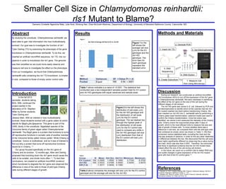
Tylen Darling and
Figure 1 to the
left shows the
average cell size
(µm) for the EV
genotype and the
rls1-KD
genotype. The
error bars for
each genotype
represent the
standard error.
Table 1 above indicates a p-value of <0.0001. The statistical test
conducted was a two-independent samples pooled t-test for EV control
and rls1-KD genotypes with equal variances and sample sizes.
Table 2 above compares the average cell size (µm) for the EV control
genotype and the average cell size for the rls1-KD.
Figure 2 to the left shows the
distribution of cell sizes (µm)
for the rls1-KD genotype and
the distribution of cell sizes
(µm) for the EV control
genotype. The red dashed line
through the highest peak of
the distribution of cell sizes
(µm) for the EV control was
used to compare any shifts in
the rls1-KD genotype cell size
(µm) distribution from that of
the EV control cell size (µm)
distribution.
Table 2
Figure 2
Figure 1
6. Create Subculture
5. DNA
Sequencing
1. Insert rls1 TD
4. DNA Elution
7. Measure Cell
Size Using Microscopy
3. Gel Electrophoresis
2. GeneJet PCR
Methods and Materials
Jessica Allen. With an interest in how multicellularity
evolved, these students worked with a gene called rls which
stands for RegA-Like-Sequence. This gene is part of the
VARL family in the unicellular, flagellated species of the
Volvocine family of green algae called Chlamydomonas
reinhardtii. The RegA gene is a protein that functions to turns
off reproductive functions in somatic cells of another member
of the Volvocine family called Volvox carteri. While Chlamy is
unicellular, Volvox is multicellular, and so it was of interest to
find out why a protein that turns off reproductive functions
would be in Chlamy. (1)
Our group focused specifically on the rls1 gene of
Chlamy and its function. 12 months ago, Allen and Darling
proposed that knocking down the rls1 gene would cause the
cells to be smaller, and divide more often (1). To test their
conclusions, we created an artificial microRNA construct
which functioned to degrade the rls1 gene and observed the
difference in size of the cells to those of wild type Chlamy
cells during different stages of growth.
Table 1