Novel Tyrosine Kinase Inhibitors for Ovarian Cancer
Mike Richardson - CVMBS Research day Poster - Draft Final
1. EFFECT OF 2-AMINOIMIDAZOLE COMPOUND 2C8
ON ADVANCED GLYCATION END-PRODUCTS
Mike A. Richardson, Brendan K. Podell, David F. Ackart, Cathy Bush, Roberta Melander, Christian Melander, Randall J. Basaraba
1Colorado State University, Department of Microbiology, Immunology and Pathology, Fort Collins, CO 2North Carolina State University, Department of Chemistry, Raleigh, NC
Background
• Advanced Glycation End-Products (AGEs) accumulate as a consequence of
uncontrolled hyperglycemia, aging and chronic inflammatory diseases.
• AGEs have been linked to numerous diabetic complications: retinopathy,
nephropathy, neuropathy, cataracts, and atherosclerosis1.
• Diabetic models concurrently infected with Mycobacterium tuberculosis (Mtb)
accumulate more AGEs due to chronic inflammation and chronic hyperglycemia.
• Currently, there are no approved AGE inhibitory compounds for humans; however, the
current gold-standard for AGE inhibition is Aminoguanidine (AG).
• High throughput screening of a library of 2-Aminoimidazole (2-AI) compounds has
identified one compound, 2c8, that has the ability to inhibit and break AGEs.
Methods
• Screening for AGE Inhibition & Breaking
• Bovine serum albumin (BSA) or bovine collagen type IV was incubated with
methylglyoxal (MGO) with or without the addition of an inhibiting compound
for 7, 14 and 28 days at 37°C.
• Samples were taken daily up to day 7, and weekly thereafter; samples were
then stored in -80°C until final analysis.
• AGE formation and inhibition was quantified by fluorescence
spectrophotometry at wavelengths λexc 370nm; λem 440nm for vesperlysine
AGEs and λexc 335nm; λem 385nm for pentosidine AGEs; quantified as
percent inhibition.
• Breaking – BSA or Collagen in the presence of MGO were allowed to
incubate for 7 days, forming AGEs. An aliquot was removed and 400uM of
inhibitory compound added. Both were allowed to incubate for an additional
24 hours, and readings taken at λexc 370nm; λem 440nm and λexc 335nm;
λem 385nm wavelengths. Quantified as percent change against the negative
control.
• Functional Inhibition Assay
• DQ Collagen, purchased from Life Technologies, is bovine type 1 collagen,
which is heavily labeled with fluorescein causing fluorescence
to be quenched.
• The addition of a collagenase breaks up the collagen, allowing it to fluoresce
at FITC wavelengths.
• DQ Collagen was incubated with MGO at 37°C with or without the addition of
an inhibitory compound for 14 days.
• Collagenase A was added to wells, incubated for 3 hours, and subsequently
read at λexc 490nm; λem 525nm.
• Percent change was then calculated.
• In-vivo granulomatous lesion comparison
• Cohorts of guinea pigs were divided into two major groups, hyperglycemic
and non-hyperglycemic, and further delineated into Mtb infected and non-
Mtb infected.
• Streptozotocin (STZ) was used to induce hyperglycemia.
• Lungs were taken for histopathology and stained with Masson Trichrome.
• Collagen surrounding granulomatous lesions was quantified via Nikon
Elements.
• Mtb lesion immunohistochemistry (IHC)
• Naïve guinea pigs infected with aerosolized Mtb.
• Sections of lung tissue taken on day 30 post infection.
• Anti-AGE (1:1000 Abcam Rb polyclonal anti-AGE) antibody was labeled with
a secondary (1:1000 Life Tech Goat Polyclonal anti-Rb Alexa Fluor 488) to
fluoresce green
• DNA fluoresce blue (1:5000 Hoechst)
• Mtb fluoresce red (1:20 Rhodamine B)
1. Ahmed N. “Advanced glycation endproducts--role in pathology of diabetic complications.” Diabetes Res Clin Pract. 2005 Jan;67(1):3-21.
2. Rachman H, Kim N, Ulrichs T, Baumann S, Pradl L, et al “Critical Role of Methylglyoxal and AGE in Mycobacteria-Induced Macrophage
Apoptosis and Activation.” PLoS ONE 2006 1(1): e29. doi:10.1371/journal.pone.0000029
Acknowledgements & References:
Conclusions
• The 2AI formulation 2c8 was found to be more effective than AG at inhibiting AGE formation.
• 2c8 was able to break 7-day pre-formed AGEs with 50% efficacy, however, 28-day pre-
formed AGEs proved more difficult to disrupt with one solitary 400uM dose.
• The presence of 2c8 restores the enzymatic functionality of Collagenase in a collagen
sample allowed to glycate for 14 days.
• Hyperglycemic animals concurrently infected with Mtb show impaired collagen deposition,
ostensibly due to excessive AGE accumulation systemically and at the granulomatous lesion.
• Non-diabetic controls, also infected with Mtb, show granulomatous lesions with organized
collagen formation and breakdown, when compared to diabetic controls.
• Tissue remodeling is an important part of the innate immune response to Mtb infection,
collagenase takes part in this remodeling by cleaving procollagen into collagen and allowing
the competent deposition of a collagen matrix. Our evidence suggests that glycation of
procollagen and collagen disrupts the organized deposition of collagen around a tuberculous
lesion.
• Further testing is necessary to illustrate repeatability.
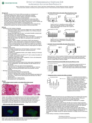
• Collagenase activity is retained when AGEs are inhibited
• 2c8 did not break 28-day pre-formed AGEs with the same efficacy as 7-day
Figures 8 & 9 (n=1): 24-hour incubation of inhibitory compound with a glycated sample revealed
the breaking ability of 2c8 and AG. In this instance, 2c8 was able to break 7-day pre-formed
AGEs with much greater efficacy when compared with AG, breaking pentosidine specific AGEs
with 50% or greater efficacy in BSA and Collagen.
Figure 10 (n=1): BSA allowed to glycate with MGO for 28
days proved more difficult to break after a 24-hour
incubation with 400uM of 2c8. Suggests a time-dependent
factor in 2c8’s ability to break pre-formed AGEs at 400uM.
• 2c8 breaks 7-day pre-formed AGEs with greater efficacy than AG
Figure 11 (n=1): Illustrates the functional effect of
glycation on enzymatic activity through collagen and
collagenase interaction. Native DQ collagen
incubated with collagenase for 3 hours shows a
large increase in RFUs when compared to a
glycated sample, suggesting that glycation of
collagen impedes collagenase activity. DQ collagen
allowed to glycate for 14 days in the presence of
2c8, also shows a large increase in RFUs when
compared with controls, further suggesting 2c8’s
anti-AGE activity. Only one concentration, 400uM,
was chosen for this assay. Further study is
necessary to assess the apparent inhibition of
collagenase by 2c8 and AG alone.
Glycated BSA spiked for 24hr
A
G
400uM
2c8
400uM
0
10
20
30
40
50
60
70
80
90
100
370:440
335:385
PercentChange
Glycated Collagen spiked for 24hr
A
G
400uM
2c8
400uM
0
10
20
30
40
50
60
70
80
90
100
370:440
335:385
PercentChange
2c8
400uM
0
1
2
3
4
5
370/440
335/385
28-day Glycated BSA spiked for 24hr
PercentChange
14-day Glycated DQ Collagen
C
ollagen
G
lycated
C
ollagen
C
ollagen
-2c8
400uM
C
ollagen
-A
G
400uM
G
lycated
C
ollagen
-2c8
400uM
G
lycated
C
ollagen
-A
G
400uM
0
5000
10000
15000
Enzymatic Activity
RFUs(490/525)
• 2c8 inhibits AGE formation with greater efficacy than AG over 7 days
Figures 5 and 6 (n=1): Quantified as percent inhibition, 2c8
was found to inhibit AGE formation in a dose-dependent
fashion. 400uM inhibited AGE formation 50% or greater at
almost all time points.
Figures 6 and 7(n=1): Represented in RFU’s, 2c8 is more
efficacious at inhibiting the formation of pentosidine specific
AGE formation in collagen.
Glycated Collagen w/ 2c8 - 335/385
0 1 2 3 4 5 6 7
0
500
1000
1500
400uM
40uM
4uM
Day
RFUs
Glycated Collagen w/ AG - 335/385
0 1 2 3 4 5 6 7
0
500
1000
1500
400uM
40uM
4uM
Day
RFUs
Glycated BSA w/ 2c8 - 370/440
1 2 3 4 5 7
0
10
20
30
40
50
60
70
80
90
100
400uM
40uM
4uM
Day
PercentInhibition
Glycated BSA w/ AG - 370/440
1 2 3 4 5 7
0
10
20
30
40
50
60
70
80
400uM
40uM
4uM
Day
PercentInhibition
• AGEs accumulate within tuberculous granulomas
Figures 3 and 4: IHC reveals AGE deposition localized in the necrotic center of a granulomatous
lesion, a direct results of the chronic inflammatory state induced by Mtb. Within the necrotic
center, MGO derivatives and macromolecules have been noted2.
Results
• Lesion collagen content is greater in non-diabetic Mtb infected animals
Figures 1 and 2: Masson Trichrome stained lesions from Mtb infected non-diabetic and diabetic
animals. The representative non-diabetic lesion depicts a well organized granulomatous lesion
with a consistent collagen matrix (in blue). The representative diabetic lesion illustrates the
negative effects of prolonged hyperglycemia on normal granuloma formation. When compared to
the non-diabetic control, the diabetic lesion has a necrotic center that is far more expansive,
macrophages are seemingly unorganized and a consistent collagen matrix is not present.
Non-Diabetic Diabetic
20x 20x