
Recommended
More Related Content
What's hot
What's hot (20)
Similar to Proton vda brochure
Similar to Proton vda brochure (20)
Recently uploaded
Recently uploaded (20)
Proton vda brochure
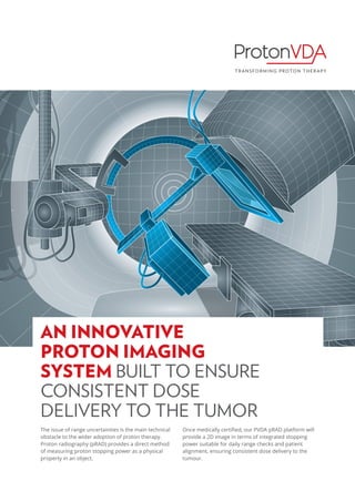
- 1. The issue of range uncertainties is the main technical obstacle to the wider adoption of proton therapy. Proton radiography (pRAD) provides a direct method of measuring proton stopping power as a physical property in an object. Once medically certified, our PVDA pRAD platform will provide a 2D image in terms of integrated stopping power suitable for daily range checks and patient alignment, ensuring consistent dose delivery to the tumour. AN INNOVATIVE PROTON IMAGING SYSTEM BUILT TO ENSURE CONSISTENT DOSE DELIVERY TO THE TUMOR
- 2. KEY FEATURES High reliability. Practical. Easy to implement in the clinical workflow. User friendly software for pRAD analysis and viewing EXAMPLES OF PROTON IMAGES AP VIEW OF PAEDIATRIC HEAD PHANTOM (LEFT). LATERAL VIEW OF PEDIATRIC HEAD PHANTOM (RIGHT) Detector systems with a mounting system for horizontal beams. • Detector positions are adjustable along the beam direction. • System geometry allows for existing beam’s-eye and horizontal x-ray systems to be used for patient alignment. • One tracker on each side of the patient, with a range detector downstream of the patient. Protons are tracked and measured individually for optimal spatial and range resolution with reduced dose to the patient relative to x-ray imaging. • Fast operational and calibration check. • Range calibration tied to facility range calibration. • Image registration to isocenter tied to facility PBS steering calibration. Automatic Image Reconstruction • Software is fully automated from the beginning: data acquisition to displaying an image. • Alignment correction algorithm automatically detects and compensates for shifts in tracker geometry.
- 3. TECHNICAL SPECIFICATIONS Excellence in engineering. Fast, lightweight, integrated. for pRAD analysis and viewing Our proton radiography system is based on using protons to image the patient before treatment. Our detectors produce an image from position and energy measurements of protons with enough energy to traverse the patient. Compared to previous projects focused on proton imaging, our design is simple, monolithic, can be easily scaled to large field sizes, and is an order of magnitude faster. Our advantage lies in our insight that proton imaging can be fast, light-weight, and seamlessly integrated into proton therapy systems. System is designed to support pCT with SW and Mechanical System Upgrade. Field size of range detector 1 40 × 40 cm 2 Event rates of data acquisition system (Readout rate) 3 MHz Data acquisition plus reconstruction time for a single 2D image 1 min Residual Range calibration accuracy 1 mm Sensitivity to range shifts comparing two pRAD images 0.3 mm Range precision per pixel 0.5 mm * Image resolution 2 5 l.p. / cm Pixel size for image 0.5 or 1 mm Dose required for 2D imaging process ~ 20 μG ** Distance to the patient Adjustable along beam direction BENEFITS A valuable tool for treatment planning, i.e. for determining the stopping power in particle therapy. Input to adaptive planning Suitable for daily range checks and patient alignment. Low absorbed dose required for a pRAD scan of a head (10 times less than X-Rays). Due to the low dose, imaging can be done for each fraction and could be used to detect anatomical changes during treatment. Proton images can be taken under the same geometrical conditions as the treatment, so the images are from a true proton-beam’s-eye view. 1 Suggested dimensions, can be customized. 2 Measured with line pair phantom placed after first tracking detector. * Dose dependent. ** Using patient specific scan patterns. Without specific scan patterns ~ 80 μG.
- 4. WHY PROTONVDA ProtonVDA, founded in December 2014, focuses on transforming the practice of proton therapy with instruments that enable facilities to efficiently and confidently deliver optimal treatments fully realizing the promise of the “Bragg peak”. Co-founders, Fritz DeJongh and Victor Rykalin, bring several decades of combined experience with detectors for particle physics, as well as several years of involvement in research projects and proposals related to Proton Computed Tomography (pCT) and particle beam therapy. With a Ph.D. in Physics from Caltech and 22 years of experience as a physicist at Fermilab, Fritz DeJongh studied the clinical needs and constraints that were not being addressed by long-term academic programs, and developed several concepts, aimed at addressing these needs in a rapid and cost-effective way, which serve as the basis for our business plan. With an M.S. in Electronics and Silicon Technology, and experience with RD and project management in both academic and industrial settings, Victor has held leading roles in the conception, management, design, RD, and construction of pCT projects at Loma Linda and Northern Illinois Universities. pRAD product have not been evaluated by the Food and Drug Administration. pRAD is not intended to diagnose, treat, cure, or prevent any disease. FRITZ DEJONGH VICTOR RYKALIN CONTACTS: Fritz DeJongh e-mail: fritz.dejongh@protonvda.com Victor Rykalin e-mail: victor.rykalin@protonvda.com Miha Ulčar e-mail: miha.ulcar@protonvda.com For general questions contact: Oksana Samnadda Phone: 312-600-7670 e-mail: oksana.samnadda@protonvda.com Address: ProtonVDA 1700 Park St Ste 208 Naperville IL 60563 Tax ID 47-2576262 www.protonvda.com