1. Shewanella oneidensis MR-1
mutants deficient in EPS
production also have affected
pellicle morphology
Megan E. Vermilion
Abstract
Shewanella oneidensis MR-1 is known to form metal-reducing pellicle biofilms. To
characterize the roles of proteins of Shewanella oneidensis MR-1 in biofilm formation,
pellicle phenotypes resulting from mutations at extracellular polymeric substances
(EPS) were investigated. Mutational analysis of Shewanella oneidensis MR-1 identified
proteins necessary for pellicle formation. The insights of this research asks for attention
to anaerobic pellicles morphology and EPS translation regulation. To better understand
the biochemical mechanisms of Shewanella oneidensis MR-1 pellicle formation,
mutants with altered EPS were evaluated for their ability to produce biofilms.
Background Information
The genus Shewanella consists of gram-negative proteobacteria that are rod-shaped, 2-
3 μm in length, and 0.4-0.7 μm in diameter. These facultative anaerobes are found in
deep sea, soil and sedimentary habitats in a planktonic state as part of a biofilm or
swimming with the aid of a single polar flagellum. In 1988, a group of scientists
discovered a species of Shewanella that respires by transferring electrons to
manganese. It was named Shewanella oneidensis MR-1 (“manganese reducer”) after
the lake in which it was discovered, Lake Oneida. This MR-1 species was the first
Shewanella genome to be sequenced and has thus become a model system of the
study of the genus.
Through further research, it became clear Shewanella oneidensis MR-1 had a way to
transfer electrons to metal outside of their cells for respiration. Due to Shewanella
oneidensis MR-1’s dissimilatory metal reducing activity, it is capable of utilizing multiple
terminal electron acceptors during anaerobic respiration including insoluble metal
oxides. Scientists have begun to deeply investigate this process. Respiring
anaerobically, Shewanella oneidensis MR-1 utilizes substrate-level phosphorylation as
a primary energy conservation mechanism to sustain growth. Major organic electron
donors for this particular bacterium are formate, lactate and pyruvate19
. The impact of
Fe(III) reduction in soil and sediment is profound. Ferrous iron is a strong reductant for a
2. variety of compounds including U(VI). The mineralized product of Fe(III) reduction,
magnetite, helps immobilize U(VI) through incorporation into the magnetite20
. The
process of metal reduction may be dependent on the formation of a biofilm.
Multilayer bacterial biofilms may form on the internal or external surfaces of another
organism, an abiotic environmental surface, a solid surface substrate such as Fe(III)
oxide or at the air-water interface2
. In research, biofilms grown as pellicles at the air and
liquid medium interface are articulated by three stages: cells touch and attach to each
other using flagella and curli fibers; an initial layer of cells produces extracellular
polymeric substance (EPS) at the air-liquid interface; and a complex three-dimensional
shape matures9,10
The EPS is an important component of biofilms. It is produced by Shewanella
oneidensis MR-1 and other biofilm-forming bacteria and is composed of
polysaccharides, nucleic acids and various proteins. Cells embedded in a biofilm are
unable to chemotaxis for respiration. The synthesis of the EPS essential for the life of
immobile cells and is regulated by complex pathways. Comparative gene studies aimed
at localizing genes coding for EPS and outer membrane proteins have provided insights
to the protein regulation and composition of biofilms5
.
During the study of bacterial biofilms, a number of genes contributing to EPS and biofilm
matrix were studied. Structural units of the biofilm matrix were identified such as curli
and flagella. Genes that code for EPS were discovered with the complete genome
of Shewanella. Analysis of their function, size and location in the Shewanella
oneidensis MR-1 genome was facilitated by a great number of scientists7, 14
.
Curli fibers, pilus-like structures extending outward from the bacterium, belong to a
group of bacterial fibers known as amyloids and are known to play a role in the
physiological and pathogenic processes of Escherichia coli and Salmonella25
. Curli
fibers confer adhesive properties to the bacterium and contribute to biofilm formation by
mediating cell-substratum and cell-to-cell contacts11
. Expression of two csg operons is
required for production of curli polymers in Shewanella oneidensis MR-112
. The csgBA
operon produces the major and minor structural subunits for curli fibers. Interestingly,
during a process called interbacterial complementation, a csgB mutant cell can secrete
soluble CsgA that can be assembled on the surface of a cell expressing only csgB.
Interbacterial complementation can work when strains are grown within a few
millimeters of each other, such as within a biofilm25
.
Shewanella oneidensis MR-1 have one polar flagella. It is composed of a motor and two
homologous flagellin fibers covered in a proteinaceous sheath. The flagella extends
away from the bacteria up to ten times the width of its outer membrane. The flagellin
subunits are encoded by the fliCDEF operon7, 14
. A fully functional flagella will propel the
bacteria and make the first contact with substrates and other cells. It is well know that
flagella play a vital role in biofilms. They are suspected of signaling EPS production and
providing structure to the biofilm matrix16
.
3. Bacteria are able to survive in a remarkably diverse collection of environments. Some of
the most overrepresented proteins found in the microbial community of S. oneidensis
involve regulatory mechanisms for physiological responses to environmental stimuli2
.
PepT is a gene from Shewanella with a VanY protein domain and it is vital to bacterial
wall synthesis and antibiotic resistance29
. The existence of antibiotic resistance in
nature suggests that it is not completely a defensive mechanism. Antibiotics may
provide a mechanism for selectively responding to environmental stimuli32
. One study
identifies PepT as a biofilm signal factor using a proteolysis and peptidolysis to regulate
the production of EPS26
.
The methodology for comprehensive profiling of the microbial proteome while
confirming the expression of a large number of hypothetical genes is crucial to creating
a better understanding of the pathways for metabolism. Metabolism of essential amino
acids is necessary to survive. Shewanella oneidensis MR-1’s metabolic pathways are
being studied using liquid chromatograpy-mass spectrometry (LC-MS)12
. Proteins
involved in EPS production require the function of the glutamine-hydrolyzing asparagine
synthase enzyme for exopolysaccharide transport, polysaccharide biosynthesis
enzymes and bacterial cell wall biosynthesis7
.
A test for metabolism of S. oneidensis MR-1 with resazurin was developed to quantify
bacterial growth rates, and calculate CFU. The resazurin assay proved especially useful
for identifying viable cells. The measure of optical density (OD) at 600 nm is indicative
of the growth phase. An OD of 0.8 is ideal as it represents the exponential growth phase
of the bacteria. Previous studies measure OD of bacteria to measure CFU in response
to environmental stimuli or growth conditions.
4. Materials and Methods
Chemicals and media
S. oneidensis MR-1 wild-type and the various mutants used in this study were routinely
cultured at 26°C in dextrose-free tryptic soy broth (TSB) and Lysogeny Broth (LB). The
media for PepT-
, Wzz-
, Wza-
, and FliC-
mutants was supplemented with kanamycin at
50 μg/mL1
.
S. oneidensis MR-1 growth
Bacterial strains and media used in this study are summarized in Table 1. CASO cultures
(9 mL) were grown aerobically for 16 h at 26°C in a Thermo Incubated Orbital Shaker at
150 rpm. Wild-type and the mutants used were also prepared micro-aerobically at room
temperature in non-shaking conditions. The optical density (OD) of S. oneidensis MR-1
cultures was captured at 573 nm by a Beckman DU530 UV/Vis spectrophotometer. Total
growth of S. oneidensis MR-1 was analyzed spectrophotometrically within 48 hours. A
standard curve of colony forming units (CFU) was created from stepwise dilutions of
wild-type S. oneidensis MR-1. CFU measurements of experimental samples were
extrapolated from the standard curve.
Pellicle formation
Strains were grown to OD of 0.8 before 1:500 transfer in LB media to sterile petri dishes.
The length of time to reach an OD of 0.8 is shown in Table 2. Plates were incubated at
26°C in non-shaking conditions in LB. Pellicles were observed for a course of five days.
Different growth phases observed include: initial surface attachment, monolayer
formation, migration to form multilayered microcolonies, production of extracellular
matrix, and biofilm maturation with characteristic three-dimensional architecture5
.
Pictures were taken at various intervals for phenotype observation, see Figure 2 & 3.
Metabolism
LB cultures (9 mL) were inoculated with S. oneidensis MR-1 wild type and mutants and
incubated at 26°C for 1 week in non-shaking lab bench conditions. Cultures were
prepared in 12-well cell culture plates. To test for the natural antibiotic resistance of
wild-type and the mutants used including WbpH-
, cultures were grown with ampicillin at
5% w/v in chemically defined Shewanella growth medium (modified M1) that contained
3 mM PIPES, 7.5 mM NaOH, 28.04 mM NH4Cl, 4.35 mM NaH2PO4, 1.34 mM KCl, 0.05
mM Fe(III)-nitrilotriacetic acid (NTA), 0.68 mM CaCl2. The bacteria were allowed to form
biofilms for one week. Cultures were then supplemented with resazurin at 10% v/v.
Plates were observed for the resazurin to resorufin oxidation which is visibly confirmed
by the blue-to-pink color change in the cultures. After two hours, the optical density of
the cultures was measured at 573 nm.
5. Results
The mutant pellicles in this study were compared to wild-type pellicles to observe
differences in incubation time and morphology. The thickness and strength of pellicles
were affected by only some of the mutations tested in this study. The mutant pellicle
phenotypes are listed in Table 2.
One of the intriguing results of this study was the finding that cultures grown below
freezing temperatures were able to form biofilms although cultures incubated at
temperatures over 30°C were unable to form biofilms. The cultures grown in shaking
conditions produced the largest pellicles. To increase the dissolved oxygen, we created
a microaerobic condition in the Thermo Orbital Shaker for the overnight cultures.
Another interesting finding was the natural resistance to ampicillin was decreased in
polysaccharide production (WzzB-
and Wza-
) and cell wall biosynthesis (WbpH-
)
mutants. This was quantified by the CFU calculated during the resazurin assay in Table
3. The introduction of ampicillin to WbpH-
and Wzz-
caused a 42% reduction in colony
formation. Also, WzzB-
mutants were affected by ampicillin, cultures reduced by 14%
once introduced to the antibiotic. These results show that wza, wzz and wbp are involved
in the mechanism for ampicillin resistance.
We focused next on the gene csg to better understand the role of curli fibers in pellicle
formation. Our results showed that in addition to loss of curlin, a CsgBA (SO0865-66)
deficiency also leads to weak pellicles. The translation of csgBA was shown to be an
important component of biofilm morphology. The csgBA operon constitutes the major
and minor subunits of curli fibers on the extracellular surface of S. oneidensis MR-1.
Since these are the only genes that code for curlin, the pMiniHimar RB1 transposon
insertion at SO0865 and SO0866 was expected to result in a complete loss of curli
fibers. This was confirmed by the inability of S. oneidensis MR-1 to adhere to substratum
and other cells. Unsurprisingly, CsgBA-
cells result in flatter colonies and a weaker
pellicle. A picture was taken of the biofilm within 24 hours of formation and is displayed in
Figure 2. The curli fibers contribute greatly to the biofilm matrix integrity in S. oneidensis
MR-1 pellicles.
In a similar functional category, we wanted to test the effect on pellicles from the loss of
flagella. The gene operon for flagella includes the protein product FliC (SO3237) which is
translated into one of the homologous flagellin of which flagella is composed. The FliC-
were unsurprisingly unable to form pellicles. Flagella have already been implicated as a
vital component for initial cell-substratum contact and cell-to-cell contact. The interesting
finding of this study was that FliC-
mutants continue to produce EPS despite lacking a
biofilm matrix. This shows that the flagella is not solely responsible for activating EPS
production or detecting an ideal location for biofilm formation.
The loss of D-alanyl-D-alanine carboxypeptidase resulted in an inability to form pellicles.
PepT-
cells mostly collected below the surface of the media. The pepT gene encodes the
enzyme necessary for the synthesis of peptidoglycan and some antibiotic resistances.
6. It confers VanA and VanB-type glycopeptide resistance by production of cell-wall
precursors ending in D-Ala-D-Lac or D-Ala-D-Ser with low binding affinity to vancomycin
and N-terminal peptide lysis of high-affinity precursors ending in D-Ala-D-Ala30
.
The genes in EPS regions were observed by comparing the EPS mutants to Shewanella
oneidensis MR-1 wild-type. The genes studied included outer membrane protein W
(ompW), aminotransferase (asnB), and polysaccharide biosynthesis proteins (wzz and
wza). We observed the mutant pellicles of AsnB-
to be thinner and slower to form
compared to wild-type using OD measurements recorded in Table 2. The mutants WzzB-
and Wza-
were unable to form pellicles. The OmpW-
mutant formed a thin pellicle with
erratic distribution of cells and EPS. The function of these genes was made clearer
through conserved domain searches and exploration of gene function in potential
orthologs.
S. oneidensis MR-1 biofilms were assessed for viability by the irreversible reduction of
resazurin to resorufin. The reduction of resazurin is only possible through the oxidation of
cytochrome c. The mechanism for metal reduction and cytochrome oxidation is outlined
in Figure 1. The change of resazurin to resorufin is observable by the color change of
blue to pink3
. S. oneidensis MR-1 mutants which failed to reduce resazurin were noted
by differences in CFU in Table 3. Triplicate runs were used to calculate CFU estimations.
Using Kaleidagraph, the growth curve of S. oneidensis MR-1 was interpolated through
the standardized absorbance measurements.
7. Conclusions
A key challenge facing researchers investigating microbial biofilm formation is to
elucidate the molecular details of the assembly and formation process. The S.
oneidensis MR-1 mutants unable to form pellicles were investigated ab initio. Tools for
proteomic analysis included annotated genomic data for S. oneidensis MR-1 on NCBI
and published genomic studies5
. A comparison of the proposed function of mutated
genes and the pellicle phenotype assisted in distinguishing integral components of
pellicle formation. S. oneidensis MR-1 metal reduction is dependent on biofilm and EPS
components. The matrix of EPS is needed to support embedded cells. EPS mutants
resulting in a failure to form a pellicle provided insights to the specific proteins regulating
pellicle formation. The rate and magnitude at which bacteria formed pellicles was altered
differently with each mutation.
Proteins associated with biofilms like curlin subunits have been identified in other
biofilm-forming bacteria such as Escherichia coli, Vibrio cholerae, Bifidobacterium
animalis and Bacillus subtilis5,6
. Adhesion to inert surfaces and development of
multilayered cell clusters is a precursor to biofilms. The existence of curli orthologs was
first identified in biofilm-forming bacteria Escherichia coli and others were discovered in
many other species including Bacillus subtilis and Bifidobacterium animalis. The
absence of a curli leads to altered colony morphology, biofilm development and
virulence in Vibrio cholerae17
. Lack of motility in Escherichia coli led to flatter
microcolonies and less biofilm in terms of thickness16
. Its importance to biofilm formation
is further supported by the results from this study. CsgBA-
mutants formed components
of the EPS under optimal pellicle growth conditions but the mutants were unable to
synthesize curli fibers and made weak pellicles that were easily disrupted, suggesting
that curli fimbriae are likely to be a structural component of the pellicles in the biofilm
matrix.
Questions still remain as to which gene products are utilized for biofilm formation. One
such hypothetical putative protein found in Shewanella oneidensis MR-1 is an outer
membrane (OM) protein OmpW. Recent research has attempted to implicate OmpW in
electron transfer systems for metal reduction but evidence thus far has only showed that
mutants deficient in OmpW affect microbial fuel cell production26
. Other studies have
shown that an OmpW deficiency can lead to altered EPS composition25
. In Escherichia
coli, the structure of OmpW was observed to be an 8-stranded beta-barrel with a long
and narrow hydrophobic channel. Single channel conductance experiments showed that
OmpW functions as an ion channel in planar lipid bilayers. Data from crystal structure
studies on Escherichia coli and Salmonella typhimurium suggest that members of the
OmpW family could be involved in the transport of small hydrophobic molecules across
the bacterial outer membrane27, 28
.
Results from several laboratories indicate that bacterial communication within the biofilm
is regulated by a number of OM proteins. The D-alanyl-D-alanine carboxypeptidase
coded by pepT is linked with bacterial cell wall production, cell-to-cell communication by
interacting with other membrane protein domains and vancomycin resistance. Mutants
lacking D,D-carboxypeptidase are unable to form pellicles, see Table 2. Initial production
8. of EPS components is rumored to regulated by a signaling cascade activated by D,D-
carboxypeptidase26
. Additionally, the loss of vancomycin resistance in PepT-
is caused
by the inability to remove high affinity targets (D-ala-D-ala) from the cell wall.
Shewanella oneidensis MR-1 has one polar flagella. It is composed of a motor and two
homologous flagellin fibers covered in a proteinaceous sheath. The flagella extends
away from the bacteria up to ten times the width of its outer membrane. The flagella is
able to sense nearby cells and surfaces by changes of resistance when using its motor
to rotate16
. The flagella is able to send the initial signals that an ideal surface has been
found. FliC- mutants unable to synthesize flagellin fibers are also unable to form
pellicles. Especially in non-motile conditions, like the pellicles grown for this study,
flagella are a strong requirement. Motility influences the biofilm architecture in
Escherichia coli as well. Eight Escherichia coli strains were studied in a continuous flow
system using confocal microscopy. BW25113 was motility-impaired and formed the
worst biofilm. The affected genes included qseB, flhD, fliA, fliC and motA. The gene
mutations resulted in flatter biofilms than those formed by DH5alpha16
. The flagella
mutants from multiple bacterial strains reveals that flagella perform a vital structural
function in all biofilms and suggest that the initial signal to produce EPS is controlled by
genes for the flagellar motor or some other process.
A mutant with a transposon insertion in asnB (SO3175), which is predicted to encode a
class II type B asparagine synthase, was recently reported to have pellicles with
deficiency in EPS which contained lipids of different composition from wild-type
Shewanella oneidensis MR-1. Asparagine synthetase B catalyses the ATP-dependent
conversion of aspartate to asparagine. Its protein domains are conserved across many
species of bacteria. The aminotransferase domain of AsnB is implicated in
exopolysaccharide-associated protein sorting33
. The pellicles formed from AsnB-
are
thin and the EPS is visibly and chemically different from wild-type. Additionally, the
growth rate of AsnB-
is slower than wild-type. To achieve an OD of 0.8, AsnB-
required
an additional 2 hours of incubation following the first dilution of overnight cultures. The
specific conserved residues in AsnB and the results from the AsnB-
mutants confirm the
involvement of aminotransferase in biofilm formation.
The O antigen is the surface polysaccharide side chain of lipopolysaccharide present in
gram-negative bacteria31
. The O-antigen genes wzzB and wza work cooperatively to
synthesize polysaccharides for the EPS during biofilm production. Recent studies of
WzzB-
biofilms showed little difference in chemical composition from Shewanella
oneidensis MR-1 wild-type biofilms. In contrast, the EPS from Wza-
biofilms contained
increased amounts of macromolecules, specifically phospholipids, proteins, and nucleic
acids25
. In the biofilm set-up from this study, no pellicles formed from either WzzB-
or
Wza-
. This may be due to a loss of antibiotic resistance conferred by the polysaccharide
chain length determinant protein WzzB.
The biofilms were prepared in a Thermo Orbital Shaker using 150 rpm and temperature
controls to test the effect that shaking cultures and temperature could have on the ability
of wild-type Shewanella oneidensis MR-1 to form pellicles below freezing temperatures.
9. The introduction of oxygen to the cultures was to assist in metabolism. We expected to
see an increase in growth rates of Shewanella oneidensis MR-1 in response to higher
DO because oxygen is the optimal terminal electron acceptor. The effect of temperature
on biofilm formation was explored from 0 to 34℃ using temperature controls on the
Orbital Shaker. It was found that pellicles grew readily from cultures grown from
temperature below 30℃. Shewanella oneidensis MR-1 can survive temperatures above
30℃, but pellicles do not form from these cultures.
Antibiotics can kill bacteria (bactericidal) or sometimes just nullify growth
(bacteriostatic). To understand how antibiotics work, and further, why they stop being
effective requires an examination of the targets for the main classes of these
antibacterial drugs. There are three targets for the main antibacterial drugs: (1) bacterial
cell-wall biosynthesis; (2) bacterial protein synthesis; and (3) bacterial DNA replication
and repair. Shewanella oneidensis MR-1 is resistant to a host of antibiotics including
vancomycin and ampicillin32
. To test for the genes responsible for antibiotic resistances
in wild-type Shewanella oneidensis MR-1, the effect of mutations at putative antibiotic
drug targets were quantified by CFU. The gene for biosynthesis cell-wall protein WbpH
(SO3176) could be connected to antibiotic resistance. The molecules that bind to WbpH
on the cellular surface should be investigated to determine the molecular mechanism.
Additionally, the mutants in polysaccharide production (Wza-
and WzzB-
), and cell wall
synthesis (WbpH-
) were found to be sensitive to Ampicillin, implicating their connection
to antibiotic resistance.
The purpose of this study was to identify how mutations in putative EPS biosynthesis
could affect pellicles. Using information it could be possible to modify the genes
necessary for heavy metal reduction to augment biofilms, and thereby enhance a
bacterium’s ability to form pellicle. We showed that cell wall biosynthesis proteins, outer
membrane proteins, flagella, curli fibers and proteins involved in polysaccharide
production had significant roles in pellicle formation.
10. Acknowledgments
This work was supported by Whitman College Biochemistry, Biophysics, and Molecular
Biology Department under the direction of Dr. Sara Mae Belchik. We thank Pacific
Northwest National Laboratories for Shewanella oneidensis MR-1 wild-type and mutant
cultures.
11. Figures
Table 1.
Bacterial strains used for this study.
Strains Description and Predicted Protein Function Reference
S. oneidensis
MR-1
wt
Manganese-reducing strain (Lake Oneida, NY). (14)
CsgBA- pMiniHimar RB1 transposon insertion in SO0865-6. Curlin
major and minor subunits
This study
OmpW- SO1673 (ompW) deletion derivative of MR-1. Outer
membrane protein of unknown function.
(25)
PepT- pMiniHimar RB1 transposon insertion in SO2472. Requires
Kma
selection. D-alanyl-D-alanine carboxypeptidase
This study
AsnB-
pMiniHimar RB1 transposon insertion in SO3175 (asnB).
Requires Kma
selection. Glutamine hydrolyzing asparagine
synthase
(25)
WbpH-
pMiniHimar RB1 transposon insertion in SO3176 (wbpH).
Requires Kma
selection. O-antigen biosynthesis glycosyl
transferase family 4.
This study
WzzB-
pMiniHimar RB1 transposon insertion in SO3191 (wzzB).
Requires Kma
selection. O-antigen chain length
determinant protein.
(25)
Wza-
pMiniHimar RB1 transposon insertion in SO3193 (wza).
Requires Kma
selection. Polysaccharide biosynthesis
protein
(25)
FliC- pMiniHimar RB1 transposon insertion in SO3237 (fliC).
Flagella subunit
This study
a
Km, kanamycin.
12. Table 2.
Shewanella oneidensis MR-1 wild-type and mutant pellicles.
S. oneidensis Pellicle Thickness Incubation Timea
MR-1 wt Normal 4
CsgBA-
Thin Pellicle 4
OmpW-
Thin Pellicle 4
PepT-
No Pellicle 4
AsnB-
Thin Pellicle 6.5
WzzB-
No Pellicle 4
Wza-
No Pellicle 4
FliC-
No Pellicle 4.5
a
Indicates average time in hours to achieve an OD of 0.6 following a 1:100 dilution from
overnight cultures (CASO)
13. Table 3.
S. oneidensis MR-1 Colony Forming Units.
S. oneidensis CFUa
CFUb
Reduction of CFU by Ampicillin
MR-1 wt 2.45 x 10⁹
1.87 x 10⁹
2%
CsgA-
, B-
2.47 x 10⁹
2.44 x 10⁹
1%
OmpW-
2.47 x 10⁹
2.44 x 10⁹
1%
AsnB-
2.47 x 10⁹
2.44 x 10⁹
1%
WbpH-
2.48 x 10⁹
1.44 x 10⁹
42%
WzzB-
2.48 x 10⁹
1.44 x 10⁹
42%
Wza-
1.75 x 10⁹
1.50 x 10⁹
14%
a
Calculated CFU in LB liquid medium 12 well plates, normal growth conditions
b
Amp is added to fully-formed biofilms at 5% w/v to LB
14. Figure 1.
Proposed Mtr extracellular electron transfer pathway for Fe(III) oxide reduction of S.
oneidensis MR-1.
The protein components identified to date for the Mtr pathway include CymA, MtrA,
MtrB, MtrC, and OmcA. Together the MtrAB facilitate the electron transfer across the
OM to the MtrC and OmcA on the bacterial surface. MtrC and OmcA are the terminal
reductases which bind the surface of Fe(III) oxides and transfer electrons directly to the
oxides via heme proteins. Flavins are used in the Mtr pathway to increase reaction
rates. The flavins are secreted by S. oneidensis MR-1 as diffusible shuttles for Fe(III)
oxide reductions. The sizes of the components depicted are not drawn to scale.
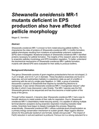
15. Figure 2.
A. Pellicle of S. oneidensis MR-1 wild-type
B. Pellicle of CsgBA-
mutant with deletion of major and minor curlin subunits
Images were taken of the pellicle after 48 hours of growth in LB. The abundance of
colonies and thickness of S. oneidensis MR-1 wild-type pellicles (A) were compared to
CsgBA-
(B), OmpW-
, PepT-
, AsnB-
, WzzB-
, and Wza-
. Biofilms were prepared as
described in Materials and Methods.
16. Sources
1. Belchik SM, Kennedy DW, Dohnalkova AC, Wang Y, Sevinc PC, Wu H, Shi L. (2011).
Extracellular Reduction of Hexavalent Chromium by Cytochromes MtrC and OmcA of
Shewanella oneidensis MR-1. Appl. Environ. Microbiol. 77(12), 4035–4041.
doi:10.1128/AEM.02463-10
2. Bencheikh-Latmani R, Williams SM, Haucke L, Criddle CS, Wu L, Zhou J, Tebo BM.
(2005). Global transcriptional profiling of Shewanella oneidensis MR-1 during Cr(VI) and
U(VI) reduction. Appl. Environ. Microbiol. 71:7453–7460 doi:10.1128/AEM.71.11.7453–
7460.2005
3. González-Pinzón R, Haggerty R, Myrold DD. (2012). Measuring aerobic respiration in
stream ecosystems using the resazurin-resorufin system. Journal of Geophysical
Research 117 (G3): G00N06. doi:10.1029/2012JG001965
4. Yin J, Sun L, Dong Y, Chi X, Zhu W, Qi SH, Gao H. (2013). Expression of blaA Underlies
Unexpected Ampicillin-Induced Cell Lysis of Shewanella oneidensis. PLoS ONE.
doi:10.1371/journal.pone.0060460
5. Lemon KP, Earl AM, Vlamakis HC, Aguilar C, Kolter R (2008). Biofilm development with
an emphasis on Bacillus subtilis. In Bacterial Biofilms, 1-16.
6. Probert HM, Gibson GR. (2002). Bacterial Biofilms In The Human Gastrointestinal Tract.
Curr. Issues Intest. Microbiol. 3: 23-27
7. Romine, MF. (2011). Genome-wide protein localization prediction strategies for gram
negative bacteria. BMC Genomics. 12(Suppl 1), S1. doi:10.1186/1471-2164-12-S1-S1
8. Sani RK, Peyton BM, Dohnalkova A. (2008). Comparison of uranium(VI) removal by
Shewanella oneidensis MR-1 in flow and batch reactors. Water Research. 42(12): 2993-
3002
9. Thormann KM, Saville RM, Shukla S, Pelletier DA, Spormann AM (2004). Initial Phases
of biofilm formation in Shewanella oneidensis MR-1. J. Bacteriol.
10.1128/JB.186.23.8096-8104.2004
10.Liang Y, Gao H, Chen J, Dong Y, Wu L, He Z, Liu X, Guanzhou Qiu, Jizhong Zhou
(2010). Pellicle formation in Shewanella oneidensis. BMC Microbiol. doi:10.1186/1471-
2180-10-29
11.Karatan E, Warnick P. (2009). Signals, Regulatory Networks, and Materials That Build
and Break Bacterial Biofilms. Microbiol. Mol. Biol. Rev. doi: 10.1128/MMBR.00041-08
12.Elias DA, Monroe ME, Marshall MJ, Romine MF, Belieav AS, Fredrickson JK, Anderson
GA, Smith RD, Lipton MS. (2005). Global detection and characterization of hypothetical
proteins in Shewanella oneidensis MR-1 using LC-MS based proteomics. Proteomics 5
(12), 3120-3130
13.Romine MF, Carlson TS, Norbeck AD, McCue LA, Lipton MS. (2008). Identification of
mobile elements and pseudogenes in the Shewanella oneidensis MR-1 genome. Appl.
Environ. Microbiol. 74 (10), 3257-3265
14.Heidelberg JF, Paulsen IT, Nelson KE, Gaidos EJ, Nelson WC, Read TD, Eisen JA,
Seshadri R, Ward NL, Methe BA, Clayton RA, Meyer T, Tsapin A, Scott J, Beanan M.J.,
Brinkac LM, Daugherty SC, DeBoy RT, Dodson RJ, Durkin AS, Haft DH, Kolonay JF,
Madupu R, Peterson JD, Umayam LA,White O, Wolf AM, Vamathevan JJ, Weidman JF,
Impraim M, Lee K, Berry KJ, Lee C, Mueller J, Khouri HM, Gill J, Utterback TR, McDonald
LA, Feldblyum TV, Smith HO, Venter JC, Nealson KH, Fraser CM. (2002). Genome
17. sequence of the dissimilatory metal ion-reducing bacterium Shewanella oneidensis. Nat.
Biotechnol. doi:20:1118-1123 10.1038/nbt749
15.Abboud R, Popa R, Souza-Egipsy V, Giometti CS, Tollaksen S, Mosher JJ, Findlay RH,
Nealson KH. (2005). Low-temperature growth of Shewanella oneidensis MR-1. Appl.
Environ. Microbiol. 71, 811–816. doi:10.1128/AEM.71.2.811-816.
16.Wood TK, Gonzalez Barrios AF, Herzberg M, Lee J. (2006).Motility influences biofilm
architecture in Escherichia coli. Appl. Microbiol. Biotech. 72, 361–367.
17.Watnick PI, Lauriano CM, Klose, KE, Croal L, Kolter R. (2001).The absence of a flagellum
leads to altered colony morphology, biofilm development and virulence in Vibrio cholerae
O139. Mol Microbiol. 39, 223–235.
18.Houry A, Briandet R, Aymerich S, Gohar M. (2010). Involvement of motility and flagella in
Bacillus cereus biofilm formation. Microbiology 156:1009–1018.
19.Burke C, Steinberg P, Rusch D, Kjelleberg S, Thomas T. (2011). Bacterial community
assembly based on functional genes rather than species. PNAS
20.Stolz, JF, Oremland RS. (2011). Microbial Metal and Metalloid Metabolism - Advances
and Applications. Amer. Soc. Microbiol. (ASM).
21.Shi L, Rosso KM, Clarke TA, Richardson DJ, Zachara JM, Fredrickson JK. (2012).
Molecular Underpinnings of Fe(III) Oxide Reduction by Shewanella oneidensis MR-1.
Frontiers in Microbiology, 3. doi: 10.3389/fmicb.2012.00050
22.Shi L, Squier TC, Zachara JM, Fredrickson JK. (2007). Respiration of metal hydroxides by
Shewanella and Geobacter: a key role for multihaem c-type cytochromes. Mol. Microbiol.
65(1):12-20. doi:10.1111/j.1365-2958.2007.05783.x.
23.Liang Y, Gao H, Guo X, Chen J, Qiu G, He Z, Zhou J, Liu X. (2012). Transcriptome
analysis of pellicle formation of Shewanella oneidensis. Archiv. Microbiol. 194(6), 473-
482. doi: 10.1007/s00203-011-0782-x
24.Belchik SM, Tucker AE, Silvia CP, Dohnalkova AC, Kennedy DW, Hirschmugl C, Marshall
MJ. (2015). Chemical Analysis of Shewanella Extracellular Polymeric Substances
Produced in Biofilms. (In Preparation).
25.Barnhart MM, Chapman MR. (2006). Curli Biogenesis and Function. Annual Rev.
Microbiol. 60, 131–147. doi:10.1146/annurev.micro.60.080805.142106
26.Bouhenni RA, Vora GJ, Biffinger JC, Shirodkar S, Brockman K, Ray R, Wu P, Johnson
BJ, Biddle EM, Marshall MJ, Fitzgerald LA, Little BJ, Fredrickson JK, Beliaev AS,
Ringeisen BR, Saffarini DA. The Role of Shewanella oneidensis MR-1 Outer Surface
Structures in Extracellular Electron Transfer. (2010). U.S. Navy Research. Paper 16. doi:
10.1002/elan.200880006
27.Hong H, Patel DR, Tamm LK, Berg BVD. (2006). The outer membrane protein OmpW
forms an eight-stranded beta-barrel with a hydrophobic channel. Jol. of Biochem.
281(11):7568-77. DOI: 10.1074/jbc.M512365200
28.Yoo AY, Yu JE, Yang J, Kim YH, Baek CH, Oh JI, Kang HY. (2008). Regulation of an
outer membrane protein, OmpW, expression and its biological function in Salmonella
typhimurium. Jol. Life Sci. 18(11):1606-1611. DOI: 10.5352/JLS.2008.18.11.1606
29.Arthur M, Molinas C, Courvalin P. (1992). Sequence of the vanY gene required for
production of a vancomycin-inducible D,D-carboxypeptidase in Enterococcus faecium
BM4147. Gene 120 (1): 111–4. doi:10.1016/0378-1119(92)90017-j
18. 30.Meziane-Cherif D1, Stogios PJ, Evdokimova E, Savchenko A, Courvalin P. (2014).
Structural basis for the evolution of vancomycin resistance D,D-peptidases. Proc. Natl.
Acad. Sci. USA. 111(16):5872-7. doi: 10.1073/pnas.1402259111
31.Fratamico, P. M., Briggs, C. E., Needle, D., Chen, C.-Y., & DebRoy, C. (2003). Sequence
of the Escherichia coli O121 O-Antigen Gene Cluster and Detection of Enterohemorrhagic
E. coli O121 by PCR Amplification of the wzx and wzy Genes. Jol. Clin. Microbiol. 41(7),
3379–3383. doi:10.1128/JCM.41.7.3379-3383.2003
32.Allen HK, Donato J, Wang HH, Cloud-Hansen KA, Davies J, Handelsman, J. (2010). Call
of the wild: antibiotic resistance genes in natural environments. Nat. Rev. Micro. 8(4),
251-259.
33.Haft DH, Paulsen IT, Ward N, Selengut JD. (2006). Exopolysaccharide-associated protein
sorting in environmental organisms: the PEP-CTERM/EpsH system. Application of a
novel phylogenetic profiling heuristic. BMC Biol. 4(29), doi: 10.1186/1741-7007-4-29