Differential Diagnosis of Neck Swellings
•
40 likes•17,796 views

For more Medical notes , visit our website: www.mediconotes.com , subscribe and get unlimited access to more than 300 notes.

Recommended
More Related Content
What's hot
What's hot (20)
Similar to Differential Diagnosis of Neck Swellings
Similar to Differential Diagnosis of Neck Swellings (20)
Recently uploaded
Recently uploaded (20)
Differential Diagnosis of Neck Swellings
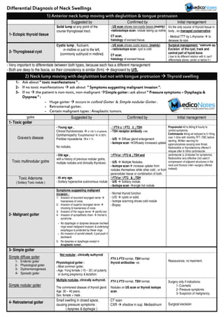
- 1. Differential Diagnosis of Neck SwellingsDifferential Diagnosis of Neck SwellingsDifferential Diagnosis of Neck SwellingsDifferential Diagnosis of Neck Swellings - Very important to differentiate between both types, because each has a different management - Both are deep to the fascia, so their consistency is similar (firm) diagnosed by U/S. 2) Neck lump moving with deglutition but not with tongue protrusion Thyroid swelling 1111---- Ask about """" toxic manifestationstoxic manifestationstoxic manifestationstoxic manifestations " ." ." ." . 2222---- If no toxic manifestations ask about """" Symptoms suggestSymptoms suggestSymptoms suggestSymptoms suggestinginginging malignant invasionmalignant invasionmalignant invasionmalignant invasion ".".".". 3333---- If no the patient is non-toxic, non-malignant Simple goiter :Simple goiter :Simple goiter :Simple goiter : ask about " Pressure symptoms" Pressure symptoms" Pressure symptoms" Pressure symptoms –––– Dysphagia &Dysphagia &Dysphagia &Dysphagia & Dyspnea "Dyspnea "Dyspnea "Dyspnea " :::: ---- Huge goiter occurs in colloid Goiter & Simple nodular Goiter . ---- Retrosternal goiter. ---- Certain malignant types : Anaplastic tumors. goitre Suggested by Confirmed by Initial management 1- Toxic goiter Graves's disease - Young age , Clinical thyrotoxicosis in 100 %of patients Ophthalmopathy "Exophthalmos" in 50% Pretibial myxoedema in 1 % No nodules. - ↑FT4 or ↑FT3 &↓TSH - TSH receptor antibody +ve. - U/S Diffuse gland enlargement - Isotope scan Diffusely increased uptake Propranolol 40 to 80mg 8 hourly to control symptoms. Carbimazole 40mg od reduced to 5-10mg over 1-3mo with monthly TFT. FBC before starting. Written warning for agranulocytosis causing sore throat. Radioiodine or thyroidectomy offered if relapse after 6-18mo carbimazole. Toxic multinodular goitre - Old age , with a history of previous nodular goitre, multiple nodules and clinically thyrotoxic. ↑FT4 or ↑FT3 &↓TSH and - U/S Multiple Nodules. - Isotope scan increase uptake from nodules themselves while other cold , or from paranodular tissue or combination of both. carbimazole (± β-blocker for symptoms). Radioiodine very effective (not used if compression of adjacent structures in the neck and thoracic inlet—surgery offered instead). Toxic Adenoma ( Solitary Toxic nodule ) - At any age, - Solitary hyperactive autonomous nodule ↑FT4 or ↑FT3 &↓TSH - U/S Solitary nodule. - Isotope scan single hot nodule. 2- Malignant goiter Symptoms suggesting malignant invasion : 1- Invasion of recurrent laryngeal nerve hoarseness of voice. 2- Invasion of superior laryngeal nerve chocking &hoarseness of voice. 3- Invasion of the vagus nerve painful ear. 4- Invasion of sympathetic chain Horner's syndrome . ---- No dysphagia or dyspnea because tracheal rings resist malignant invasion &underlying esophagus is protected by these rings. ---- No invasion of carotid sheath, it just push it backward. 5- No dyspnea or dysphagia except in Anaplastic tumor. - Normal thyroid function - U/S cystic or solid. - Isotope scanning shows cold nodule - Biopsy 3- Simple goiter Simple diffuse goiter 1- Endemic goiter 2- Physiological goiter 3- Dyshormonogenesis 4- Sporadic goiter Not nodular ,clinically euthyroid Physiological goiter : ----Most common goiter. ----Age: Yong female ( 15 – 20 ) at puberty or during pregnancy &lactation. FT4 &FT3 normal, TSH normal thyroid antibodies -ve. Reassurance, no treatment. Simple nodular goiter Multiple nodules,clinically euthyroid. The commonest disease of thyroid gland Age: 30 – 40 years. Sex: female > male . FT4 &FT3 normal, TSH normal. Nodules on US scan or thyroid isotope scan. Surgery only if indications: 1- Cosmetic 2- Pressure symptoms 3- Suspicion of malignancy. 4- Retrosternal goiter Small swelling in closed space, causing pressure symptoms : ( dyspnea &dyphagia ) CT scan CXR shadow in sup. Mediastinum Surgical excision 1) Anterior neck lump moving with deglutition & tongue protrusion Suggested by Confirmed by Initial management 1- Ectopic thyroid tissue Solid lump at any point of the course thyroglossal tract. - US scan shows non-cystic lesion,(mainly) -radioisotope scan: nodule taking up iodine CT scan, histology of excised tissue. It's the only source of thyroid tissue in body , so managed conservative : - Medical TTT by L-thyroxine to decease its size 2- Thyroglossal cyst Cystic lump , fluctuant, in midline or just to the left, (commonly subhyoid in midline) - US scan shows cystic lesion, (mainly) - radioisotope scan: cyst is cold CT scan, histology of excised tissue. Surgical management: "sistrunk op." Excision of the cyst,track and central part of hyoid bone ( due to its different relation with it, can't differentiate above, center or below it )
- 2. 3) Neck lump doesn't move with deglutition nor tongue protrusion Other neck swellings Parotid region swellings A- Localized : The pathognomonic signs of parotid malignancy are late signs: 1- Facial nerve palsy 2- Fixity of mandible From skin & subcutaneous tissue: - Sebaceous cyst - Subcutaneous abcess - Lypoma - hemangioma - lymphangioma From margins : - Masseter hypertrophy - Zygomatic tumor - Mastoiditis LNs : Multiple - Pre-auricular LNs - Parotid LNs - Buccinator LNs LN is diagnosed by two items : 1- Anatomical site 2- Multiplicity ( but can be single ) . Single - Pre-auricular LNs or - Parotid gland neoplasm - Both are deep to parotid fascia ( which is strong deep fascia ). - To differentiate between both by either : 1- U/S, is there line of cleavage ? If yes LNs If no Parotid neoplasm. 2- CT, is there line of cleavage ? We ask for images because the least biopsy in parotid gland swelling is superficial parotidectomy because of branches of facial nerve !(it might be just LN ). If Parotid gland neoplasm Pathological differentiation of parotid neoplasm : ( can't be assessed by clinician ). A) Benign : 1- Pleomorphic adenoma (most common 85%) It's pleomorphic adenoma till proven otherwise. 2- Monomorphic adenoma "Adenolymphoma" , "Warthin's Tumor" , " papillary cystadenoma lymphomatosum" B) Malignant : 1- Adenocarcinoma on top of pleomorphic adenoma ( most common malignancy ). 2- Adenocarcinoma fromthe start 3- Adenoid cystic carcinoma 4- Acinic cell tumor. 5- Epidermoid carcinoma 6- Mucoepidermoid carcinoma Clinical estimation of type of tumor : ---- If old male ,with history of remission & exacerbation think of "adenolymphoma" Ask for technetium scan Hot spot adenolymphoma (only hot spot tumor) Cold spot other benign &malignant tumors TTT of adenolymphoma: The only tumor will be treated by evacuation ( not by superficial parotidectomy), because it's very localized tumor. ---- If history of pain before swelling because tumor spread along sheaths of facial nerve branches think about "adenoid cystic carcinoma" Ask for CT or MRI ( not felt because parotid is covered by very dense fascia). The pathognomonic signs are early in adenoid cystic carcinoma because the tumor spread along myelin sheaths of facial nerve branches so if you neglected the pain of the patient and didn't diagnose the neoplasm, the patient might come early with facial nerve palsy. B- Diffuse : Predisposing factors of acute bacterial parotitis : 1- Immunosupressive 2- Local irradiation 3- Chemotherapy 4- Diabetic,neglected,poor control 5- Bad oral hygiene. It's difficult for parotid to get inflamed because it's highly vascular, so there should be predisposing factors ,, TTT of these predisposing factors Diffuse Parotid swellings Acute Chronic (all bilateral) 1- Endemic parotitis - Bilateral. - ttt: reassurance & conservative. 2- Sialosis (Sialadenosis): conservative. - Better seen (inspection) than felt - Associated with : • Acromegaly • Diabetes (controlled or not) 3- Lipomatous pSeudohypertrophy - Exaggerated form of sialosis - Sagging of the enlargement. 4- Sialectasis(ectatic duct) conservative. by x-ray: Sand ground appearance. 5- Sarcoidosis : - Generalized lymphadenopathy with hilar shadow except submental LNs - Renal calcinosis & Renal stones 6- Sjogren's syndrome Lympho-epithelial disease complex - Rheumatoid arthritis - Dry eye - Dry mouth ( due to chronic diffuse parotitis ) Obstructive : 1- Stone 2- Stricture - C/P : colicky facial pain - Can't be differentiated clinically so ask for X-ray : • If radiopaque stone • If no Stricture. TTT: - Proximal (near gland) superficial Parotidectomy - Distal (near duct) meatomy - Intermediate expectant ttt : by dilating duct every month by dilator till we found the stone or relieve stricture. Non-Obstructive : - Acute inflammation • Viral : 1- Mumps - Usually bilateral (may start unilateral ) - Occurs in children . - we scared of 3 complications 2ry encephalitis, Pancreatitis. Orchitis - Require isolation , bed rest ,antibiotics & vitamins 2- Coxsackie virus • Bacterial ( usually unilateral ) TTT: 1- TTT of predisposing factors 2- Analgesia & Massive antibiotics 3- Hilton incision (pre-auricular longitudinal incision & open the fascia transversely to avoid injury of facial nerve) & evacuation - Don't wait for fluctuation because of dense parotid fascia.
- 3. Anterior Triangle swellings 1- Submandibular Triangle Cystic Ranula - It's retention cyst arising from sublingual salivary gland ( cyst in mouth floor ). -It may extend down to the neck over post. margin of mylohyoid "plunging ranula" - Suggested by: translucent cyst lateral to midline, with domed, bluish discoloration in floor of mouth lateral to frenulum presents itself as swelling in submandibular or submental triangle. management: 1- Marsupilization (deroofing) &suture cyst wall to oral mucous m. 2- Excision(difficult) in recurrent cases. Solid Multiple LNs Single 1- LNs *Inflammation "Suggested: tender,solid,nodular swelling especially <20y of age." *Neoplastic or metastatic 2- Submandibular gland : *Submandibular sialadenitis or *Submandibular tumor To differentiate, roll the swelling: - If rolled LNs - If not rolled - Fixed LNs ( long stand neglected inflamed LNs ) - Salivary ( due to floor muscle: mylohyoid muscle ) Bimanual examination : - if 2 lobes are palpable Salivary gland - if Not Fixed LNs or superficial parotid tumor Excision &pathology to differentiate. 2- Submental Triangle Cystic Ranula Solid Single or Multiple LNs only ( no salivary gland ) N.B. Sarcoidosis has generalized lymphadenopathies except Submental LNs . Branchial cyst dangerous because it passes between the carotid bifurcation to the glossopharyngeal &vagus nerve 3- Carotid Triangle Cystic Branchial cyst "Congenital" Presents partially deep to sternomastoid muscle and extends to the upper 1/3 of its ant. border. Suggested by: fluctuant swelling at anterior border of sternomastoid muscle, Confirmed by: US scan, CT scan Initial management:excision it till lateral wall of pharynx with segment of lat. Wall. Tuberculosis (‘cold’) abscess Suggested by: fluctuant(cystic) swelling with low grade or no fever. Confirmed by: acid-fast bacilli (AFB), on microscopy or culture and sensitivity of aspirate. Carotid aneurysm Pulsatile swelling coincides with carotid pulsation. – Carotid angiography Pharyngeal diverticulum It's herniation of pharyngeal mucosa through Killian's dehiscence Suggested by: intermittent, fluctuant, compressible, swelling (usually on left) under sternomastoid muscle , and dysphagia. confirmed by: barium swallow fills pouch. Initial management: surgical referral for excision. Laryngocele It's herniation of the mucous membrane through a weak point in the thyrohyoid membrane. Suggested by: fluctuant swelling in neck which becomes prominent on straining. Swelling is resonant &compressible Occurs in musicians . Confirmed by: MRI &laryngoscope. Solid Multiple LNs Single Carotid body tumor(potato Tumor) ---- Occurs in high attitude.(pero) ---- Differentiation by size because no pathological differentiation : >4cm is malignant <4 cm is benign. Suggested by: Very slowly growing mass with history of mass for years - mobile : fromside to side but not vertical - arising from chemoreceptors at carotid bifurcation (upper third of sternomastoid), - pulsatile , bruit maybe heard by stethoscope. - My extend to parapharyngeal space present in oropharynx. Confirmed by: 1- Angiography: widening of carotid artery bifurcation ( characteristic sign) management: Surgical excision with preservation of ICA. Posterior Triangle swellings Cystic Cystic hygroma (Cavernous Lympangioma) "Congenital" - It's dilated cavernous lymph spaces. - Due to failure of sequestration of part of jugular lymph sac of the fetus. -Presents superficial to Sternomastoid muscle Suggested by: Single , large , irregular, ill-defined, swelling that transilluminates well (only translucent neck swelling) , appears at birth or <20y of age. Confirmed by: US scan,CT scan, Complications: Recurrent infection , respiratory distress due to compression of trachea, increases in size on coughing or crying. management: Surgical excision at about the age of 3 years. Tuberculosis (‘cold’) abscess ……………….. ( most common site ) Suggested by: fluctuant(cystic) swelling with low grade or no fever. Confirmed by: acid-fast bacilli (AFB), on microscopy or C&S of aspirate. Esophageal diverticulum Confirmed by: barium swallow fills pouch. Pnematocele Herniation of the lung apex through weak suprapleural membrane (Sibson's fascia), which extends from transverse process of C7 to 1st rib. Suggested by : - Cystic swelling in the supraclavicular region -Become prominent on straining - Resonant &compressible management : 1- Correct straining factors 2- plication of Sibson's fascia. Solid Multiple LNs Single LNs Cervical rib is a supernumerary (or extra) rib which arises fromthe seventh cervical vertebra The presence of a cervical rib can cause a formof thoracic outlet syndrome due to compression of the lower trunk of the brachial plexus or subclavian artery Compression of the brachial plexus may be identified by weakness of the muscles around the muscles in the hand, near the base of the thumb "neurovasculardeficient" . (diagnosed by x-ray or CT) Sternomastoid tumor - Mostly seen in newborns due to birth trauma - Rare condition in which ischemia of the muscle fibrosis &mass