
Recommended
More Related Content
Similar to Body fluid , analytical chemistry
Similar to Body fluid , analytical chemistry (20)
Recently uploaded
Recently uploaded (20)
Body fluid , analytical chemistry
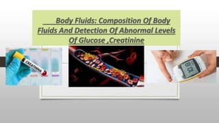
- 1. Body Fluids: Composition Of Body Fluids And Detection Of Abnormal Levels Of Glucose ,Creatinine
- 2. CONTENTS COMPOSITION OF BODY FLUIDS TYPES OF BODY FLUIDS. DETECTION OF ABNORMAL LEVELS OF GLUCOSE. DETECTION OF ABNORMAL LEVELS OF CREATININE.
- 3. Composition of body fluids “Body fluids are the fluids such as blood, lymph, and saliva which are produced in the body and then either circulated within the body or secreted outside It. Blood and lymph are the two most important body fluids in the human body. Blood comprises plasma, white blood cells, red blood cells, and platelets. Lymph is a colorless fluid that circulates inside the lymphatic vessels
- 4. TYPES OF BODY FLUIDS 1.Blood THE MAIN COMPONENTS OF BLOOD INCLUDE: Plasma: Plasma is the liquid component of blood. It is a thick fluid .Plasma is 91% water and the rest of the total volume is made up of ions, proteins, gases, nutrient molecules and waste products. . Albumin is the major protein in plasma. Red Blood Cells: 40% of the blood contains red blood cells. RBCs contain protein hemoglobin that gives a red colour to the blood.
- 5. • White Blood Cells: The white blood cells are very few in number. They mainly protect the body against infections. Many WBCs cross the walls of the vessels and penetrate other tissues. • Platelets: These are fewer in number than the red blood cells. The platelets help in the clotting of blood at the site of a wound. Lymph Lymph is a colourless fluid present in the interstitial tissues. It circulates throughout the lymphatic system.. The exchange of nutrients, hormones, and gases occurs through this fluid. It consists of lymphocytes that play a major function in the immune responses of the body.
- 6. INTRODUCTION • Blood sugar, or glucose, is the main sugar found in your blood. • It comes from the food you eat, and is your body's main source of energy. • Diabetes is a disease in which your blood sugar levels are too high. • Over time, having too much glucose in your blood can cause serious problems.
- 7. Normal values of blood glucose : • Normal fasting level = 60-90mg/100ml of blood • Glucose level half an hour (after meal) (post prandial) = 120- 150mg/100ml of blood • In normal healthy individuals the peak glucose level (at any time of the day) = 60-110 mg/100ml of blood is considered normal.
- 8. Clinical significance of blood sugar level • Blood glucose level increases in diabetes mellitus, acute stress, hyperthyroidism and chronic liver disease. • Blood glucose level decreases in Addison’s disease, hypothyroidism and cancer of the pancreas. • The increase in the blood glucose level is called hyperglycaemia and decrease in blood glucose level as hypoglycaemia. • People suffering from diabetes mellitus need to get their blood glucose tested frequently.
- 9. Methods used to measure blood glucose level. • Although a number of methods are used for glucose determination, commonly used two methods are discussed here. • These can be grouped into two categories- chemical and enzymatic. Chemical method • Folin-Wu method • Ortho-Toluidine method Enzymatic method • GOD-POD method. (Glucose oxidase method)
- 10. Chemical method to estimate blood glucose; Folin-Wu method: • It is based on the principle that glucose when heated with an alkaline copper solution, reduces cupric ions to cuprous ions. • The cuprous ions are then measured photometrically (colorimetry) by adding phosphomolybdic acid which gets reduced to molybdenum blue. • In this method, whole blood is used and the blood glucose value is determined by the intensity of blue color. Ortho-Toluidine method: • This is an ideal manual method used for its rapidity, sensitivity, accuracy, and relative simplicity. • It is based on the principle that the aldose sugar i.e. glucose on condensation with ortho-toluidine in glacial acetic acid gives a green color that can be measured spectrophotometrically.
- 11. Enzymatic method- GOD-POD method to estimate blood glucose • Enzymatic methods provide maximum degree of glucose specificity, hence are very good in estimating true blood glucose. • For this method, only blood plasma or serum is used. • The glucose remains stable for 24 hours if serum or plasma is prepared within 30 minutes after collection. • The enzyme peroxidase catalyses the following reaction. The hydrogen peroxide formed reacts with phenol to a red-violet dye as indicator. • The intensity of the colour formed is measured colorimetrically (or spectrophotometrically) which is directly proportional to the blood glucose level. • This test is not influenced by the pressure of uric acid and ascorbic acid
- 12. WHAT IS CREATININE ..? Creatinine is a nitrogenous waste product formed from the metabolism of creatine in skeletal muscle. It has no useful function so excreted in urine after it has been filtered by the glomerulus from the blood. . With normal kidney function, then, the amount of creatinine in the blood remains relatively constant and normal. For this reason, and because creatinine is affected very little by liver function, an elevated blood creatinine is a more sensitive indication of impaired kidney function than the BUN.
- 13. • A BUN test may be done with a blood creatinine test. Blood urea nitrogen (BUN) and creatinine tests can be used together to find the BUN-to- creatinine ratio (BUN: creatinine). • BUN-to-creatinine ratio 10:1–20:1 • High BUN-to-creatinine ratio occur with sudden (acute) kidney failure. A blockage in the urinary tract (such as a kidney stone) can cause a high BUN-to-creatinine ratio. • A very high BUN-to-creatinine ratio may be caused by bleeding in the digestive tract or respiratory tract. • A low BUN-to-creatinine ratio may be caused by a diet low in protein, a severe muscle injury and others.
- 14. Normal Value Of Creatinine Serum creatinine is reported as milligrams of creatinine to a deciliter of blood (mg/dL) or micromoles of creatinine to a liter of blood (micromoles/L). The typical range for serum creatinine is: For adult men, 0.74 to 1.35 mg/dL (65.4 to 119.3 micromoles/L) For adult women, 0.59 to 1.04 mg/dL (52.2 to 91.9 micromoles/L)
- 15. METHOD USED TO MEASURE CREATININE • The principal of this assay is based on the reaction of creatinine with sodium picrate as described by Jaffe. • Creatinine reacts with alkaline picrate forming a red complex. The time interval chosen for measurements avoids interferences from other serum. • The intensity of the color formed is proportional to the creatinine concentration in the sample.
- 16. REFERENCES • https://byjus.com/biology/body-fluids-and-circulation/ • https://www.onlinebiologynotes.com/blood-urea-normal-value-clinical- significance-and-methods-of-estimation/ • https://www.mayoclinic.org/tests-procedures/creatinine-test/about/pac- 20384646