
Recommended
More Related Content
What's hot
What's hot (20)
Similar to Chest Auscultation Guide for Nurses
Similar to Chest Auscultation Guide for Nurses (20)
More from MURUGESHHJ
More from MURUGESHHJ (14)
Recently uploaded
Recently uploaded (20)
Chest Auscultation Guide for Nurses
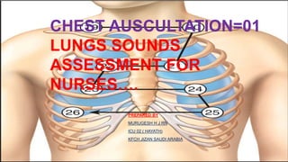
- 1. PREPARED BY MURUGESH H J RN ICU 02 ( HAYATH) KFCH JIZAN SAUDI ARABIA CHEST AUSCULTATION=01 LUNGS SOUNDS ASSESSMENT FOR NURSES….
- 2. CHEST AUSCULTATION…. Auscultation is the important component in Physical examination, using stethoscope hearing different sounds , type & Tone… What is chest auscultation? Vesicular breath sounds occur when the vocal cords vibrate during inspiration and expiration, when the vibrations are transmitted to the trachea and bronchi. These sounds are audible when auscultation is performed using a stethoscope. “Chest auscultation involves listening to these internal sounds to assess airflow through the trachea and the bronchial tree” (Sarkar et al, 2015). “vesicular breath sounds is Normal breathing sounds”
- 3. LOCATIONS OF VESICULAR BREATH SOUNDS … “The bell of the stethoscope is generally used to detect high-pitched sounds – at the apex of the lungs above the clavicle; its diaphragm is used to detect low-pitched sounds in the rest of the chest” (Dougherty and Lister, 2015) As a Nurse its important To know the specific locations of lungs sounds for assessment ….. *** Apical zone: above the clavicles; *** Upper zone: below the clavicles and above the cardiac silhouette (HEART SORROUNDINGS) *** Mid zone: level of the hilar structures;( ENTRY HOLE) *** Lower zone: bases.( BELOW LUGS)
- 4. POSITION.…… ***provide comfortable position as patient requests eg: 45 Degree elevation, in chair , side of the bed… ***chest & back need to be exposed follow patient privacy & orient the procedure completely…
- 5. THE PROCEDURE…. The procedure 1. Ensure your stethoscope has been cleaned following local infection prevention and control guidance. 2. Discuss the procedure with the patient and gain informed consent. 3. Check that the patient is kept warm and the area is free from drafts. 4. Screen the bed to maintain patient privacy and dignity. 5. Decontaminate your hands according to local policy.
- 6. Cont….. 6. Position the patient comfortably so you can access their chest. 7. Remove or rearrange the patient’s clothing as necessary to enable you to see the chest. 8. See whether the stethoscope feels cold. Warm it between your hands if necessary 9. Position the ear tips in your ears so they point slightly forward towards the nose;this will help to create a seal and will reduce external noise. 10. Holding it between the index and middle finger of your dominant hand, place the chest piece of the stethoscope flat on the patient’s chest using gentle pressure. 11. Using a ‘stepladder’ approach listen to breath sounds on the anterior chest. This technique allows you to compare one side of the chest with the other in a systematic manner and detect any asymmetry. The stethoscope should be in before applying it to the chest to avoid discomfort for the patient contact with the chest for a full cycle of inspiration and expiration at each point on the stepladder
- 7. Cont….. 12. Use the step ladder approach for the posterior chest avoid the scapula as lung sounds cannot be heard through bone (Ferns and West, 2008). 13. Ask the patient to move their right arm to the side so the right lateral chest can be assessed .Starting with the upper lobe move to the middle lobe, and finally the lower lobe at the bottom (Ferns and West, 2008). 14. Repeat on the left side where the lung is made up of an upper lobe and lower lobe. 15. Replace the patient’s clothing and make them comfortable. 16. Explain your findings to the patient and check whether they have any questions. 17. Decontaminate your stethoscope. 18. Decontaminate your hands. 19. Record findings in the patient’s notes
- 8. NORMAL BREATHING SOUNDS… Bronchovesicular sounds Normal findings on auscultation include: Loud, high-pitched bronchial breath sounds over the trachea. Medium pitched bronchovesicular sounds over the mainstream bronchi, between the scapulae, and below the clavicles. Soft, breezy, low-pitched vesicular breath sounds over most of the peripheral lung fields.
- 10. ABNORMAL LUNG SOUNDS …… There are several adventitious sounds but the main ones to be aware of are snoring , crackles, wheeze , absent breath sounds, and pleural friction rub. SNORING… ITS IS AN MOST COMMONNEST IN OBESE,OBSTRUCTIVE SLEEP APNOEA ( OSA) “VIBRATING OR HARSH LIKE LOUD SOUND AUDIBLE BECAUSE OF NOSE, THROAT ITS GET OBSTRUCTED BECAUSE OF THICK MUCOSAL SECRETIONS ,THICK MUCOSAL TISSUE, UNDERLYING INFECTIONS LIKE SINUSITSIS, LARYNIGITIS”….
- 11. ABNORMAL LUNG SOUNDS …… Crackle OR CREPITATIONS OR CREPS Crackles are generated within the small airways( because fluid in the airway); they predominantly occur during the inspiratory phase but can happen on expiration. Clinical conditions where crackles may be present include pneumonia, pulmonary fibrosis, chronic obstructive pulmonary disease (COPD), lung infection and heart failure……. Crackles can be categorised as coarse or fine; distinguishing between these can be significant – coarse crackles may indicate pneumonia, while fine crackles may suggest pulmonary oedema...
- 13. ABNORMAL LUNG SOUNDS …… Wheeze Wheeze often occurs on expiration, but can also occur on inspiration. Wheezing is often louder than usual breath sounds and in some patients it is audible from some distance or when the patient breathes through the mouth. With a stethoscope you may also be able to hear a wheeze over the patient’s trachea (Sarkar et al, 2015). Clinical conditions such as asthma are associated with a high-pitched musical wheeze that may be more evident on expiration. An inspiratory wheeze (stridor) usually results from an upper airway obstruction such as laryngeal oedema or the presence of a foreign body. A wheeze on both inspiration and expiration could be due to secretions in the airways (Welch and Black, 2017) and the patient may need to be advised how to clear their chest of secretions.
- 15. ABNORMAL LUNG SOUNDS …… Absent breath sounds Absent breath sounds This describes a lack of audible breath sounds on auscultation. It could be caused by lung disorders that inhibit the transmission of sounds, for example, a pneumothorax, pleural effusion or areas of lung consolidation, Atelectasis All these conditions prevent air flow reaching parts of the lung due to a pathological change in the function of the lung. Rhonchi, Rhonchi,or “large airway sounds,” are continuous gurgling or bubbling sounds typically heard during both inhalation and exhalation. These sounds are caused by movement of fluid and secretions in larger airways (asthma, viral URI). Rhonchi, unlike other sounds, may clear with coughing. Rhonchi occur due to conditions that block airflow through the large airways, including the bronchi. There may also be inflammation and fluid in these airways. Conditions such as acute bronchitis and COPD may cause rhonchi…… Pleural rub or pleural friction- Heard primarily on inspiration over an area of pleural inflammation;may be describes as a grating sound ….
- 18. NANDA NURSING DIAGNOSIS ASSOCETED WITH LUNG CONDITIONS…. Ineffective Airway Clearance RELATED To Increased Secretions Interventions- ***Often Chest Physio & Positioning The Patient ***Administer The Nebs As Per Physician Advice Impaired Gas Exchange RELATED TO Altered Lung Physiology Or Alveolar Function Interventions- ***Often Chest Physio & Positioning The Patient Ineffective Breathing Pattern RELATED TO Decreased Lung Expansion Or Lung Damage Interventions- ***Often Chest Physio & Positioning The Patient ***Administer The Nebs As Per Physician Advice
- 19. NANDA NURSING DIAGNOSIS ASSOCETED WITH LUNG CONDITIONS…. Imbalanced Nutrition: Less Than Body Requirements RELATED To Poor Intake Interventions- *** Encourage Oral Intake *** Prefer Parenteral Nutrition Supplementation Risk for Infection RELATED to altered immune system Interventions- *** follow strict hand hygiene & hand washing technique . * *** minister medications or antibiotics as prescribed Deficient Knowledge RELATED to lack of awareness Interventions- *** patient education & disease orientation *** patient & family counselllig
- 20. NURSING RESPONSIBILITIES…. ***Explain the procedure in detail before assessing, respect patient privacy *** follow the correct manner while assessing I mean , start from observation , auscultation , percussion … **** provide appropriate position & avoid errors …… *** inform & documents the findings in nurses notes…..
- 21. References….. References Cedar SH (2018) Every breath you take: the process of breathing explained. Nursing Times; 114: 1, 47-50. Dougherty L, Lister S (2015) The Royal MarsdenManual of Clinical Nursing Procedures. Chischester: Wiley.Ferns T, West S (2008) The art of auscultation evaluating a patient’s respiratory pathology.British Journal of Nursing; 1: 6, 772-777. Longtin Y et al (2014) Contamination ofstethoscopes and physician’s hands after a physical examination. Mayo Clinic Proceedings; 89:291-299. Nursing and Midwifery Council (2018) FutureNurse: Standards of Proficiency for Registered Nurses. Bit.ly/NMCFuture Royal College of Nursing (2018) Tools of the Trade:Guidance for Health Professionals on Glove Useand the Prevention of Contact Dermatitis. London:RCN. Sarkar M (2015) Auscultation of the respiratorysystem. Annals of Thoracic Medicine; 10: 3, 158-168. sy mpson H (2015) Respiratory assessment. British Journal of Nursing; 15: 9, 484-488. Welch J, Black C (2017) Respiratory problems. In: Adam S et al (eds) Critical Care Nursing Science and Practice. Oxford: Oxford University Pres Abnormal breath sounds: Causes and treatment - Medical News Today https://www.medicalnewstoday.com › article
- 22. THANK YOU ALL