
Recommended
More Related Content
What's hot
What's hot (20)
Similar to Bone marrow density (BMD)
Similar to Bone marrow density (BMD) (20)
Recently uploaded
Recently uploaded (20)
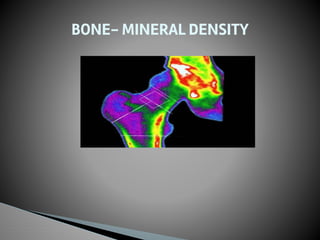
Bone marrow density (BMD)
- 2. BONE DENSITOMETRY A bone densitometry scan is an enhanced form of X-ray technology used to measure the bone mineral (calcium) content or bone loss and density of specific skeletal sites or whole body.
- 4. ▶ The examination is also called a DEXA scan, QDR scan or BMD measurement. ▶ THE MOST VERSATILE AND WIDELY USED TECHNIQUE : DXA or DEXA (Dual energy X-Ray Absorptiometry) ▶ DEXA is most often performed on the lower spine and hips. Portable Dexa devices, including some that use ultrasound waves rather than Xrays measure the wrist fingers or heel and sometimess used for screening purposes. ▶ A bone densitometry scan measures the calcium content in the bones, which can’t be evaluated in an ordinary XRAY. An Xray is more reliable when it comes to detecting a recent bone fracture.
- 5. ▶ Osteoporosis undetected and overlooked until 1920. ▶ The first publication about bone mass appeared in 1930s. ▶ Radiographic absorptiometery Xray taken and compared to a standard radiograph. ▶ Radiogammetry introduced in the 1960s in response to the measurement of the bone loss in astronauts. ▶ In 1970 CT was used through the specialized software - Quantitative CT. ▶ In 1970s and 1980s brought the first dedicated scanners. First SPA then DPA used radioisotope as a source of radiation. ▶ First commercial DXA scanner introduced in 1987 first used radioisotope as the Xray source then replaced by the Xray tube. HISTORY
- 6. ▶ DEXA bone densitometery is used to diagnose osteoporosis and to assess risk of bone fracture within the next few years. ▶ Osteoporosis or thin bones is a condition that often affects women after menopause but may also be found in men. In osteoporosis, gradual loss of calcium causing the bones to become thinner, more fragile and more likely to break. Its not a form of arthritis. ▶ DEXA is also effective in tracking the effects of treatment for osteoporosis and other conditions that cause bone loss. If the patient receives medical treatment for bone loss then the scan can be repeated after 2 years to assess their progress. ▶ RISK FACTORS FOR OSTEOPOROSIS IN WOMEN: - menopause - family history - small bone fracture - advanced age - low calcium diet ↖ inactive lifestyle - Cigarette smoking -GI malabsorption problem - certain medication use (corticosteroids) USES
- 8. ▶ Post menopausal women who is not taking estrogen ▶ Having personal or maternal history of hip fracture or smoking ▶ A man with clinical condition associated with bone loss ▶ A person use medications that are known to cause bone loss, including corticosteroids such as prednisone, antiseizure medicines like dilantin or high dose thyroid replacement drugs. ▶ Patient having type 1 diabetes, liver disease, kidney disease or family history of osteoporosis. ▶ Thyroid condition such as hyperthyroidism. ▶ Parathyroid condition like parathyroidism. ▶ Patients experienced a fracture after mild trauma. INDICATIONS FOR BONE DENSITOMETRY
- 9. ▶ The scan will take between 10 and 30 minutes depending on the equipment used and the parts of the body examined. ▶ Patients can eat normally before the test but they should not take calcium supplements for atleast 24 hours before your examination. ▶ Patients should wear loose, comfortable clothing avoiding garments that have zippers, belts or buttons made of metal. ▶ Patient may be asked to remove some or all of your clothes and to wear a gown during the exam. ▶ Remove jewellery, eye glasses and any metal objects or clothing that might interfere with the Xray images ▶ notify the technologist if you may be pregnant ▶ Inform your physician if you recently had a barium examination or have injected with a contrast material for a computed tomography scan. You may have to wait 10 – 14 days before undergoing a DEXA test. Procedure For Bone Densitometry
- 10. ▶ Two types: Central device Peripheral device Central DEXA device are usually located in hospitals and medical offices ↖ Measure bone density in the hip and spine. ↖ Central dexa devices have a large flat table and an arm suspended overhead Peripheral DEXA are often available in drug stores and on mobile health vans. ↖ Measure bone density in the wrist, heel or finger. DEXA Equipment
- 11. ▶ DEXA machine send a thin invisible beam of low dose Xray with two distinct energy peaks through the bones being examined. 0ne peak is absorbed mainly by soft tissue and the other by bone. The soft tissue amount can be subtracted from the total and what remains is a patient’s bone mineral density. ▶ DEXA machines feature special software that compute and display the bone density measurements on a computer monitor. How does the Procedure work
- 12. ▶ In Central DEXA examination patients lies on a padded table. An Xray generator is located below the patient and an imaging device is positioned above. ▶ To assess the spine, the patient’s legs are supported on a padded box to flatten the pelvis and lumbar spine. To asses the hip the patients foot is placed in a brace that rotates the hip inward. In both cases, the detector is slowly passed over the area, generating images on a computer monitor. ▶ The patient must hold very still and may be asked to keep from breathing for a few seconds while the Xray picture is taken to reduce the possibility of a blurred image. ▶ The PERIHERAL DEXA: test is simpler. The patient finger, hand, forearm or foot is placed in a small device that obtain a bone density reading within a few minutes. ▶ Lateral Vertebral Assessment : (LVA) is now being done at many centres. LVA is a low dose XRAY examination of spine to screen for vertebral fractures that is performed on the DEXA machine. ▶ In these procedures, under the table a photon generator will pass beneath you, while an Xray detector camera will pass above the table parallel to the table parallel to the photon generator beneath, projecting images of the lumbar and hip bones onto a computer monitor ▶ The computer will calculate the amount of photons that are not absorbed by Procedure
- 13. ▶ the bones to determine the bone mineral content. ▶ Bone densitometry testing can determine decreasing bone density and strength at a much earlier stage when treatment of the bone weakness can be beneficial.
- 14. Central DEXA Peripheral DEXA
- 15. ▶ There is no special type of care following bone densitometry testing. You may resume to usual diet and activities unless your physician advises you differently. ▶ TEST RESULTS: ▶ BMD is compared to two norms: Healthy young adults (T-score) and age mated (Z-score) ▶ T-score: the standard deviation is the difference between your BMD and that of the healthy young adults . This result is your T score. Positive T scores indicate the bone is stronger than normal, negative Tscores indicate the bone is weaker than normal ▶ A score above -1 is considered normal. A score between -1 to -2.5 is classified as osteopenia, the first stage of bone loss. A score below -2.5 is defined as osteoporosis. ▶ The risk for bone fracture doubles with every SD below normal (T score of -1 ) has twice the risk for bone fracture as a person with a normal BMD. A person with a (T score of -2) has 4 times the risk of bone fracture as a person with normal BMD After the procedure
- 17. Report of T score
- 18. ▶ Z score : This number reflects the amount of bone you have compared with other people in your age group and of same gender, race, height and weight. If the score is unusually high or low it may indicate a need for further medical tests. ▶ Advantages of bone densitometry or DEXA: - it is simple quick and non invasive procedure. -Low radiation dose less than 1/10th of chest Xray - no anesthesia is required - most accurate method for the diagnosis of osteoporosis and is also considered an accurate estimator of fracture risk. - WIDE availability - ease of use - short scan time - high resolution images - good precision RISK OF DEXA OR BONE DENSITOMETRY: slight chances of cancer
- 19. ▶ LIMITATION OF DEXA SCAN: ↖ Centeral Dexa devices are more sensitive than pDEXA devices but they are also somewhat more expensive ↖ A DEXA test cannot predict who will experience a fracture but can provide indications of relative risk. ↖ A test done on a peripheral location, such as the heel or wrist, may help predict the risk of fracture in the spine or hip. But because bone mass tends to vary from one location to the other, measuring the heel is not as accurate as measuring spine or hip
- 20. THANK YOU