Dr. A Sumathi - LINEARITY CONCEPT OF SIGNIFICANCE.pdf
6 slides.pptx
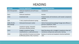
1. HEADING
Antibody Common Applications Staining Characteristics
Bcl-2 Oncoprotein Follicular lymphoma and soft tissue
tumors
Cytoplasmic
Bcl-6 Follicular lymphoma Nuclear
CD31 Endothelial Cells Predominately cell membrane, with weaker cytoplasmic
staining.
CD3 T-cells, lymphoma/leukemia typing Membrane
CD68, PG-M1,
Macrophage
Histiocytic/monocytic marker Cells of monocyte/macrophage lineage stain diffuse or
granular cytoplasmic. Mast
CD10 Lymphoma typing, metastatic
carcinoma unknown primary
Cytoplasmic and Membrane
CD15 Hodgkin Lymphoma Typing,
mesothelioma vs. adenocarcinoma
Reed-Sternberg cells in Hodgkin’s lymphoma show cell
membrane and granular paranuclear staining.
CDX2 Colon and other GI cancers are strong
& diffusely positive. Also, mucinous
Nucleus in normal and neoplastic intestinal epithelial
cells.
2. Antibody Common Applications Staining Characteristics
CD138 Syndecan-1 Plasma Cells (also stains endothelial cells,
fibroblasts, keratinocytes, and
Cell Membrane, pre-B cell and plasma cell marker,
but is absent from mature B
CD30 Anaplastic large cell lymphoma, Hodgkin
lymphoma
Membrane and/or a dot like cytoplasmic staining
CD34 Soft tissue tumor classification, leukemia
typing
Membrane/cytoplasmic
CD99, SEE MIC2
CD117 (c-kit) Gastrointestinal Stromal Tumors (GIST),
Mast Cells, Stains approximately 75% of
mesenteric fibromatosis tumors.
Membrane and/or cytoplasmic
Cytokeratin, AE1/AE3 Epithelial tumors, hepatoma vs.
adenocarcinoma
Cytoplasmic
C-Myc
CD1a Langerhan cells, thymic T-cells, thymoma Membrane and weakly cytoplasmic
Calcitonin Medullary Thyroid Carcinoma Cytoplasmic
C-erb-2 (Her2neu)
Oncoprotein
HER-2/neu overexpression for invasive
breast and gastric cancers.
Follow CAP and ASCO guidelines for interpretation.
Laboratory Developed Test (LDT).
3. Antibody Common Applications Staining Characteristics
Desmin Smooth and skeletal muscle differentiation Cytoplasmic, may show a fibrillary aspect.
E-Cadherin Lobular vs. ductal breast carcinoma Cellular membrane, some cytoplasmic
Estrogen Receptor( ERA) Breast carcinoma prognostic marker,
metastatic carcinoma of unknown primary
Nuclear, cytoplasmic is considered nonspecific
Occasional lymphoid tumors and non-lymphoid
neoplasms such as melanomas are labeled. Follow CAP
& ASCO guidelines.
GATA-3 Urothelial carcinoma, breast ductal
epithelium, and transitional cells
Must be nuclear, strong or moderate intensity, and non-
focal in urothelial carcinoma.
HMB-45, Melanosome HMB-45, Melanosome Cytoplasmic. Order DAB or RED
Inhibin, Alpha Adrenal cortical, sex-cord stromal tumors Cytoplasmic
CD45 (LCA) Lymphohematopoetic tumors Membrane, but cytoplasmic may also occur
MyoD1 Rhabdomyosarcoma Nuclear. Results of a study suggest that expression in
rhabdomyosarcomas is inversely related to the degree
of cellular differentiation of the tumor cells.
Myogenin Rhabdomyosarcoma Nuclear. Nuclear e xpression has been reported to be
inversely related to the degree of cellular differentiation
of rhabdomyosarcoma tumor cells
4. Antibody Common Applications Staining Characteristics
Melan-A (A103) Melanocyte marker, adrenal cortical, sex-
cord stromal tumors
Cytoplasmic The Melan-A gene is also called MART-1.
Order DAB or RED
Napsin A Pulmonary Adenocarcinoma Cytoplasmic
NKX 2.2
Neuron Specific Enolase
NSE
Can be found in virtually any type of
neoplasm
Cytoplasmic. Neurons are labeled in both cytoplasm
and processes.
Prostate Specific
Antigen PSA
Prostate carcinoma Cytoplasmic. Staining is predominantly
intracytoplasmic and secretions are also frequently
stained positively.
PAX-5 B-cell, lymphoma/leukemia typing Nuclear
Progesterone Receptor,
PRA
Breast prognostic marker Nuclear, cytoplasmic is considered nonspecific.
Follow CAP & ASCO guidelines.
P63 Antibody Basal cells in the prostate gland,
myoepithelial cells in breast
Nuclear
p63/CK5/CK14 Stains basal cells of normal and benign
prostate glands, and myoepithelial cells of
breast.
p63 nuclear DAB, CK5/CK14 cytoplasmic DAB.
5. Antibody Common Applications Staining Characteristics
Synaptophysin Neuroendocrine differentiation Cytoplasmic pattern, occasionally revealing a
punctuate or granular pattern
S100 Melanoma, neural marker Cytoplasmic. Order DAB or RED
Thyroglobulin Thyroid carcinomas Staining is confined to the lumen of thyroid
follicles and the apical surface of thyrocytes.
TTF-1, Thyroid
Transcription
Factor 1
Lung & thyroid marker, also some
neuorendocrine
Nuclear
Vimentin Metastatic carcinoma of unknown
primary, sarcomas
Cytoplasmic
Wilms’ Tumor (W
T1)
Wilms’ Tumor, serous carcinoma, & other
tumors
Nuclear
6. Helps to prevent
Elution
Degradation
Modification
Preserves the position of the Ag
Preserves the secondary and tertiary structure to a possible extent
Provides target of Ab molecules
Formaldehyde is the preferred fixative
Most of the Ab available are optimized for use with formaldehyde
TISSUE PREPARATION
1. Fixation
Editor's Notes
PLP- periodate-lysine-paraformaldehyde
TEM- Transmission electron microscopy